
Case Report | DOI: https://doi.org/10.31579/2768-2757/072
1 Visceral surgery department, CHU Hassan II, Morocco, Fes.
2 Gynecology department, CHU Hassan II, Morocco, Fes.
*Corresponding Author: Saad Slaiki, Visceral surgery department, CHU Hassan II, Morocco, Fes.
Citation: Slaiki S., Jamor J., (2023), The Hidden Breast: A Compelling Case of Supernumerary Breast, Journal of Clinical Surgery and Research, 4(2); DOI:10.31579/2768-2757/072
Copyright: © 2023, Saad Slaiki. This is an open access article distributed under the Creative Commons Attribution License, which permits unrestricted use, distribution, and reproduction in any medium, provided the original work is properly cited.
Received: 10 March 2023 | Accepted: 24 March 2023 | Published: 25 April 2023
Keywords: polymastia; supernumerary; breast
Supernumerary breast is a rare condition in which one or more additional breasts develop along the mammary line. Although it is often asymptomatic, it can cause discomfort or lactation problems in some women. This article with a review of literature provides a comprehensive overview of supernumerary breast, including its definition, prevalence, symptoms, diagnosis, treatment, consequences, and prevention.
Axillary supernumerary breast tissue, also known as polymastia, is a rare condition characterized by the presence of extra breast tissue in the axilla [1]. While diagnosing supernumerary breasts is relatively straightforward in the presence of a nipple and lactating discharge, it becomes challenging in their absence or when there is a significant amount of adipose tissue [2]. In this article, we will provide a brief overview of axillary supernumerary breast tissue, its clinical presentation, and its diagnosis and management, based on the available literature. We will also discuss the importance of recognizing this condition and the need for further research to better understand its prevalence and clinical significance.
A Young 28-year-old woman with no notable medical history, who sought medical attention due to the presence of a left axillary mass that had been gradually increasing in size for a year, particularly around the time of her menstrual cycle. Physical examination revealed a well-defined, soft, painless, and mobile mass measuring 9 cm in diameter that was adherent to the skin but not to the deep plane. There were no visible skin changes around the mass, and the initial diagnosis was an axillary lipoma (figure 1).
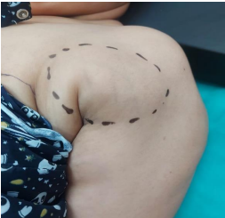
Figure 1 : left supernumerary breast
The rest of the physical exam was unremarkable. An ultrasound was performed and revealed ectopic glandular tissue surrounded by fatty tissue in the left axillary fossa without any detectable nodular or cystic lesion (figure 2).
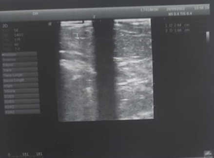
Figure 2: ultrasound picture suspecting Supernumerary Breast
Surgical excision was performed (figure 3), and the anatomopathological study confirmed the presence of a supernumerary breast with no evidence of tumor cells. The patient had an uneventful postoperative recovery.
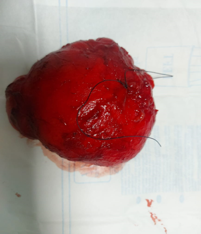
Figure 3: Resected Specimens
Supernumerary breast, also known as polymastia or accessory breast, is a rare congenital condition characterized by the development of one or more additional breasts along the milk line [3]. This condition occurs in approximately 1-5% of the population, with the highest prevalence in females [1]. It’s generally considered to be benign, but it can sometimes cause discomfort or embarrassment for the patient. Furthermore, axillary supernumerary breast tissue can be confused with other benign or malignant conditions, such as lipomas or cysts, making diagnosis and management challenging [2].
The etiology of supernumerary breast is not fully understood, but it is believed to be caused by the persistence of embryonic mammary ridges, which give rise to breast tissue during fetal development [4].
The diagnosis of supernumerary breast is made through physical examination and imaging studies, such as mammography, ultrasound, and magnetic resonance imaging (MRI) [5]. In some cases, a biopsy may be necessary to confirm the diagnosis [6]. Once a diagnosis has been made, the management of axillary supernumerary breast tissue depends on the symptoms and clinical presentation of the patient. If the condition is causing discomfort or cosmetic concerns, surgical excision may be considered [5].
Although axillary supernumerary breast tissue is a rare condition, it is important for clinicians to be aware of its existence and the potential for misdiagnosis. Misdiagnosis can lead to unnecessary procedures and delayed treatment. Therefore, accurate diagnosis and appropriate management of axillary supernumerary breast tissue can improve patient outcomes and avoid unnecessary interventions.
Further research is needed to better understand the prevalence and clinical significance of axillary supernumerary breast tissue. Studies have shown that the condition can be difficult to diagnose and may be misdiagnosed as other benign or malignant conditions [7]. More research is needed to improve diagnostic accuracy and develop evidence-based management guidelines.
Axillary supernumerary breast tissue is a rare condition that can present diagnostic challenges. However, with appropriate diagnosis and management, patients can be effectively treated and their symptoms alleviated. Healthcare providers should be aware of this condition and consider it in the differential diagnosis of patients presenting with axillary masses.
Dear Editorial Team, Clinical Medical Reviews and Reports. My experience with the journal was highly positive. The peer-review process was rigorous, constructive, and completed in a timely manner. The reviewers provided valuable comments that helped improve the quality and clarity of our manuscript. The editorial office was professional, responsive, and supportive throughout all stages of the publication process. Communication was clear and efficient, and any questions were addressed promptly. Overall, I found the journal to maintain high scientific standards and an excellent publication workflow. I would be pleased to consider submitting future work to this journal. Best wishes from, Elena Popa.

It was my pleasure to submit my testimonial concerning the Reviewer Board of our Scientific Journal “Brain and Neurological Disorders”. The Reviewers focused on some modifications and their contribution was helpful. The ladies of our Editorial Office were also supported my efforts. It was my honor to have such a co-operation and I am looking forward for more collaboration.

Dear Grace Pierce, Editorial Coordinator of Journal of Clinical Research and Reports, Thank you for the speedy and efficient peer review process. I appreciate the fact that your peer reviewers do not take months to respond like with some other journals. I would also like to thank the editorial office for responding quickly to my questions. It is an excellent journal. I plan to submit more manuscripts in the future. Best wishes from, Robert W. McGee

Dear Grace Pierce, Editorial Coordinator of Journal of Clinical Research and Reports, Working with you and your team on our recent publication in JCRR has been a truly wonderful and enjoyable experience. The responses were prompt, and the reviewers were patient, constructive, and highly professional. One reviewer in particular gave me the feeling that a professor was carefully reading and commenting on my coursework, which was deeply touching. The entire process was straightforward and hassle‑free, with no tedious online forms to complete. I highly recommend this journal. Best wishes from, DR Aibing Rao, Head of R&D

I Appreciate the Opportunity to Share my Experience with the Journal of Clinical Research and Reports. The peer review process was timely and constructive, and the feedback provided helped improve the quality of our manuscript. The editorial office was professional, responsive, and supportive throughout the process, ensuring smooth communication and efficient handling of the submission. Overall, it was a positive experience collaborating with your team.

Dear Mercy Grace, Editorial Coordinator of Obstetrics Gynecology and Reproductive Sciences, We would like to express our gratitude for your help at all stages of publishing and editing the article. The editors of the magazine answer all the necessary questions and help at every stage. We will definitely continue to cooperate and publish other works in the Obstetrics Gynecology and Reproductive Sciences! Best wishes from, Alla Konstantinovna Politova,
