
Case Report | DOI: https://doi.org/10.31579/2693-4779/200
1Department of Maxillofacial SurgeryHospital of Specialities Rabat, Morocco.
2Department of Otolaryngology and neck surgeryHospital of Specialities Rabat, Morocco.
3Faculty of Medicine and Pharmacy of Rabat. MohammedV University in Rabat, Rabat, Morocco.
*Corresponding Author: Rajaa El Azzouzi, Department of Maxillofacial Surgery Hospital of Specialities; CHU Ibn Sina, Av. Abderrahim Bouabid, Rabat-Morocco. BP 1382 RP. 10001 Rabat, Maroc
Citation: Rajaa El Azzouzi, Kawtar Ayyad, Bouchra Dani, Malik Boulaadas, Houssayni L. Essakalli, (2024), Rare Tumor of Oral Cavity: soft Palate Hemangioma: Case Report and Literature Review, Clinical Research and Clinical Trials, 9(5); DOI:10.31579/2693-4779/200
Copyright: © 2024, Rajaa El Azzouzi. This is an open access article distributed under the Creative Commons Attribution License, which permits unrestricted use, distribution, and reproduction in any medium, provided the original work is properly cited.
Received: 27 March 2024 | Accepted: 02 April 2024 | Published: 08 April 2024
Keywords: case report; hemangioma; soft palate; oral cavity tumors
Hemangiomasare benign tumors of bloodvessel characterized by hyperplasia of venous and capillary structures embedded in the submucosal connective tissue. Most hemangiomas are observed in the head and neck region; however, the oral cavity is a rare location and palatal hemangiomas are uncommon with an occurrence of less than 3% of oral hemangiomas.
We report a rare location of hemangioma on the soft palate in a 25-year-old man. The lesion was diagnosed by histopathology after a biopsy and the patient underwent a total surgical excision. And by a literature review we will describe the clinical, histological, therapeutic features and the prognosis of this rare tumor.
Hemangioma is a benign tumor of vaso-formative origin that is characterized by abnormal expansion of veins and capillaries settled in the submucosal connective tissue. Commonly seen in a younger population and represent the most common head and neck neoplasm at the pediatric age. Generally, 60%-70% of hemangiomas are observed in the head and neck; however, the oral cavity is still an uncommon location [1]. They may be observed in lips, buccal mucosa and tongue but the hard, soft palates and uvula are rarely affected. There occurrence on the palatal mucosa is extremely rare. The present case report is an extremely rare site of hemangioma involving the soft palate.
We report the case of a 25-year-old patient, with psychomotor retardation and no other medical history, admitted to our department for management of a soft palate mass, discovered 3 years ago by the family, which has since, progressively increased in volume with dysphagia, phonation disorders and no associated dyspnea. On clinical examination, the mass was soft, painless, with no-thrilling and normal-looking mucosal covering, no bleeding on contact, measuring approximately 4 cm long (Figure 1).
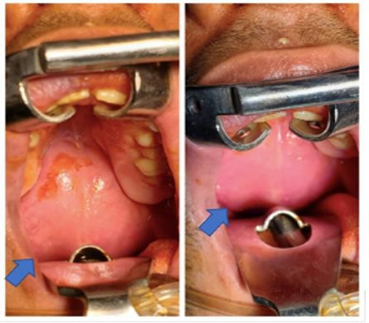
Figure 1: Intraoral view of the soft palate mass.
Radiological examination was carried out: Facial MRI: Massof the soft palate, regular and homogeneous, discreetly hyperintense in T1 and T2, with moderate contrast in late arterial time without consequent on venous return, partially obstructing the oro-pharyngeal tract (Figure 2)
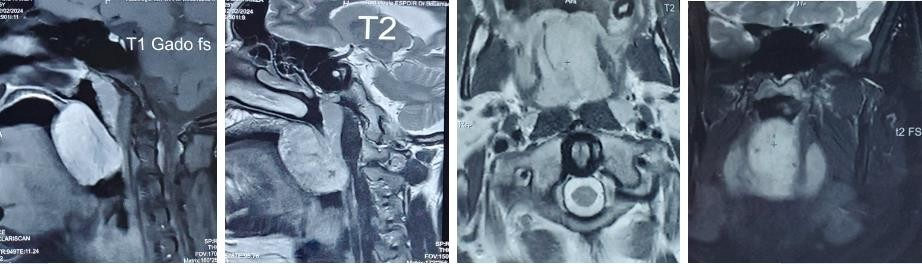
Figure 2: IMR in axial coronal and sagittal sectionsshowing the soft palate hemangioma
Cranio-facial CT: Soft palatemass measuring 30 x 30 x 54 mm, well limited, with no associated bone lysis (Figure 3).
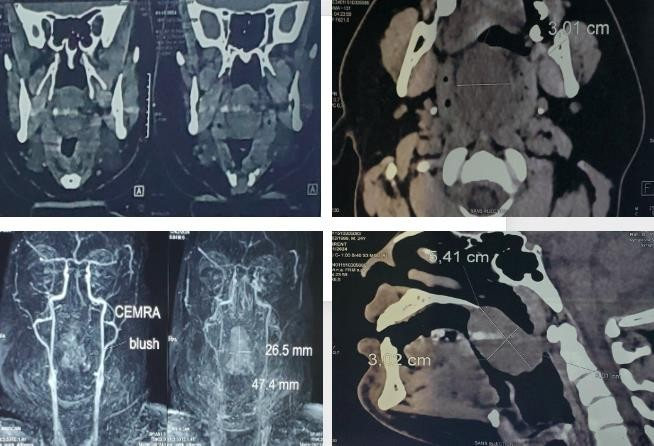
Figure 3: Cervico-facial CT scan in coronal, axial, sagittal sectionshowing the soft palate hemangiom
A biopsy was first performed, anatomopathological study revealed a velar hemangioma diagnosis; the patient subsequently underwent a complete removal of the mass under general anesthesia (Figure 4), the final anatomopathological result confirmed the diagnosis of soft palate hemangioma.
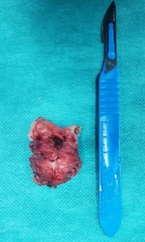
Figure 4: Macroscopic appearance of soft palatehemangioma.
Hemangioma is a benign tumor of vaso-formative origin that is characterized by abnormal expansion of veins and capillaries settled in the submucosal connective tissue with no malignant potential. Hemangioma is further sub classified based on their histological appearance as: cavernous lesions, capillary lesions and mixed lesions. A sclerosing variety also occurs that tends to undergo spontaneous fibrosis.
They are considered to be the most common tumor of childhood, occurring in about 5–10% of children <1>40 years of age. Generally, 60%-70% of hemangiomas are observed in the head and neck [1]. The lips, buccal mucosa and tongue are the most common sites of occurrence, while the hard and soft palates and uvula are rarely affected. Palatal hemangioma is uncommon, with fewer than 3% of the cases occurring in the site [4]. In the oral cavity they may occur in any area, at any age, without any racial or gender predilection.
Clinical examination reveals a well circumscribed, smooth, or lobulated blue red colored, painless and compressible swelling that refill slowly after releasing the compression, of variable volume, that can even cause airway obstruction and may bleed profusely in some cases, spontaneously or following trauma. [5]. Usually, congenital. These growths may not manifest themselves for years and spontaneous remission is unlikely during adulthood.
Radiographic imaging as CT scan and MRI are useful for analyzing exact features of the lesion, such as the size, location, extension, and relation with surrounding vital structures. They are also used for differential diagnosis. The varied appearance of soft palate hemangioma, mimic other lesions such as pyogenic granuloma, epulis granulomatosa, telegenctesia, angiosarcoma or squamous cell carcinoma [3,6] histopathological examination is important for a final diagnosis.
MRI is superior to CT for evaluating soft tissue masses. In MRI, these tumors typically show low-signal intensity on T1-weighted images and high-signal intensity on T2-weighted images. The angiography shows hemangioma as well-circumscribed lesions with intense tissue staining and demonstrates afferent and efferent vascular supply.
Management of hemangioma depends on several factors, the size, location, extent, clinical characteristics of the lesion, the age and patient oriented issues. Most of the hemangiomas are managed conservatively and require no intervention; The watch and wait policy.
However, 10–20% requires treatment. The range of treatment includes intralesional or systemic corticosteroid therapies, surgical excision, immunomodulatory therapies, embolization techniques and electrocoagulation while cryosurgery are the most popular, sclerotherapy is favored because of its efficiency and ability to secure the surrounding structures [7]. Indications for surgery are no evidence for involution, very large tumors with involvement of the adjacent structures, symptomatic (hemorrhage and infection) tumors, or cosmetic risk [7]. In the present case, surgery was carried out on the basis of size, location, the tumor's evolutionary profile, its extension and the association of dysphagia and phonation disorders.
Hemangiomas are benign tumors of blood vessel, most frequently observed in the head and neck region, however the soft palate localization is very rare and can be confused with other diagnosis which require a careful clinical and radiological evaluation. Several treatment modalities exist, that should be patient specific and depend on the tumor features, In the case of surgery option, an adequate measure to control bleeding should be encouraged.
Dear Editorial Team, Clinical Medical Reviews and Reports. My experience with the journal was highly positive. The peer-review process was rigorous, constructive, and completed in a timely manner. The reviewers provided valuable comments that helped improve the quality and clarity of our manuscript. The editorial office was professional, responsive, and supportive throughout all stages of the publication process. Communication was clear and efficient, and any questions were addressed promptly. Overall, I found the journal to maintain high scientific standards and an excellent publication workflow. I would be pleased to consider submitting future work to this journal. Best wishes from, Elena Popa.

It was my pleasure to submit my testimonial concerning the Reviewer Board of our Scientific Journal “Brain and Neurological Disorders”. The Reviewers focused on some modifications and their contribution was helpful. The ladies of our Editorial Office were also supported my efforts. It was my honor to have such a co-operation and I am looking forward for more collaboration.

Dear Grace Pierce, Editorial Coordinator of Journal of Clinical Research and Reports, Thank you for the speedy and efficient peer review process. I appreciate the fact that your peer reviewers do not take months to respond like with some other journals. I would also like to thank the editorial office for responding quickly to my questions. It is an excellent journal. I plan to submit more manuscripts in the future. Best wishes from, Robert W. McGee

Dear Grace Pierce, Editorial Coordinator of Journal of Clinical Research and Reports, Working with you and your team on our recent publication in JCRR has been a truly wonderful and enjoyable experience. The responses were prompt, and the reviewers were patient, constructive, and highly professional. One reviewer in particular gave me the feeling that a professor was carefully reading and commenting on my coursework, which was deeply touching. The entire process was straightforward and hassle‑free, with no tedious online forms to complete. I highly recommend this journal. Best wishes from, DR Aibing Rao, Head of R&D

I Appreciate the Opportunity to Share my Experience with the Journal of Clinical Research and Reports. The peer review process was timely and constructive, and the feedback provided helped improve the quality of our manuscript. The editorial office was professional, responsive, and supportive throughout the process, ensuring smooth communication and efficient handling of the submission. Overall, it was a positive experience collaborating with your team.

Dear Mercy Grace, Editorial Coordinator of Obstetrics Gynecology and Reproductive Sciences, We would like to express our gratitude for your help at all stages of publishing and editing the article. The editors of the magazine answer all the necessary questions and help at every stage. We will definitely continue to cooperate and publish other works in the Obstetrics Gynecology and Reproductive Sciences! Best wishes from, Alla Konstantinovna Politova,
