
Research Article | DOI: https://doi.org/10.31579/2642-9756/171
1 National Institute of Cancerology. Bogota, DC, Colombia.
2 ECCI University. Bogota, DC, Colombia.
*Corresponding Author: Devi Nereida Puerto-Jiménez, National Institute of Cancerology. Bogota, DC, Colombia.
Citation: Harley A. Martínez, Alexandra P. Rodríguez, Andrés C. Sevilla-Moreno, Emeterio C. Salazar, Puerto-Jiménez DN. (2023), Levels of Diagnostic Reference in Radiography Teams Dental in Bogota, Colombia, J. Women Health Care and Issues. 6(7); DOI:10.31579/2642-9756/171
Copyright: © 2023, Devi Nereida Puerto-Jiménez. This is an open access article distributed under the Creative Commons Attribution License, which permits unrestricted use, distribution, and reproduction in any medium, provided the original work is properly cited.
Received: 13 November 2023 | Accepted: 30 November 2023 | Published: 14 December 2023
Keywords: values of reference; radiography dental; protection radiology; optimization
Introduction: the ace radiographs dental her una of the ace exhibitions medicines more frequent has there radiation ionizing. The use of ionizing radiation is associated with a probable risk of adverse biological effects and possible harm to the patient's health. To avoid that patients receive unnecessarily high doses during this time exhibitions, there Composition International of Protection Radiológica recommended there use of the bone levels reference para diagnostic, like una herramienta effective of ayuda has there optimization of there protection radiology of patients. Purpose: to estimate the reference levels for diagnostic intraoral and panoramic dental radiography in the city of Bogotá, DC Methodology: to evaluate the radiographic exposure parameters of the teams and the image quality of 68 periapical dental radiography teams and 23 dental radiography teams panoramic radiography. Estimate the dosimetric magnitudes of incident kerma in area (K a,i ) in intraoral equipment for the radiography of an adult's maxillary molar and the producto kerma aire-area (P KA ) in panoramic radiography equipment in A exam of A adult standard.
Results: el tercer cuartil of there distribution of kerma incident in area para radiography intraoral fire of 3.3 mGy y of the product kerma aire-area, para radiography panoramic view of 103.9 mGy·cm 2. In the frequency distribution of incident heat in the area for intraoral radiography, the highest frequency of equipment is in the range of 2.0–3.0 mGy. In the frequency distribution of the product range-area for the panoramic radiography equipment, the highest percentage of equipment is in the range of 60 to 80 mGy cm 2.
Discuss: the institutions considered to establish diagnostic reference levels in this studio have an adequate quality of the image evaluated with a dental clinician, pero the ace variations in the ace dose of radiation between institutions señalan there necessary of implement herramientas que contributoryan has there optimization of the ace prácticas.
Conclusions: se recommended user the bone values of the bone levels of reference for diagnosis encountered in this investigation to optimize the radiological protection in the radiological dental exposures, and we hope that this studio will be a basic one for new investigations in the next cities of the country.
Dental radiographs are fundamental for the diagnosis of oral diseases, planning and supervision of dental treatments. Informs the Scientific Committee of the National Unions for the study of the boneeffects of thereradiation ionizing classifies dental radiography as one of the most frequent radiological procedures [1,2] . The dose of radiation received during a radiological dental examination is relatively lower in comparison with other radiographic techniques, and is generally lower than the natural radiation received by a person during One day, however, exposure to ionizing radiation is associated with a probable risk of adverse biological effects and possible harm to the health of the patient [1,3,4] . The International Radio
Protection Commission (ICRP in English) endorses the linear model sin umbral (LNT of the English). Este model asume that, in the range of low ionizing radiation doses, any dose different has cero increase el riesgo of induction of cancer o effects geneticos heredables 5. The ICRP sostiene that el model LNT are el more cautious to use in relationship idiot el uso seguro of the ace ionizing radiations 6. Lo mismo señala el Council National Radiological Protection and Mediciones of United States of America (NCRP of the English). El NCRP, Read a critical review of recent epidemiological studies of populations exposed to ionizing radiation, concluding that the majority of these studies are relevant evidence that apoya el uso of the model LNT in protection radiológica 7. Por esta razón, are necessary that health services adopt formal radiological protection methods that can log the maximum benefit possible idiot el minimo riesgo para el patient. In knew mayoria, the ace pathologies secundarias asociadas has Radiation exposure may decrease with adequate preventive and protective intervention [8.9].
Acknowledgment of the recommendations of the ICRP, no Dosage limits and restrictions are recommended for individual patients, as this may reduce the effectiveness of the diagnosis and is more harmful than that. beneficio. Por consistent, el enfasis se debe focus on the justification of radiological examinations, the optimization of protection and the use of levels of Reference para Diagnostic (DRLs, por sus siglas in English) 5. The DRLs are used in the clinical diagnosis to indicate whether in routine conditions the dose levels are paced in a specific image procedure, it is exceptionally high or low for this procedure, considering that the quality the image has not been compromised 10. DRLs do not apply to an individual or to a population group. Various studies have demonstrated that DRLs have an effective herramienta that helps optimize radiological protection in medical exposure of patients for diagnosis and intervention procedures 11. Sin embargo, su application en Colombia are recent y knew determination are A required to obtain the license to use ionizing radiation generators for toma and dental radiography interpretation services from Colombia 12.
In publication No. 135 13, the ICRP defined a magnitude DRL like una magnificence easily medible y that allowed evaluate there cantidad of radiation Used to carry out a concrete clinical test. The ICRP recommends the cantidates to be used as DRLs in different image modalities: for intraoral dental radiography equipment the recommended cantidad. particular in the ace modalities of radiography intraoral and panoramic sound relatively independent of the patient's size. The intraoral dental equipment has its voltage and current intensity and has an adjustable timer. Previously, the best option is to keep the medidos values during the temperature control of the K a, i at the safety of the separator cone to the intraoral equipment and the P KA Medido a la salida del tubo of the bone team of radiography panorama, like the value típico of cada team. There ICRP stablece el Local DRL for dental radiography as the third quarter the distribution of typical dose values from each institution. This calculation form can be useful in identifying the units of X-rays which require more attention for optimization.
The object of this studio is to evaluate the exposure parameters by dental radiography teams and determine the bone DRLs local para there city of Bogota in exams para pacientes adults in team intraoral and panoramic radiography. In actual literature no hay postpone of DRLs in there mayoria of the ace X-ray imaging methods in Colombia, for this reason, This investigation represents a basis for comparison The usual clinical practice in the institutions that provide dental radiography services with international DRLs is established.
Materials methods
There city of Bogota cuenta approximately con el 22 % of the ace institutions of radiography dental of are el kerma incident in area (K ) y para radiography Colombia, between the ace that se encuentran clinics dental, have dental panorámica el product kerma aire-area ( PKA). El consultorios odontologists y centros of radiology oral.
“DRL typical value” refers to the DRL of a dental center that tengan una o miscellaneous salads of rays X. In this case the DRL is calculated using the median of the selected magnitude. The DRL of the dental centers of una locality o una city se denomina “value local of DRL”; of multiple installations in todo el country se llama “value national of DRL” y of multiple countries in one region of the world are called “regional value of DRL”, using the value of the median of the bone values national available). In These cases are stable at the 75th percentile of the distribution of las medianas of there magnificence DRL elegida. The bone DRLs define para the bone different tips of team y examinations in groups of patients of acuerdo with the range of edad y fundamentalmente el peso. Sin embargo, all parameters of exhibition in radiology dental y in The study was carried out between 2016 and 2018 and included the voluntary participation of 68 institutions, among which 101 intraoral and panoramic radiography teams were evaluated. Each team will carry out one evaluation of calidad that included the ace kilovoltage meters pico del tube de x-rays (kVp), capa hemirreductora (HVL), tiempo of exhibition, calidad of imagen y there medicine of K a, i para radiography periapical y P KA para radiography panorámica. In el calculation solo se will include the bone team cuyas radiographs tenian a sufficient image quality for diagnosis, the fuel evaluated with a maniquí TOR DEN (Leeds Test Objects, USA), Figure 1 (derecha). Por end, solo fuiron considered 91 unidades, 68 team intraoral and 23 panoramic radiographs.
For the medidas of the exposure parameters to the salt of the spacer for intraoral equipment and on the front of the image receptor for panoramic radiography equipment, Figure 1 (izquierda) y Figura 2 (derecha), se used el sensor R/F of the Raysafe X2 (Fluke Biomedical, USA). Para the ace medidas of the K a, i In intraoral equipment we use the R/F sensor and to measure the P KA in panoramic radiography equipment we use the Vacup DAP ionization camera (Vacudec, Germany) colocada has there salida of the colimador of the tube X-rays intercept all hazards, Figura 2 (izquierda). esta metodología se used porque no requires that a patient was present during the medication and was based on technical report No. 457 of the International Organization of Energy Atómica (IAEA, por sus siglas in English) 14. (mA) values, select the exposure time for an adult's tooth from the information given by the dentist or the manufacturer's configured time for the equipment. For the case of the team panoramic radiography depends on the configuration of parameters of exhibition of A patient adult standard.
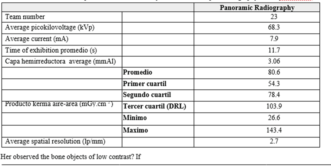
The quality of the image will be considered acceptable if the resolution space era of al menos 5 pares of linea per milímetro (lp/mm) for intraoral radiographs and 2 lp/mm para radiographs panoramas. Asimismo, todas the basic structures contrast with a maniquí TOR DEN fireon observed. The statistical analysis is carried out with the data of the dosimetric magnitude of the medida in the bone team para the bone procedures especificados. The values minimum, medios, maximums, of the primer (Q 1 ), second ( Q2 ) y tercer cuartil ( Q3 ) se calcularon para las dos modalidades de imagen dental.
The results of the exposure parameters and the quartiles of the distribution of the dose medications in the intraoral teams are presented in Table 1 . El Q3 of the K a, i in the bone team digital (CR/DR) are of 2.55 mGy y 4.84 mGy para the bone team analogues of movie type E (fast movie). The Graphic 1 show the distribution of frequencies K a, i spans of A histogram for intraoral equipment. The highest percentage of teams está in el rango of 2.0 – 3.0 mGy. El 50 % of K values is between 1.59 and 3.3 mGy. The distribution tends to be positive, the data is concentrated in there leaves inferior of there distribution. El

Figure 1. Montajes experimental para the ace medidas of parameters of exhibition (quierda) y calidad of imagen in 25 % of the bone values of K have more violas estan more dispersed team intraoral (derecha).
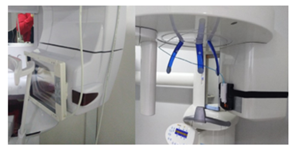
Figure 2: Experimental settings for the product medidas kerma aire-area, PKA, (izquierda) and exposure parameters in panoramic radiography equipment (derecha).
Para las medidas en los intraoral equipos, los cuales tienen normally there tension (kVp) y there corriente that el 25 % of the bone values more bajos. Hay very team intraoral that presentan values atípicos mayores at 6 mGy. The DRL for intraoral, analog and digital radiography equipment in the city of Bogotá is 3.3 mGy.
The results of the exposure parameters and the quartiles of the distribution
of the dose medications in the panoramic radiography teams are presented in Table 2. The Graphic 2 shows the frequency distribution of the P KA a histogram for panoramic radiography teams. The highest percentage of equipment is in the range of 60 to 80 mGy·cm 2 . The bone values of the P KA varian between 26.6 y 143.4 mGy cm. For the 23 panoramic radiography equipment included in the analysis, the Q3 of the P KA es of 103.9 m Gy·cm 2.

has. CR: digitalized.
b. TL;DR: digital directly.
vs. lp/mm: pares of lines por milimeter.
d DRL: levels of reference diagnostics, por sus siglas in English.
Table 1: Results of the boneparameters of exhibition y estimate of dose para radiography dental intraoral para el molar maxilar of a standard adult.
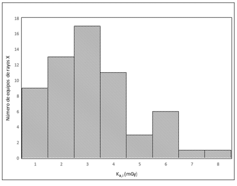
Graphic 1. Histograma of there distribution of frequencies of kerma incident in area in radiography intraoral.
has lp/mm: pares of linespor milimeter.
b DRL: levelsof reference diagnostics, por sus siglas in English.
Table 2: Results of the boneparameters of exhibition y estimate of dose para radiography dental panorámica.
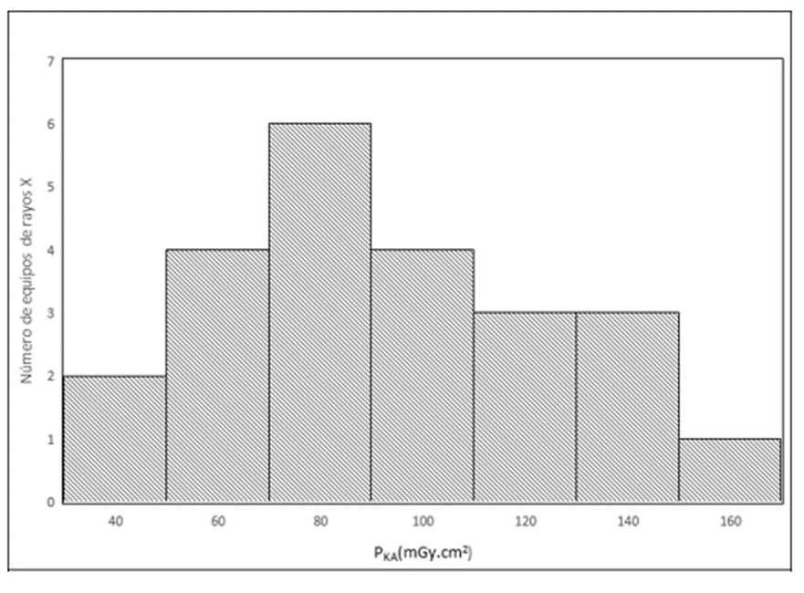
Graphic 2. Histograma of there distribution of frequencies of theproduct kerma aire-areain radiography panorámica.
El number of institutions that presta dental radiology services in Colombia, registered with the Registry Special of Providers of Services of Health (REPS), is approximately 3700 15 . Sin embargo, en Colombia do not report DRLs for practical purposes of radiology odontology, neither has level national nor local. There estimation of the bone DRLs THE permit has the ace
Odontological institutions compare the dose that receives groups of patients from different localities, cities or countries, identify the groups with the dose that is received systematically by the start or by the reference value, take into account the causes and apply corrective medications. For this reason, the determination of diagnostic reference levels is a necessity in the country because it represents the most important guide para optimize the ace dose in studios of radiography dental, apoyada in there mejora continued of the quality of the images.
Para the bone team intraoral, there Table 1 muestra that the exposure of patients to ionizing radiation is 90 % higher in institutions that have analog equipment, compared with those that have digital equipment (CR/DR). Average exposure time for intraoral analogue equipment is 68 % higher than in digital intraoral equipment. For equipment with nominal kVp less than igual to 70 kV, the HVL that is mida should be greater or igual to 1.5 mm of aluminum 16 , the intraoral, digital and analog equipments are in this condition. The HVL of digital intraoral equipment is 6 % greater than the los analogos.
The distribution of doses among the different teams, mostradas in the ace graphics 1 y 2, muestran that exists variations substantial in there practical, between the ace institutions of there city, para el mismo tipo of exploration diagnosis. There variability of the results between the different participating institutions may be related to the type of image processing technology (film or digital), idiot el tiempo of life useful of the team (year of manufacture), with the different models and manufacturers of the equipment and with the exposure parameters used in each institution, among other factors.
So it is accepted that the institutions considered to establish the diagnostic reference levels in este studio, contaron idiot una adequate quality of the image evaluated with a ghost dental, variations in radiation dose señalan there necessary of implement herramientas that contribute to the optimization of practices. This includes the implementation of periodic quality control procedures for our teams, an adequate maintenance program, and data management auditors. there dose of radiation, el uso of técnicas of exhibition
adecuadas has the bone different tamaños of the bone patients and a continued capacity of the personal involucrado in the practice. It's important to be rewarded by many institutions the bone parameters of exhibition se maintain the bones, independent of the patient's anatomy. For example, use the same exposure times in an adult patient than in a pediatric patient.
As for the results in other international studies, Tabla 3, the evolution of dosimetric magnitudes shows a decrease with time of the bone levels of reference para diagnostic (no its statics) for the types of dental radiography modalities. The diagnostic reference levels obtained in Bogotá in this studio, compared with the results at an international level, show that intraoral radiography is the result of the most recent results obtained in countries like this Spain, Emirates Arabs, Reino Unido e Ireland. In the case of panoramic radiography, we see that there is a major fluctuation of the reference levels for diagnostics depends on the country, because of its value obtained in este studio are comparable idiot The countries of India, South and Republic of Korea. The value encountered by the DRL for intraoral dental radiography is found in the range recommended by the European countries in 2014 (5 – 7 mGy) 17 . Sin embargo, se debe hold cuidado al compare of will direct the results of this studio to other countries, ya that the bone studios her affected por differences in the size of the picture, type of projection and dosage, technical exposure parameters such as kVp or exposure time, the parameters in there we realized the studies, among others.
All the previous reasons require you to download the programs of protection radiology in there dental radiology practice, provided for individual justification of the bone studios y there optimization of dose form _ that se getgan images idiot there calidad necessary for the diagnosis and with the lowest possible dose.
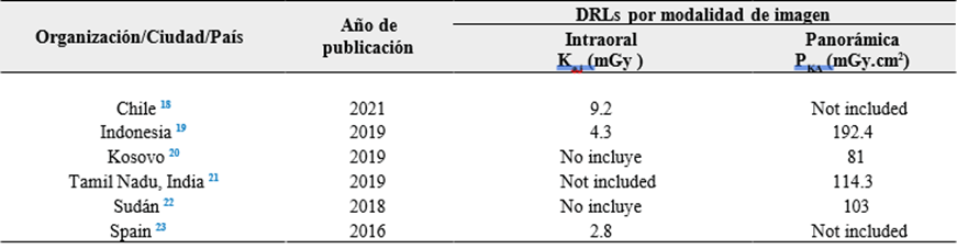
Table 3: Niveles of reference para diagnostic (tercer cuartil of the bone values of kerma in area in there area of entrance) para
dental radiography in adults, include the literature published and for this studio.

In this study a first estimate of the losses was made levels of reference para diagnostic in dental radiography for the city of Bogota obtained from the tercer cuartil in una muestra of 68 team intraoral y 23 of radiography panorámica. The bone results get suggestions that el level of reference of diagnostic are of 3.3 mGy for the incident kerma in the area for a higher molar of A adult y 103.9 mGy.cm 2 para A standard panoramic radiography exam. Estos valores pueden ser used por the bone providers of the service of radiology dental like una guide para there practical clinica. If in practice the values are presented at the beginning or significantly below these values, you must have a revision at the end of checking the conditions particularities of there practical that contribute to these dose values and implement optimization strategies.
Please note that in the country we do not know the reference levels for diagnostics in any type of exam o procedure diagnostic idiot team rays _ X, este studio pretend serve of base para new studies in two cities of the country that allow you to drive has there optimization of dose in the bone pacientes y to create a reference mark for practice. Of the same form, before the increment of conical tomography units computed from conical radiography to dental radiography, the country requires the establishment of reference levels for diagnosis for this image mode and updating of periodical format for different radiodiagnostic examinations.
The results of this study can guide future investigations in this field and provide information on the radiation level in these procedures, with which we hope to promote a radioprotection culture and motivate the optimization of the recorded doses.
Agradecemos has there Vicerectoría of Investigations of the ECCI University for technical and financial sources. También agradecemos has the ace 68 health institutions that accept participation in the study.
This study is adjusted to international standards, particularly to the Helsinki Declaration and has established parameters for biomedical investigation defined by the Consejo de Organizaciones Internacionales de las Ciencias Médicas (CIOMS) and has stable parameters in the national ambito for the Resolution 8430 from 1993.
The bone autores declare no hold no conflict of interest.
Este studio fire financed idiot recursos of the Institute
National Cancer Center and ECCI University .
Dear Editorial Team, Clinical Medical Reviews and Reports. My experience with the journal was highly positive. The peer-review process was rigorous, constructive, and completed in a timely manner. The reviewers provided valuable comments that helped improve the quality and clarity of our manuscript. The editorial office was professional, responsive, and supportive throughout all stages of the publication process. Communication was clear and efficient, and any questions were addressed promptly. Overall, I found the journal to maintain high scientific standards and an excellent publication workflow. I would be pleased to consider submitting future work to this journal. Best wishes from, Elena Popa.

It was my pleasure to submit my testimonial concerning the Reviewer Board of our Scientific Journal “Brain and Neurological Disorders”. The Reviewers focused on some modifications and their contribution was helpful. The ladies of our Editorial Office were also supported my efforts. It was my honor to have such a co-operation and I am looking forward for more collaboration.

Dear Grace Pierce, Editorial Coordinator of Journal of Clinical Research and Reports, Thank you for the speedy and efficient peer review process. I appreciate the fact that your peer reviewers do not take months to respond like with some other journals. I would also like to thank the editorial office for responding quickly to my questions. It is an excellent journal. I plan to submit more manuscripts in the future. Best wishes from, Robert W. McGee

Dear Grace Pierce, Editorial Coordinator of Journal of Clinical Research and Reports, Working with you and your team on our recent publication in JCRR has been a truly wonderful and enjoyable experience. The responses were prompt, and the reviewers were patient, constructive, and highly professional. One reviewer in particular gave me the feeling that a professor was carefully reading and commenting on my coursework, which was deeply touching. The entire process was straightforward and hassle‑free, with no tedious online forms to complete. I highly recommend this journal. Best wishes from, DR Aibing Rao, Head of R&D

I Appreciate the Opportunity to Share my Experience with the Journal of Clinical Research and Reports. The peer review process was timely and constructive, and the feedback provided helped improve the quality of our manuscript. The editorial office was professional, responsive, and supportive throughout the process, ensuring smooth communication and efficient handling of the submission. Overall, it was a positive experience collaborating with your team.

Dear Mercy Grace, Editorial Coordinator of Obstetrics Gynecology and Reproductive Sciences, We would like to express our gratitude for your help at all stages of publishing and editing the article. The editors of the magazine answer all the necessary questions and help at every stage. We will definitely continue to cooperate and publish other works in the Obstetrics Gynecology and Reproductive Sciences! Best wishes from, Alla Konstantinovna Politova,
