AUCTORES
Globalize your Research

Research Article | DOI: https://doi.org/10.31579/CIC-2021/007
1 Department of Cardiology, Istanbul University Cerrahpasa, Institute of Cardiology, Istanbul, Turkey.
2 Laboratory of Clinical Microbiology, Istanbul University Cerrahpasa, Institute of Cardiology, Istanbul, Turkey.
*Corresponding Author: Nazmi Gultekin, Istanbul Universty-Cerrahpasa, Institute of Cardiology, Istanbul, Turkey. Tel: +90 212 4592000; Fax: +90 2124592069.
Citation: Anil Tanki, Nazmi Gultekin, and Emine Kucukates (2021). Evaluation of autophagy and microtubules inhibition through blood beclin-1 levels in subgroups with Heart Failure reduced Ejection Fraction. Clinical and Interventional Cardiology. 1(1); DOI: 10.31579/CIC-2021/007
Copyright: © 2021, Nazmi Gultekin. This is an open access article distributed under the Creative Commons Attribution License, which permits unrestricted use, distribution, and reproduction in any medium, provided the original work is properly cited.
Received: 02 October 2021 | Accepted: 25 October 2021 | Published: 06 November 2021
Keywords: heart failure; dilated cardiomyophathy; cell death; autophagy; beclin-1; microtubules
Background and Aim: In this study, we aimed to compare beclin-1, one of the factors and moderators of autophagy activity, in the serum of heart failure patients with low ejection fraction (HFrEF) with those in the serum of healthy individuals. Also, we investigated serum beclin-1 levels according to etiological classifications (ischemic/non-ischemic subgroups). Additionally, the subset of patients using colchicine as a microtubule inhibitor for at least three months due to HFrEF was included.
Methods: This study included 50 patients with HFrEF (25 with ischemic etiology, 25 with non-ischemic etiology) and 30 healthy subjects between January 2018 and December 2019 in Istanbul University Cardiology Institute. Serum beclin-1 levels were determined by using the ELISA method by the ELISA Kit.
Results: Although serum beclin-1 levels of all HFrEF group compared to the control group did not reach statistical significance, increased serum beclin-1 levels were found (p:0.64). However, NT-proBNP levels were found significantly higher (p:0.01) .Serum beclin-1 levels correlated with ejection fraction in 50 patient with HFrEF (p: 0.018, R²: 0.088). In the non-ischemic etiology subgroup with HFrEF especially had higher serum beclin-1 levels (p:0.01). There was also no significant correlation between creatinine and eGFR levels and autophagic activity (p:0.482). Also, we found lower levels of NT-proBNP that did not reach statistical significance and higher beclin-1 levels to reach statistical significance (p: 0.015 ) in the colchicine using patient subset.
Conclusıons: Beclin-1 levels especially increased in the HFrEF with non-ischemic etiology group. Low dose colchicine affects autophagy, microtubules inhibition, and vesicle trafficking in HFrEF.
The autophagic response has been described in various pathophysiological situations, including neurobiology, cancer, cardiovascular disease, and infectious diseases [1-3]. Membrane trafficking, i.e. endocytic/exocytic, endo-lysosomal and autophagic pathways are well studied. “New era of membrane trafficking diseases” as emerged and de-regulation of vesicle trafficking pathways were shown to be related to disease syndromes. However, their roles in diseases were not fully understood as they can act in different conditions to work towards cell survival or induce cell death [1,4].
As the mammalian ortholog of the yeast Atg6 gene, beclin-1 is an essential mediator of autophagy [5-10]. Beclin-1 forms a multimeric complex with vacuolar protein sorting 34 (Vps34) and class 3 phosphatidylinositol 3-kinase (PI3k), which is necessary for the formation of autophagosome. Component of various PI(3)K complexes beclin-1 interacts with PIK3C3/VPS34 to signal the onset of autophagy. Atg6/beclin-1 Inhibited by binding to Bcl2 [5-10] (Figure 1).
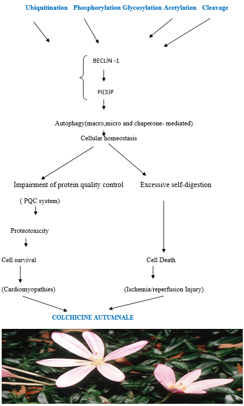
•Post-translational modifications of Beclin 1 affect protein stability, confirmation, activity, and its interactome and can be used as a molecular rheostat to fine-tune autophagic activity.
•Targeting Beclin 1 modifiers to regulate Beclin 1 post-translational modifications could provide a possible therapeutic intervention for upregulatin autophagy.
•Low dose colchicine accelerates the physiological clearance of misfolded proteins from cells through the protein quality control (PQC) system and cytoprotective autophagy and inhibits NLRP3) inflammasome, antigen presentation to T lymphocyte and the lysozymes of cardiomyocytes with acquired autoimmunity.
The mechanism of action of colchicine is through the inhibition of tubulin polymerization and potentially also through effects on cellular adhesion molecules and inflammatory chemokines. Colchicine may also have direct anti-inflammatory effects by inhibiting key inflammatory signaling networks known as the inflammasome and pro-inflammatory cytokines. Through the disruption of the cytoskeleton, colchicine is believed to suppress the secretion of cytokines and chemokines as well as in vitro platelet aggregation. Colchicine interferes with neutrophil adhesion and recruitment to inflamed tissue and antigen presentation to T lymphocyte and the lysozymes of cardiomyocytes with acquired autoimmunity.. Also, when misfolded proteins cannot be efficiently removed by degradative pathways. Misfolded proteins may accumulate and block the ubiquitin-proteasome system (UPS) and autophagy. Colchicine modulates regulation of the protein quality control (PQC) system via chaperone-mediated autophagy and post-translational modification and also by effective mitochondrial dynamics and homeostasis. These modifications have a role in multiple cellular functions, ranging from cell motility, cell cycle progression or cell differentiation to intracellular trafficking and signaling [1,10-12,] (Figure1).
Colchicine is a medication with a relatively low therapeutic index [13]. Because high doses, especially in muscle cells can lead to vacuolization and myopathy should be avoided [11,13]. Considerable researches have highlighted the potential of colchicine in the treatment of cardiovascular diseases mediated by pro-inflammatory processes [13-15]. The sustained benefit of short-term colchicine treatment on survival, cardiac function, and ventricular remodeling after myocardial infarction(MI )seems to be beneficial [13-15]. Whereas in previous studies, the colchicine treatment of Heart Failure with Reduced Ejection Fraction (HrEF) has been reported to be conflicting [16,17].
In this study, it was aimed to compare the serum beclin-1 levels which is one of the markers of autophagic activity in the patients with heart failure reduced ejection fraction (HFrEF), and those healthy subjects. Besides, in the study, variations of serum beclin-1 levels will be investigated in patients who were followed-up due to HFrEF according to etiological classification (ischemic/non-ischemic subgroup), and also in the colchicine-using subset.
Patient and public involvement statement
The patients and public were not involved in designing of, recruitment to, or conduct of the present study.
The sample size was calculated by the formula: n=Z121 α/2S2÷d2(n=80=1.962×(1-0.55/2)×0.502÷0.052)(N= Population Size; n=Sample Size; S= Standard Error;0.05 (5%) (Confidence Interval [CI] 95%); Confidence Interval: upper=95%; Lower: 5%; The Z-values for confidence levels were: 1.645=90 percent confidence level; 1.96= 95 percent confidence level; 2.576 = 99 percent confidence level (Z for p=0.05, 0.01, 0.001 are1.96, 2.58 and 3.28 Z values respectively); d= RelativeStandard Error) and it was confirmed by automatic sample size calculator [18].
Design and definitions
Our study was designed as a prospective cross-sectional observational. Based on ESC 2016 heart failure guidelines, symptoms and findings, biochemical and echocardiographic parameters, and etiology of the disease were evaluated. A total of 80 patients with 50 HFrEF (25 with ischemic etiology and 25 with non-ischemic etiology) and 30 healthy subjects were included in the study. Half of the HFrEF patients included in the study were selected from ischemic dilated cardiomyopathy patients with ischemic etiology, while the other half were selected from non-ischemic dilated cardiomyopathy patients without ischemic etiology [19].
For the control group, healthy individuals without heart failure with similar demographic characteristics were included in the study. In addition, low-dose colchicine (0.5-1 mg) was added to the treatment for at least three months in our 13 patients HFrEF with non-ischemic etiology for various reasons such as pericardial effusion accompanying HFrEF, and/or who developed hyperuricemia as a result of intensive diuretic therapy.
Exclusion criteria
Patients with the following characteristics were excluded from the study: under 18 old, being older than 80, having a history of hospitalization due to acute coronary syndrome in the last 30 days, end-stage renal disease patients, To have an active infection, having a life expectancy of less than 1 year (such as malignancies) due to noncardiac reasons, to have advanced valve disease.
Study population characteristics
The demographic characteristics of all patients included in the study and all individuals included in the control group; Age, gender was recorded in appropriate forms in the study forms, and symptom status was classified according to the NYHA symptom evaluation system. Comorbid diseases; hypertension, Diabetes Mellitus, hyperlipidemia, ischemic heart disease history were recorded in the study form. Heart rhythms (sinus rhythm / atrial fibrillation/battery rhythm), presence of the device (Pacemaker/ICD/CRT-D), and habits of the patients and control group; cigarettes, alcohol, and medications they used were questioned and written to the appropriate places in the study forms. In the study group and control group, the diagnosis of DM was based on at least one history of antidiabetic drug use or HbA1c level> 6.5%.
Recent biochemical tests of all patients and control subjects included total cholesterol, LDL cholesterol, HDL cholesterol, triglycerides, glucose, HbA1c, creatine, eGFR, total protein, albumin, ALT, AST, NT-ProBNP levels and the hemogram parameters (hemoglobin, leukocyte, neutrophil, monocyte, lymphocyte, basophil, platelet, MPV) were written in the appropriate places. No additional tests were performed to determine the necessary blood results of the patients. Glomerular filtration rates (GFR) of the patients were calculated using the MDRD formula. Also, echocardiography parameters of all patients and control groups were examined in detail. EF, left atrium (LA), left ventricular end-diastolic diameter (LVD), right ventricular diameter (RVD), septum thickness (IVS), segmenter or global where the study forms were recorded in the appropriate places (Table1-3).


Parameters B) NT-proBNP and Beclin-1 values of patients with heart failure according to ischemic (N: 25) and non-ischemic (N: 25) etiology included in the study(mean ± SD).,
Parameters C) Differences in NT-ProBNP and Beclin-1 Levels and echocardiographic parameters of colchicine treated(N:13) and non-colchicine treated patients in the HFrEF patient group (N: 37) included in the study(mean ± SD).
There was no significant correlation between creatinine and eGFR levels and beclin-1 (ATG6 analog in yeast cells), an autophagy marker in mammals (p: 0.482) ( Table 3 A). Additionally, in the subgroup analysis of the heart failure group, statistically significant serum beclin-1 and low NT-proBNP levels were found in the non-ischemic etiology group according to the ischemic etiology group (respectively p:0.01:p:0.01) (Table 3 B ). Also, we found lower levels of NT-proBNP that did not reach statistical significance and higher beclin-1 levels to reach statistical significance (p: 0.015 ) in colchicine- using patient group (Table 3 C). Moreover, EFs increased, LVDD decreased.
* Continuous variables are presented as mean ± SD and dichotomous variables as percentages. A two-tailed t-test was used for comparison of means, and an x2-test for percentages in statistical analysis at table1,2,3..
Plasma beclin-1 levels determination
The consent form was obtained from the patients and the control group at the time of admission. Blood samples were taken into 8-ml K-EDTA-tube with Monovette brand vacutainer and centrifuged at 3000 rpm for 10 minutes. Serum samples obtained were stored at -80 ° C for further study. After all the samples were completed, they were studied in the microbiology laboratory of Haseki Training and Research Hospital.
Plasma beclin-1 levels were determined using the human beclin-1 ELISA Kit (Bioassay Technology Laboratory). Each well was previously prepared according to the kit package insert. All reagents, standard solutions, and samples were prepared as instructed. 50μl Standard solution was added to a standard well, 40 μl sample to sample wells and then 10μl anti-BECN1 antibody to sample wells, subsequently 50 μl streptavidin-HRP to sample wells and Standard wells. The plate with a sealer was covered and incubated 60 minutes at 37°C. The plate was washed 5 times with wash buffer. For automated washing, someone was by aspirated all wells and wash by 5 times with wash buffer, overfilling wells with wash buffer. The plates onto paper towels or other absorbent material were blotted. 50 μl substrate solution A to each well and then add 50μl substrate solution B was added to each well and incubated by plate covered with a new sealer for 10 minutes at 37°C in the dark. Subsequently, 50 μl stop solution was added to each well, the blue color will change into yellow immediately. The optical density (OD value) of each well immediately using a microplate reader set to 450 nm within 10 minutes after adding the stop solution. The blank value (incubation buffer + stop solution), was subtracted from all other values obtained.
If the summarized; in serum samples, beclin-1 was measured immunoturbidimetrically by turbidity caused by the antigen-antibody complex formed by binding of monoclonal anti-BECN1 coated latex microparticles with beclin-1 in serum. The curve of the optical densities formed 10 minutes after the addition of the stopping solution was calculated with a compatible computer-based software system.
Statistical analysis was performed by using SPSS (Statistical Package for Social Science) for Windows 23. 0 program. The student's t-test was used when the parameters providing normal distribution conditions were compared according to two independent groups. In the comparison of categorical values, independence control was performed by using Chi-Square (χ2) analysis. Beclin-1 levels of healthy individuals, ischemic dilated cardiomyopathy and non-ischemic dilated cardiomyopathy patients were compared with the Mann-Whitney U test. Statistical significance was accepted as p≤0.05.
All data and statistical comparisons are shown in the legends of (Tables 1, 2, 3) and (Figure 1, 2).
Beclin-1, the mammalian orthologue of yeast Atg6, has a central role in autophagy, a process of programmed cell survival, which is increased during periods of cell stress and extinguished during the cell cycle. It interacts with several cofactors (Atg14L, UVRAG, Bif-1and survivin) to regulate the lipid kinase Vps-34 protein and promote the formation of beclin 1-Vps34-Vps15 core complexes, thereby inducing autophagy [10,20]. In contrast, the BH3 domain of belcin-1 is bound to and inhibited by Bcl-2 or Bcl-XL.This interaction can be disrupted by phosphorylation of Bcl -2 and belcin-1 or ubiquitination of belcin-1. Interestingly, caspase-mediated cleavage of belcin-1 promotes crosstalk between apoptosis and autophagy.Beclin-1 dysfunction has been implicated in many disorders, including cancer and neurodegeneration. IL-6 could inhibit starvation-induced autophagy by the STAT3/Bcl2/beclin-1 pathway in cells [1-8,10,20]. Beclin-1 and PIK3C3 are involved in the complex which signals the onset of autophagy and could be used to monitor autophagy [1,6-10](Figure 1).
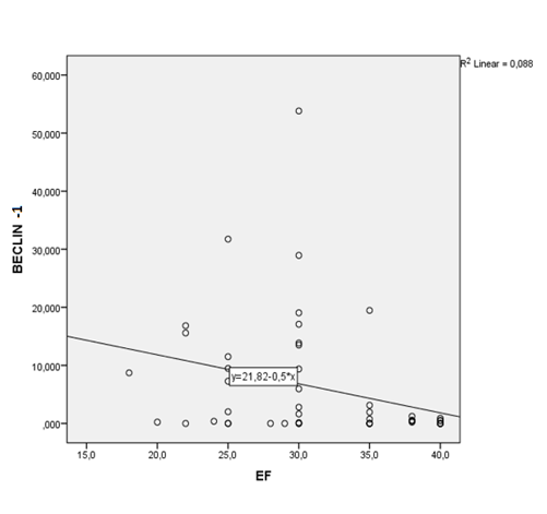
In our study, beclin-1 levels were higher in the HFrEF patient group compared to the control group, but not statistically significant (p:0.64); however, NT-proBNP levels were found significantly higher (p:0.01). When the relationship between ejection fraction (EF) and beclin-1 levels of HFrEF group in the study was examined, we found that patients with lower EF had correlated higher beclin-1 levels (p: 0.018, R²: 0.088) (Figure 2; Table 3 A).
In the observational and experimental studies to determine the relationship between heart failure and autophagy, increased autophagic activity was found in individuals with heart failure [21-25]. Although there are many molecular studies, biopsy and autopsy studies and animal experiments to determine the relationship between cardiovascular diseases and autophagy [4,21-25]. We could not find any study in the literature based on the determination of the level of autophagy markers in serum in patients with Heart Failure (HF).
Furthermore, in our study, higher creatinine and decreased eGFR levels were found in the heart failure group with low EF compared to the control group (p:0002;0,05). When HFrEF groups with ischemic and non-ischemic etiology were compared, renal functions were similar. There was also no significant correlation between creatinine, eGFR values and beclin-1levels (ATG6 analog in yeast cells), an autophagy marker in mammals in the patient groups (p: 0.482) (Table 3 B). This proves that other mechanisms such as dehydration, electrolyte imbalance, and hypotensive condition are responsible for the deterioration of renal function in patients with HFrEF rather than autophagy [26].
Also, in the subgroups analysis of the HFrEF group, serum beclin-1 levels were statistically significantly increased in non-ischemic etiologysubgroup but not ischemic subgroup. But, NT-proBNP levels were found low in the non-ischemic etiology subgroup according to the ischemic etiology subgroup (respectively, p:0.01:p:0.01) (Table 2,3 B ). We consider that serum beclin-1 levels were significantly found low in the patients with HFrEF subgroup with ischemic etiology, because of the ischemia /reperfusion process was completed and autophagic activity decreased in the scar tissue. Changes in autophagic flux are seen in essentially all forms of heart disease and all cell types. It can either be beneficial or aid to disease progression. Ischemic heart disease is characterized by lack of nutrients and oxygen to the myocardium [14,15]. Studies looking at models of ischemia have shown that autophagy functions as a pro-survival mechanism by adapting to changing metabolic needs and eliminate damaged mitochondria, which can release ROS and initiate apoptosis to prevent any tissue damage [27,28 ].Inhibition of autophagy can exacerbate cardiac dysfunction and remodeling [1,2,4,20]. Once oxygen and nutrients are restored, autophagy can either be adaptive or detrimental to the tissue. Some studies have shown that reperfusion following an ischemic attack leads to an increase in autophagy but an impairment of autophagosome clearance leading to cell death [13-15.] Also, recent studies have demonstrated that nucleotide-binding oligomerization domain-like receptors, pyrin domain -containing 3 (NLRP3) inflammasome, is associated with endogenous sterile inflammation after Myocardial infarction. NLRP3 is a cytosolic multiprotein complex activated by tissue danger signals and associated with the production of activated interleukin (IL)-1β and IL-18 [14,15]. Colchicine was reported to reduce inflammatory cytokines (including IL-1β and IL-18) and infarction size in MI [13,21]. Therefore, the sustained benefit of short-term colchicine treatment on survival, cardiac function, and ventricular remodeling after MI is to be beneficial [13].
Non-ischemic etiology with heart failure may also be associated with an increased vacuolization [14,15,25]. Because, high dose colchicine can lead to vacuolization and myopathy especially in muscle cells should be avoided [26,29]. Therefore, in our study low-dose colchicine (0.5-1 mg) exclusively was added to the treatment for at least three months in 13 patients with non-ischemic etiology with HFrEF for various reasons. In this subset, we found lower levels of NT-proBNP that did not reach statistical significance (p:0.69) However, statistical significance was reached in the beclin-1 levels of this subset (p:0.015) (Table 3 C). At baseline, low-dose colchicine was used in 13 patients with terminal non-ischemic cardiomyopathy with severe left ventricular dysfunction (p = 0.009) and an enlarged left ventricle (p = 0.002). After 3 months, these patients were detected low NT-proBNP levels and higher beclin-1 levels. Moreover, EFs increased, LVDD decreased. Actually, we did not expect these paradoxical results. Low levels of NT-proBNP and higher beclin-1 levels discordances suggests another mechanism that low dose colchicine probably demonstrates cytoprotective autophagic clearance and regeneration activities via chaperone-mediated autophagy [27,28]. Thus; colchicine effects regulation of the protein quality control (PQC), chaperone-mediated autophagy and post-translational modification and also by effective mitochondrial dynamics and homeostasis [1,11,27,28]. Therefore,increased autophagy in our cases subgroup using colchicine did not correspond to cell death, but to selective cytoprotective autophagic clearance and regeneration activities that enabled cell survival, and reduced autoimmunity of cardiomyocytes according to our opinion.
Frankly; colchicine accelerates misfolded protein are physiologically cleared from the cells by the protein quality control (PQC) system. The PQC system is composed of two main arms: Firstly the molecular chaperone, mainly represented by the heat shock proteins (HSPs), and secondly the degradative pathways, including the proteasome, the autophagic response and the unfolded protein response (UPR). A specific degradative pathway for a given misfolded protein is selected by a defined class of chaperones, with the assistance of co-chaperones. These pathways may determine whether misfolded proteins have to be refolded or degraded. So, the PQC system controls dynamic selective autophagy [1,11].
As is known, autophagic vacuoles have been described in the heart tissue of patients with idiopathic dilated cardiomyopathy [25]. Cardiac hypertrophy is characterized by an increase in cardiomyocyte size, disarrangement of sarcomeric structure, enhanced protein synthesis and re-expression of fetal genes [24]. Cardiac hypertrophy is accomplished by the dysfunction of numerous signaling pathways that are involved in autophagy. PI3K/AKT, GSK3, MAPKs, and mTOR pathways have all been associated with the regulation of cardiomyocyte autophagy [2-10,20]. The deficiency of the autophagy gene LAMP-2 was shown to cause Danon disease [25]. Autophagy acts as a defense mechanism against the of cardiomyocytes. Furthermore, the heart develops cardiac hypertrophy and diastolic dysfunction with aging when autophagy is inhibited [22]. Recent studies have shown that miRNAs play an important role in regulating cardiac autophagy and there is a growing number of studies targeting autophagy regulating miRNAs as a potential strategy in the treatment of cardiac disorders [23].
Under the light of the literature data by we have explained above, this increase in autophagic activity in our study can also be explained by low-dose colchicine’s induction or upregulation of macro and microautophagy and especially selective chaperone-mediated autophagy for the clearance and regeneration from dysfunctional cardiomyocytes which are thought to contribute to heart failure [1,13,28]. On the other hand, in contrast to the high dose, low dose colchicine probably cleans bad vacuolization in HFrEF cases with non-ischemic etiology [1,25]. According to our opinion, regulation of autophagy , miRNAs and vesicle trafficking pathways in microenvironment can either be a cell– protective mechanism or in fact, an alternative cell–death mechanism, given that in cardiomyocytes in heart failure the apoptosis pathway is commonly altered. In this respect, colchicine or similar drugs treatment needs to be tried in more extensive studies.
Limitations of the study were non-randomized prospective cross-sectional observational design, relatively small number of patients, and whether there was not investigated the correlation between serum beclin-1 levels and beclin-1 levels measured in cardiac biopsy materials.
Our study reveals that HFrEF had higher serum Beclin-1 levels, especially in the non-ischemic etiology subgroup with HFrEF because of higher autophagic activity. Low-dose colchicine (0.5-1 mg) probably induces selective and cytoprotective autophagic clearance and regeneration activities in HFrEF and seems to be manifestly beneficial. On the other hand, in contrast to the high dose, low dose colchicine probably cleans bad vacuolization in HFrEF cases with non-ischemic etiology.There was also no significant correlation between creatinine and eGFR levels and autophagic activity. Further study of the autophagic and vesicle trafficking pathways and miRNAs will lead to an increased understanding of the regulation of this critical homeostatic pathway and identify new therapeutic potentials for cardiac diseases.
Clearly Auctoresonline and particularly Psychology and Mental Health Care Journal is dedicated to improving health care services for individuals and populations. The editorial boards' ability to efficiently recognize and share the global importance of health literacy with a variety of stakeholders. Auctoresonline publishing platform can be used to facilitate of optimal client-based services and should be added to health care professionals' repertoire of evidence-based health care resources.

Journal of Clinical Cardiology and Cardiovascular Intervention The submission and review process was adequate. However I think that the publication total value should have been enlightened in early fases. Thank you for all.

Journal of Women Health Care and Issues By the present mail, I want to say thank to you and tour colleagues for facilitating my published article. Specially thank you for the peer review process, support from the editorial office. I appreciate positively the quality of your journal.

Journal of Clinical Research and Reports I would be very delighted to submit my testimonial regarding the reviewer board and the editorial office. The reviewer board were accurate and helpful regarding any modifications for my manuscript. And the editorial office were very helpful and supportive in contacting and monitoring with any update and offering help. It was my pleasure to contribute with your promising Journal and I am looking forward for more collaboration.

We would like to thank the Journal of Thoracic Disease and Cardiothoracic Surgery because of the services they provided us for our articles. The peer-review process was done in a very excellent time manner, and the opinions of the reviewers helped us to improve our manuscript further. The editorial office had an outstanding correspondence with us and guided us in many ways. During a hard time of the pandemic that is affecting every one of us tremendously, the editorial office helped us make everything easier for publishing scientific work. Hope for a more scientific relationship with your Journal.

The peer-review process which consisted high quality queries on the paper. I did answer six reviewers’ questions and comments before the paper was accepted. The support from the editorial office is excellent.

Journal of Neuroscience and Neurological Surgery. I had the experience of publishing a research article recently. The whole process was simple from submission to publication. The reviewers made specific and valuable recommendations and corrections that improved the quality of my publication. I strongly recommend this Journal.

Dr. Katarzyna Byczkowska My testimonial covering: "The peer review process is quick and effective. The support from the editorial office is very professional and friendly. Quality of the Clinical Cardiology and Cardiovascular Interventions is scientific and publishes ground-breaking research on cardiology that is useful for other professionals in the field.

Thank you most sincerely, with regard to the support you have given in relation to the reviewing process and the processing of my article entitled "Large Cell Neuroendocrine Carcinoma of The Prostate Gland: A Review and Update" for publication in your esteemed Journal, Journal of Cancer Research and Cellular Therapeutics". The editorial team has been very supportive.

Testimony of Journal of Clinical Otorhinolaryngology: work with your Reviews has been a educational and constructive experience. The editorial office were very helpful and supportive. It was a pleasure to contribute to your Journal.

Dr. Bernard Terkimbi Utoo, I am happy to publish my scientific work in Journal of Women Health Care and Issues (JWHCI). The manuscript submission was seamless and peer review process was top notch. I was amazed that 4 reviewers worked on the manuscript which made it a highly technical, standard and excellent quality paper. I appreciate the format and consideration for the APC as well as the speed of publication. It is my pleasure to continue with this scientific relationship with the esteem JWHCI.

This is an acknowledgment for peer reviewers, editorial board of Journal of Clinical Research and Reports. They show a lot of consideration for us as publishers for our research article “Evaluation of the different factors associated with side effects of COVID-19 vaccination on medical students, Mutah university, Al-Karak, Jordan”, in a very professional and easy way. This journal is one of outstanding medical journal.

Dear Hao Jiang, to Journal of Nutrition and Food Processing We greatly appreciate the efficient, professional and rapid processing of our paper by your team. If there is anything else we should do, please do not hesitate to let us know. On behalf of my co-authors, we would like to express our great appreciation to editor and reviewers.

As an author who has recently published in the journal "Brain and Neurological Disorders". I am delighted to provide a testimonial on the peer review process, editorial office support, and the overall quality of the journal. The peer review process at Brain and Neurological Disorders is rigorous and meticulous, ensuring that only high-quality, evidence-based research is published. The reviewers are experts in their fields, and their comments and suggestions were constructive and helped improve the quality of my manuscript. The review process was timely and efficient, with clear communication from the editorial office at each stage. The support from the editorial office was exceptional throughout the entire process. The editorial staff was responsive, professional, and always willing to help. They provided valuable guidance on formatting, structure, and ethical considerations, making the submission process seamless. Moreover, they kept me informed about the status of my manuscript and provided timely updates, which made the process less stressful. The journal Brain and Neurological Disorders is of the highest quality, with a strong focus on publishing cutting-edge research in the field of neurology. The articles published in this journal are well-researched, rigorously peer-reviewed, and written by experts in the field. The journal maintains high standards, ensuring that readers are provided with the most up-to-date and reliable information on brain and neurological disorders. In conclusion, I had a wonderful experience publishing in Brain and Neurological Disorders. The peer review process was thorough, the editorial office provided exceptional support, and the journal's quality is second to none. I would highly recommend this journal to any researcher working in the field of neurology and brain disorders.
Dear Agrippa Hilda, Journal of Neuroscience and Neurological Surgery, Editorial Coordinator, I trust this message finds you well. I want to extend my appreciation for considering my article for publication in your esteemed journal. I am pleased to provide a testimonial regarding the peer review process and the support received from your editorial office. The peer review process for my paper was carried out in a highly professional and thorough manner. The feedback and comments provided by the authors were constructive and very useful in improving the quality of the manuscript. This rigorous assessment process undoubtedly contributes to the high standards maintained by your journal.

International Journal of Clinical Case Reports and Reviews. I strongly recommend to consider submitting your work to this high-quality journal. The support and availability of the Editorial staff is outstanding and the review process was both efficient and rigorous.

Thank you very much for publishing my Research Article titled “Comparing Treatment Outcome Of Allergic Rhinitis Patients After Using Fluticasone Nasal Spray And Nasal Douching" in the Journal of Clinical Otorhinolaryngology. As Medical Professionals we are immensely benefited from study of various informative Articles and Papers published in this high quality Journal. I look forward to enriching my knowledge by regular study of the Journal and contribute my future work in the field of ENT through the Journal for use by the medical fraternity. The support from the Editorial office was excellent and very prompt. I also welcome the comments received from the readers of my Research Article.

Dear Erica Kelsey, Editorial Coordinator of Cancer Research and Cellular Therapeutics Our team is very satisfied with the processing of our paper by your journal. That was fast, efficient, rigorous, but without unnecessary complications. We appreciated the very short time between the submission of the paper and its publication on line on your site.

I am very glad to say that the peer review process is very successful and fast and support from the Editorial Office. Therefore, I would like to continue our scientific relationship for a long time. And I especially thank you for your kindly attention towards my article. Have a good day!

"We recently published an article entitled “Influence of beta-Cyclodextrins upon the Degradation of Carbofuran Derivatives under Alkaline Conditions" in the Journal of “Pesticides and Biofertilizers” to show that the cyclodextrins protect the carbamates increasing their half-life time in the presence of basic conditions This will be very helpful to understand carbofuran behaviour in the analytical, agro-environmental and food areas. We greatly appreciated the interaction with the editor and the editorial team; we were particularly well accompanied during the course of the revision process, since all various steps towards publication were short and without delay".

I would like to express my gratitude towards you process of article review and submission. I found this to be very fair and expedient. Your follow up has been excellent. I have many publications in national and international journal and your process has been one of the best so far. Keep up the great work.

We are grateful for this opportunity to provide a glowing recommendation to the Journal of Psychiatry and Psychotherapy. We found that the editorial team were very supportive, helpful, kept us abreast of timelines and over all very professional in nature. The peer review process was rigorous, efficient and constructive that really enhanced our article submission. The experience with this journal remains one of our best ever and we look forward to providing future submissions in the near future.

I am very pleased to serve as EBM of the journal, I hope many years of my experience in stem cells can help the journal from one way or another. As we know, stem cells hold great potential for regenerative medicine, which are mostly used to promote the repair response of diseased, dysfunctional or injured tissue using stem cells or their derivatives. I think Stem Cell Research and Therapeutics International is a great platform to publish and share the understanding towards the biology and translational or clinical application of stem cells.

I would like to give my testimony in the support I have got by the peer review process and to support the editorial office where they were of asset to support young author like me to be encouraged to publish their work in your respected journal and globalize and share knowledge across the globe. I really give my great gratitude to your journal and the peer review including the editorial office.

I am delighted to publish our manuscript entitled "A Perspective on Cocaine Induced Stroke - Its Mechanisms and Management" in the Journal of Neuroscience and Neurological Surgery. The peer review process, support from the editorial office, and quality of the journal are excellent. The manuscripts published are of high quality and of excellent scientific value. I recommend this journal very much to colleagues.

Dr.Tania Muñoz, My experience as researcher and author of a review article in The Journal Clinical Cardiology and Interventions has been very enriching and stimulating. The editorial team is excellent, performs its work with absolute responsibility and delivery. They are proactive, dynamic and receptive to all proposals. Supporting at all times the vast universe of authors who choose them as an option for publication. The team of review specialists, members of the editorial board, are brilliant professionals, with remarkable performance in medical research and scientific methodology. Together they form a frontline team that consolidates the JCCI as a magnificent option for the publication and review of high-level medical articles and broad collective interest. I am honored to be able to share my review article and open to receive all your comments.

“The peer review process of JPMHC is quick and effective. Authors are benefited by good and professional reviewers with huge experience in the field of psychology and mental health. The support from the editorial office is very professional. People to contact to are friendly and happy to help and assist any query authors might have. Quality of the Journal is scientific and publishes ground-breaking research on mental health that is useful for other professionals in the field”.

Dear editorial department: On behalf of our team, I hereby certify the reliability and superiority of the International Journal of Clinical Case Reports and Reviews in the peer review process, editorial support, and journal quality. Firstly, the peer review process of the International Journal of Clinical Case Reports and Reviews is rigorous, fair, transparent, fast, and of high quality. The editorial department invites experts from relevant fields as anonymous reviewers to review all submitted manuscripts. These experts have rich academic backgrounds and experience, and can accurately evaluate the academic quality, originality, and suitability of manuscripts. The editorial department is committed to ensuring the rigor of the peer review process, while also making every effort to ensure a fast review cycle to meet the needs of authors and the academic community. Secondly, the editorial team of the International Journal of Clinical Case Reports and Reviews is composed of a group of senior scholars and professionals with rich experience and professional knowledge in related fields. The editorial department is committed to assisting authors in improving their manuscripts, ensuring their academic accuracy, clarity, and completeness. Editors actively collaborate with authors, providing useful suggestions and feedback to promote the improvement and development of the manuscript. We believe that the support of the editorial department is one of the key factors in ensuring the quality of the journal. Finally, the International Journal of Clinical Case Reports and Reviews is renowned for its high- quality articles and strict academic standards. The editorial department is committed to publishing innovative and academically valuable research results to promote the development and progress of related fields. The International Journal of Clinical Case Reports and Reviews is reasonably priced and ensures excellent service and quality ratio, allowing authors to obtain high-level academic publishing opportunities in an affordable manner. I hereby solemnly declare that the International Journal of Clinical Case Reports and Reviews has a high level of credibility and superiority in terms of peer review process, editorial support, reasonable fees, and journal quality. Sincerely, Rui Tao.

Clinical Cardiology and Cardiovascular Interventions I testity the covering of the peer review process, support from the editorial office, and quality of the journal.

Clinical Cardiology and Cardiovascular Interventions, we deeply appreciate the interest shown in our work and its publication. It has been a true pleasure to collaborate with you. The peer review process, as well as the support provided by the editorial office, have been exceptional, and the quality of the journal is very high, which was a determining factor in our decision to publish with you.

The peer reviewers process is quick and effective, the supports from editorial office is excellent, the quality of journal is high. I would like to collabroate with Internatioanl journal of Clinical Case Reports and Reviews journal clinically in the future time.

Clinical Cardiology and Cardiovascular Interventions, I would like to express my sincerest gratitude for the trust placed in our team for the publication in your journal. It has been a true pleasure to collaborate with you on this project. I am pleased to inform you that both the peer review process and the attention from the editorial coordination have been excellent. Your team has worked with dedication and professionalism to ensure that your publication meets the highest standards of quality. We are confident that this collaboration will result in mutual success, and we are eager to see the fruits of this shared effort.

Dear Dr. Jessica Magne, Editorial Coordinator 0f Clinical Cardiology and Cardiovascular Interventions, I hope this message finds you well. I want to express my utmost gratitude for your excellent work and for the dedication and speed in the publication process of my article titled "Navigating Innovation: Qualitative Insights on Using Technology for Health Education in Acute Coronary Syndrome Patients." I am very satisfied with the peer review process, the support from the editorial office, and the quality of the journal. I hope we can maintain our scientific relationship in the long term.

Dear Monica Gissare, - Editorial Coordinator of Nutrition and Food Processing. ¨My testimony with you is truly professional, with a positive response regarding the follow-up of the article and its review, you took into account my qualities and the importance of the topic¨.

Dear Editorial Coordinator of the Journal of Nutrition and Food Processing! "I would like to thank the Journal of Nutrition and Food Processing for including and publishing my article. The peer review process was very quick, movement and precise. The Editorial Board has done an extremely conscientious job with much help, valuable comments and advices. I find the journal very valuable from a professional point of view, thank you very much for allowing me to be part of it and I would like to participate in the future!”

Dealing with The Journal of Neurology and Neurological Surgery was very smooth and comprehensive. The office staff took time to address my needs and the response from editors and the office was prompt and fair. I certainly hope to publish with this journal again.Their professionalism is apparent and more than satisfactory. Susan Weiner

My Testimonial Covering as fellowing: Lin-Show Chin. The peer reviewers process is quick and effective, the supports from editorial office is excellent, the quality of journal is high. I would like to collabroate with Internatioanl journal of Clinical Case Reports and Reviews.

My experience publishing in Psychology and Mental Health Care was exceptional. The peer review process was rigorous and constructive, with reviewers providing valuable insights that helped enhance the quality of our work. The editorial team was highly supportive and responsive, making the submission process smooth and efficient. The journal's commitment to high standards and academic rigor makes it a respected platform for quality research. I am grateful for the opportunity to publish in such a reputable journal.

My experience publishing in International Journal of Clinical Case Reports and Reviews was exceptional. I Come forth to Provide a Testimonial Covering the Peer Review Process and the editorial office for the Professional and Impartial Evaluation of the Manuscript.

I would like to offer my testimony in the support. I have received through the peer review process and support the editorial office where they are to support young authors like me, encourage them to publish their work in your esteemed journals, and globalize and share knowledge globally. I really appreciate your journal, peer review, and editorial office.

Dear Agrippa Hilda- Editorial Coordinator of Journal of Neuroscience and Neurological Surgery, "The peer review process was very quick and of high quality, which can also be seen in the articles in the journal. The collaboration with the editorial office was very good."

I would like to express my sincere gratitude for the support and efficiency provided by the editorial office throughout the publication process of my article, “Delayed Vulvar Metastases from Rectal Carcinoma: A Case Report.” I greatly appreciate the assistance and guidance I received from your team, which made the entire process smooth and efficient. The peer review process was thorough and constructive, contributing to the overall quality of the final article. I am very grateful for the high level of professionalism and commitment shown by the editorial staff, and I look forward to maintaining a long-term collaboration with the International Journal of Clinical Case Reports and Reviews.

To Dear Erin Aust, I would like to express my heartfelt appreciation for the opportunity to have my work published in this esteemed journal. The entire publication process was smooth and well-organized, and I am extremely satisfied with the final result. The Editorial Team demonstrated the utmost professionalism, providing prompt and insightful feedback throughout the review process. Their clear communication and constructive suggestions were invaluable in enhancing my manuscript, and their meticulous attention to detail and dedication to quality are truly commendable. Additionally, the support from the Editorial Office was exceptional. From the initial submission to the final publication, I was guided through every step of the process with great care and professionalism. The team's responsiveness and assistance made the entire experience both easy and stress-free. I am also deeply impressed by the quality and reputation of the journal. It is an honor to have my research featured in such a respected publication, and I am confident that it will make a meaningful contribution to the field.

"I am grateful for the opportunity of contributing to [International Journal of Clinical Case Reports and Reviews] and for the rigorous review process that enhances the quality of research published in your esteemed journal. I sincerely appreciate the time and effort of your team who have dedicatedly helped me in improvising changes and modifying my manuscript. The insightful comments and constructive feedback provided have been invaluable in refining and strengthening my work".

I thank the ‘Journal of Clinical Research and Reports’ for accepting this article for publication. This is a rigorously peer reviewed journal which is on all major global scientific data bases. I note the review process was prompt, thorough and professionally critical. It gave us an insight into a number of important scientific/statistical issues. The review prompted us to review the relevant literature again and look at the limitations of the study. The peer reviewers were open, clear in the instructions and the editorial team was very prompt in their communication. This journal certainly publishes quality research articles. I would recommend the journal for any future publications.

Dear Jessica Magne, with gratitude for the joint work. Fast process of receiving and processing the submitted scientific materials in “Clinical Cardiology and Cardiovascular Interventions”. High level of competence of the editors with clear and correct recommendations and ideas for enriching the article.

We found the peer review process quick and positive in its input. The support from the editorial officer has been very agile, always with the intention of improving the article and taking into account our subsequent corrections.

My article, titled 'No Way Out of the Smartphone Epidemic Without Considering the Insights of Brain Research,' has been republished in the International Journal of Clinical Case Reports and Reviews. The review process was seamless and professional, with the editors being both friendly and supportive. I am deeply grateful for their efforts.

To Dear Erin Aust – Editorial Coordinator of Journal of General Medicine and Clinical Practice! I declare that I am absolutely satisfied with your work carried out with great competence in following the manuscript during the various stages from its receipt, during the revision process to the final acceptance for publication. Thank Prof. Elvira Farina

Dear Jessica, and the super professional team of the ‘Clinical Cardiology and Cardiovascular Interventions’ I am sincerely grateful to the coordinated work of the journal team for the no problem with the submission of my manuscript: “Cardiometabolic Disorders in A Pregnant Woman with Severe Preeclampsia on the Background of Morbid Obesity (Case Report).” The review process by 5 experts was fast, and the comments were professional, which made it more specific and academic, and the process of publication and presentation of the article was excellent. I recommend that my colleagues publish articles in this journal, and I am interested in further scientific cooperation. Sincerely and best wishes, Dr. Oleg Golyanovskiy.

Dear Ashley Rosa, Editorial Coordinator of the journal - Psychology and Mental Health Care. " The process of obtaining publication of my article in the Psychology and Mental Health Journal was positive in all areas. The peer review process resulted in a number of valuable comments, the editorial process was collaborative and timely, and the quality of this journal has been quickly noticed, resulting in alternative journals contacting me to publish with them." Warm regards, Susan Anne Smith, PhD. Australian Breastfeeding Association.

Dear Jessica Magne, Editorial Coordinator, Clinical Cardiology and Cardiovascular Interventions, Auctores Publishing LLC. I appreciate the journal (JCCI) editorial office support, the entire team leads were always ready to help, not only on technical front but also on thorough process. Also, I should thank dear reviewers’ attention to detail and creative approach to teach me and bring new insights by their comments. Surely, more discussions and introduction of other hemodynamic devices would provide better prevention and management of shock states. Your efforts and dedication in presenting educational materials in this journal are commendable. Best wishes from, Farahnaz Fallahian.

Dear Maria Emerson, Editorial Coordinator, International Journal of Clinical Case Reports and Reviews, Auctores Publishing LLC. I am delighted to have published our manuscript, "Acute Colonic Pseudo-Obstruction (ACPO): A rare but serious complication following caesarean section." I want to thank the editorial team, especially Maria Emerson, for their prompt review of the manuscript, quick responses to queries, and overall support. Yours sincerely Dr. Victor Olagundoye.

Dear Ashley Rosa, Editorial Coordinator, International Journal of Clinical Case Reports and Reviews. Many thanks for publishing this manuscript after I lost confidence the editors were most helpful, more than other journals Best wishes from, Susan Anne Smith, PhD. Australian Breastfeeding Association.

Dear Agrippa Hilda, Editorial Coordinator, Journal of Neuroscience and Neurological Surgery. The entire process including article submission, review, revision, and publication was extremely easy. The journal editor was prompt and helpful, and the reviewers contributed to the quality of the paper. Thank you so much! Eric Nussbaum, MD

Dr Hala Al Shaikh This is to acknowledge that the peer review process for the article ’ A Novel Gnrh1 Gene Mutation in Four Omani Male Siblings, Presentation and Management ’ sent to the International Journal of Clinical Case Reports and Reviews was quick and smooth. The editorial office was prompt with easy communication.

Dear Erin Aust, Editorial Coordinator, Journal of General Medicine and Clinical Practice. We are pleased to share our experience with the “Journal of General Medicine and Clinical Practice”, following the successful publication of our article. The peer review process was thorough and constructive, helping to improve the clarity and quality of the manuscript. We are especially thankful to Ms. Erin Aust, the Editorial Coordinator, for her prompt communication and continuous support throughout the process. Her professionalism ensured a smooth and efficient publication experience. The journal upholds high editorial standards, and we highly recommend it to fellow researchers seeking a credible platform for their work. Best wishes By, Dr. Rakhi Mishra.

Dear Jessica Magne, Editorial Coordinator, Clinical Cardiology and Cardiovascular Interventions, Auctores Publishing LLC. The peer review process of the journal of Clinical Cardiology and Cardiovascular Interventions was excellent and fast, as was the support of the editorial office and the quality of the journal. Kind regards Walter F. Riesen Prof. Dr. Dr. h.c. Walter F. Riesen.

Dear Ashley Rosa, Editorial Coordinator, International Journal of Clinical Case Reports and Reviews, Auctores Publishing LLC. Thank you for publishing our article, Exploring Clozapine's Efficacy in Managing Aggression: A Multiple Single-Case Study in Forensic Psychiatry in the international journal of clinical case reports and reviews. We found the peer review process very professional and efficient. The comments were constructive, and the whole process was efficient. On behalf of the co-authors, I would like to thank you for publishing this article. With regards, Dr. Jelle R. Lettinga.

Dear Clarissa Eric, Editorial Coordinator, Journal of Clinical Case Reports and Studies, I would like to express my deep admiration for the exceptional professionalism demonstrated by your journal. I am thoroughly impressed by the speed of the editorial process, the substantive and insightful reviews, and the meticulous preparation of the manuscript for publication. Additionally, I greatly appreciate the courteous and immediate responses from your editorial office to all my inquiries. Best Regards, Dariusz Ziora

Dear Chrystine Mejia, Editorial Coordinator, Journal of Neurodegeneration and Neurorehabilitation, Auctores Publishing LLC, We would like to thank the editorial team for the smooth and high-quality communication leading up to the publication of our article in the Journal of Neurodegeneration and Neurorehabilitation. The reviewers have extensive knowledge in the field, and their relevant questions helped to add value to our publication. Kind regards, Dr. Ravi Shrivastava.

Dear Clarissa Eric, Editorial Coordinator, Journal of Clinical Case Reports and Studies, Auctores Publishing LLC, USA Office: +1-(302)-520-2644. I would like to express my sincere appreciation for the efficient and professional handling of my case report by the ‘Journal of Clinical Case Reports and Studies’. The peer review process was not only fast but also highly constructive—the reviewers’ comments were clear, relevant, and greatly helped me improve the quality and clarity of my manuscript. I also received excellent support from the editorial office throughout the process. Communication was smooth and timely, and I felt well guided at every stage, from submission to publication. The overall quality and rigor of the journal are truly commendable. I am pleased to have published my work with Journal of Clinical Case Reports and Studies, and I look forward to future opportunities for collaboration. Sincerely, Aline Tollet, UCLouvain.

Dear Ms. Mayra Duenas, Editorial Coordinator, International Journal of Clinical Case Reports and Reviews. “The International Journal of Clinical Case Reports and Reviews represented the “ideal house” to share with the research community a first experience with the use of the Simeox device for speech rehabilitation. High scientific reputation and attractive website communication were first determinants for the selection of this Journal, and the following submission process exceeded expectations: fast but highly professional peer review, great support by the editorial office, elegant graphic layout. Exactly what a dynamic research team - also composed by allied professionals - needs!" From, Chiara Beccaluva, PT - Italy.

Dear Maria Emerson, Editorial Coordinator, we have deeply appreciated the professionalism demonstrated by the International Journal of Clinical Case Reports and Reviews. The reviewers have extensive knowledge of our field and have been very efficient and fast in supporting the process. I am really looking forward to further collaboration. Thanks. Best regards, Dr. Claudio Ligresti

Dear Chrystine Mejia, Editorial Coordinator, Journal of Neurodegeneration and Neurorehabilitation. “The peer review process was efficient and constructive, and the editorial office provided excellent communication and support throughout. The journal ensures scientific rigor and high editorial standards, while also offering a smooth and timely publication process. We sincerely appreciate the work of the editorial team in facilitating the dissemination of innovative approaches such as the Bonori Method.” Best regards, Dr. Matteo Bonori.

I recommend without hesitation submitting relevant papers on medical decision making to the International Journal of Clinical Case Reports and Reviews. I am very grateful to the editorial staff. Maria Emerson was a pleasure to communicate with. The time from submission to publication was an extremely short 3 weeks. The editorial staff submitted the paper to three reviewers. Two of the reviewers commented positively on the value of publishing the paper. The editorial staff quickly recognized the third reviewer’s comments as an unjust attempt to reject the paper. I revised the paper as recommended by the first two reviewers.

Dear Maria Emerson, Editorial Coordinator, Journal of Clinical Research and Reports. Thank you for publishing our case report: "Clinical Case of Effective Fetal Stem Cells Treatment in a Patient with Autism Spectrum Disorder" within the "Journal of Clinical Research and Reports" being submitted by the team of EmCell doctors from Kyiv, Ukraine. We much appreciate a professional and transparent peer-review process from Auctores. All research Doctors are so grateful to your Editorial Office and Auctores Publishing support! I amiably wish our article publication maintained a top quality of your International Scientific Journal. My best wishes for a prosperity of the Journal of Clinical Research and Reports. Hope our scientific relationship and cooperation will remain long lasting. Thank you very much indeed. Kind regards, Dr. Andriy Sinelnyk Cell Therapy Center EmCell

Dear Editorial Team, Clinical Cardiology and Cardiovascular Interventions. It was truly a rewarding experience to work with the journal “Clinical Cardiology and Cardiovascular Interventions”. The peer review process was insightful and encouraging, helping us refine our work to a higher standard. The editorial office offered exceptional support with prompt and thoughtful communication. I highly value the journal’s role in promoting scientific advancement and am honored to be part of it. Best regards, Meng-Jou Lee, MD, Department of Anesthesiology, National Taiwan University Hospital.

Dear Editorial Team, Journal-Clinical Cardiology and Cardiovascular Interventions, “Publishing my article with Clinical Cardiology and Cardiovascular Interventions has been a highly positive experience. The peer-review process was rigorous yet supportive, offering valuable feedback that strengthened my work. The editorial team demonstrated exceptional professionalism, prompt communication, and a genuine commitment to maintaining the highest scientific standards. I am very pleased with the publication quality and proud to be associated with such a reputable journal.” Warm regards, Dr. Mahmoud Kamal Moustafa Ahmed

Dear Maria Emerson, Editorial Coordinator of ‘International Journal of Clinical Case Reports and Reviews’, I appreciate the opportunity to publish my article with your journal. The editorial office provided clear communication during the submission and review process, and I found the overall experience professional and constructive. Best regards, Elena Salvatore.

Dear Mayra Duenas, Editorial Coordinator of ‘International Journal of Clinical Case Reports and Reviews Herewith I confirm an optimal peer review process and a great support of the editorial office of the present journal

Dear Editorial Team, Clinical Cardiology and Cardiovascular Interventions. I am really grateful for the peers review; their feedback gave me the opportunity to reflect on the message and impact of my work and to ameliorate the article. The editors did a great job in addition by encouraging me to continue with the process of publishing.

Dear Cecilia Lilly, Editorial Coordinator, Endocrinology and Disorders, Thank you so much for your quick response regarding reviewing and all process till publishing our manuscript entitled: Prevalence of Pre-Diabetes and its Associated Risk Factors Among Nile College Students, Sudan. Best regards, Dr Mamoun Magzoub.

International Journal of Clinical Case Reports and Reviews is a high quality journal that has a clear and concise submission process. The peer review process was comprehensive and constructive. Support from the editorial office was excellent, since the administrative staff were responsive. The journal provides a fast and timely publication timeline.

Dear Mayra Duenas, Editorial Coordinator of the journal IJCCR, I write here a little on my experience as an author submitting to the International Journal of Clinical Case Reports and Reviews (IJCCR). This was my first submission to IJCCR and my manuscript was inherently an outsider’s effort. It attempted to broadly identify and then make some sense of life’s under-appreciated mysteries. I initially had responded to a request for possible submissions. I then contacted IJCCR with a tentative topic for a manuscript. They quickly got back with an approval for the submission, but with a particular requirement that it be medically relevant. I then put together a manuscript and submitted it. After the usual back-and-forth over forms and formality, the manuscript was sent off for reviews. Within 2 weeks I got back 4 reviews which were both helpful and also surprising. Surprising in that the topic was somewhat foreign to medical literature. My subsequent updates in response to the reviewer comments went smoothly and in short order I had a series of proofs to evaluate. All in all, the whole publication process seemed outstanding. It was both helpful in terms of the paper’s content and also in terms of its efficient and friendly communications. Thank you all very much. Sincerely, Ted Christopher, Rochester, NY.

Dear Grace Pierce, Editorial Coordinator of the journal IJCCR, I had a very positive experience with Auctores - Journal throughout the publication process. The Editorial Team was highly responsive, professional, and supportive at every stage. I would like to extend my sincere thanks to the Editor: Grace Pierce, for her guidance and assistance. The peer-review process was smooth and constructive, helping improve the quality of my work. I would gladly recommend Auctores Journal to fellow researchers and authors. Dr. SABITA SINHA, Medical Oncologist, MD (Electro Homeopathy).

Dear Maria Emerson, Editorial Coordinator of - Journal of Clinical Research and Reports. ''I am pleased to provide this testimonial following the publication of our recent case report in this journal. The peer review process was rigorous, constructive, thorough, and conducted in a timely manner. The reviewers’ comments were thoughtful, detailed, and highly constructive, contributing substantially to the refinement, clarity, and scientific robustness of our manuscript. The process was conducted with professionalism and academic integrity throughout. The support provided by the editorial office was exemplary. Communication was consistently prompt, clear, and courteous at all stages of the submission and publication process. The editorial team demonstrated a high level of organization and responsiveness, ensuring that all queries were addressed efficiently and that the process remained transparent and well-coordinated. The overall quality of the journal is reflected in its strong editorial standards, commitment to scientific excellence, and dedication to publishing clinically meaningful research. It has been a privilege to publish our work in this journal, and we would welcome the opportunity to contribute further in the future.'' Best wishes from, Dr. Efstratios Trogkanis, Cardiologist.

Dear Reader: We have published several articles in the Auctores Publishing, LLC, journal, Clinical Medical Reviews and Reports in recent years (CMRR). This is an ‘open access’ journal and the following are our observations. From the initial invitation to submit an article, to the final edits of galley proofs, we have found CMRR personnel to be professional, responsive, rapid and thorough. This entire process begins with Catherine Mitchell, Editorial Coordinator. She is simply outstanding, and, I believe, unparalleled in her capacity. I cannot imagine a more responsive and dedicated Editorial Coordinator. As I read the dates and timing of her correspondence with us, it seems that she never sleeps. I hope Auctores Publishing, LLC, appreciates her efforts as much as these authors do. Thank you to Auctores Publishing, LLC, to the Editorial Staff/Board, and to Catherine Mitchell from a grateful author(s).

Dear Maria Emerson, Editorial Coordinator of International Journal of Clinical Case Reports and Reviews, What distinguishes International Journal of Clinical Case Report and Review is not only the scientific rigor of its publications, but the intellectual climate in which research is evaluated. The submission process is refreshingly free of unnecessary formal barriers and bureaucratic rituals that often complicate academic publishing without adding real value. The peer-review system is demanding yet constructive, guided by genuine scientific dialogue rather than hierarchical or authoritarian attitudes. Reviewers act as collaborators in improving the manuscript, not as gatekeepers imposing arbitrary standards. This journal offers a rare balance: high methodological standards combined with a respectful, transparent, and supportive editorial approach. In an era where publishing can feel more burdensome than research itself, this platform restores the original purpose of peer review — to refine ideas, not to obstruct them Prof. Perlat Kapisyzi, FCCP PULMONOLOGIST AND THORACIC IMAGING.

Dear Mercy Grace, Editorial Coordinator of Obstetrics Gynecology and Reproductive Sciences, We would like to express our gratitude for your help at all stages of publishing and editing the article. The editors of the magazine answer all the necessary questions and help at every stage. We will definitely continue to cooperate and publish other works in the Obstetrics Gynecology and Reproductive Sciences! Best wishes from, Alla Konstantinovna Politova,
