
Research Article | DOI: https://doi.org/10.31579/CCOR-2021/013
*Corresponding Author: Richmond Ronald Gomes, Associate Professor Medicine, Ad din Women’s Medical College Hospital, Dhaka.
Citation: FM Monjur Hasan, Richmond R. Gomes. (2021) Demographic Profile & Correlation between Retinal Hemorrhages and Hematological Parameters in Leukemic Patients. Clinical Cancer and Oncology Research 1(2) DOI:10.31579/CCOR-2021/013
Copyright: © 2021, Richmond Ronald Gomes, This is an open access article distributed under the Creative Commons Attribution License, which permits unrestricted use, distribution, and reproduction in any medium, provided the original work is properly cited.
Received: 21 July 2021 | Accepted: 14 August 2021 | Published: 31 August 2021
Keywords: leukemic retinopathy; retinal hemorrhage; thrombocytopenia; anemia; flame hemorrhage
Background: Leukemia is frequently associated with fundoscopic abnormalities. However, no organized effort has been made for analyzing leukemic retinopathy in our country. This study was done to observe the demographic profile and correlation between fundoscopic findings of retinal hemorrhages and hematological parameters in leukemic patients.
Materials and Methods:The study was a hospital- based descriptive cross-sectional study among 50 leukemic patients in Medicine and Oncology departments of Bangladesh Medical College and Hospital (BMCH) from May, 2020 to October, 2020. Fundoscopic examination was done which was reviewed by an ophthalmologist. Collected data was analyzed statistically by using SPSS-17 (Chicago, Illinois).
Results: Among 50 leukemic patients, fundal lesion was detected in 32 patients (64%), retinal hemorrhages being the most common lesion (90.63%). Among hemorrhages, the majority (25, 86.2%) had flame shaped hemorrhage and 4(13.8%) had white centered hemorrhage. Hemorrhage was found in majority of acute lymphoblastic leukemia (ALL) and acute myeloid leukemia (AML) patients, 48.28% and 51.28% respectively. Hemorrhage was less frequently found in chronic lymphocytic leukemia (CLL) and chronic myeloid leukemia (CML) patients, 38.10% and 28.57% respectively. Hence hemorrhages were frequently seen in acute than chronic leukemia (p<0.001). Retinal hemorrhages were significantly associated with thrombocytopenia (p value<0.001). There was no statistically significant association between the fundal hemorrhages and high white cell count (p=0.25) or low hemoglobin level (p=0.08).
Conclusion: This study has identified retinopathy occurring frequently in leukemic patients. Therefore, an adequate attention should be paid at fundoscopic evaluation while treating leukemic patients.
Leukemia is a malignant proliferative disorder of leukopoitic bone marrow stem cells characterized by over-crowding of the bone marrow by immature neoplastic leukocytes and widespread infiltration of organs, tissues and peripheral blood by immature leukocytes [1]. The resultant displacement of normal haemopoitic stem cells from the bone marrow leads to secondary hematologic complications such as erythropenia, thrombocytopenia and leukostasis. Although there is peripheral leukocytosis, the circulating leukocytes are immature and dysfunctional. The secondary hematologic alterations are responsible for tissue ischemia, bleeding diathesis, immunosuppression, and hyperviscosity state which are the cardinal pathological features of leukaemia [2].
Based on cell types, differentiation, morphology, cytogenetic characteristics and immunophenotyping, leukemia is classified into acute and chronic subgroups. Each subgroup further sub-divided into myeloblastic and lymphoblastic variants such as acute lymphoblastic leukemia (ALL), acute myeloblastic leukemia (AML), chronic lymphocytic leukemia (CLL) and chronic myeloid leukemia (CML) [1-3]. In a study, reports of patients with acute lymphoblastic leukemia presenting with visual symptoms as the initial sign of the disease are rare. However, the ocular changes in acute lymphoblastic leukemia (ALL) are common. It has been reported to occur in up to 90% of patients with this disease. It was observed in 48 of the 50 patients in a study[4]. Leukemic relapses are often diagnosed after the ocular presentation. The ocular involvement has been associated with higher frequency of bone marrow relapses and central nervous system compromise weeks or months later, which means a poor prognosis and low survival rate.
A lot of lesions are asymptomatic and patients are diagnosed during routine examination by ophthalmologist [5]. Therefore it is important to consider fundoscopic evaluation at the time of diagnosis of leukemia. Ophthalmic involvement in acute lymphoblastic leukemia can be classified into two major categories: primary or direct leukemic infiltration of the ocular structures and secondary or indirect involvement. These secondary changes may be the result of hematological abnormalities such as anemia, thrombocytopenia, leucopenia, and hyperviscosity. Likewise, opportunistic infections due to immunosuppression particularly viral, protozoal and fungal infections and the leukemia treatment itself may secondarily involve the ocular system [6]. Previous studies revealed optic disc edema, dilated and tortuous retinal vessels, retinal blot and flame shaped hemorrhages, Roth’s spot, perivascular sheathing and exudates during fundoscopic examination of leukemia patients [7].
This descriptive, cross-sectional study was carried out in Medicine and Oncology departments of Bangladesh Medical College Hospital from May, 2020 to October 2020. Total 50 known cases of leukemia were selected. The diagnosis was made by bone marrow study in case of acute leukemia’s and chronic myeloid leukemia and by peripheral blood film with immunophenotyping in case of chronic lymphocytic leukemia. Fundoscopic findings and hematological parameters of these diagnosed cases were collected in this study. Fundoscopic examination was reviewed by an ophthalmologist. Collected data was analyzed statistically by using SPSS-17 (Chicago, Illinois).
Inclusion criteria
Exclusion criteria
This descriptive cross-sectional study on 50 Leukemic patients was carried out in the in-patients departments of medicine and oncology of Bangladesh Medical College and Hospital (BMCH), Dhaka from May, 2020 to October, 2020 to find out the demographic profile and correlation between fundoscopic findings and hematological parameters of leukemia patients in the tertiary level hospital in Bangladesh.
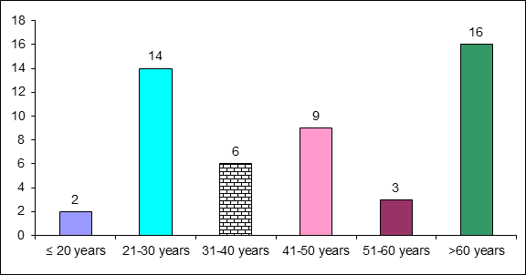
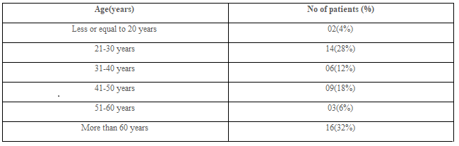
Figure and table 1 showing the age distribution of 50 leukemic patients. It was obvious that more than 60 years persons were affected more (total 16, 32%) than that of any age group followed by age between 21-30 years (total 14, 28%), between 41-50 years (total 9, 18%), between 31-40 years (total 6, 12%),between 51-60 years(total 3, 6%). Ages less than 20 years were the least affected (total 2, 4%).
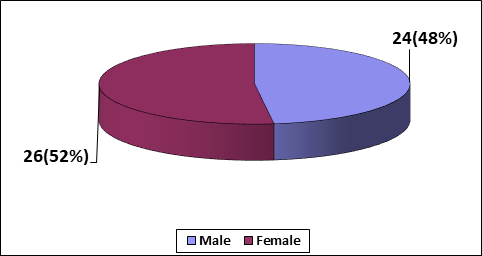
Figure 2 showing females were affected more (total 26, 52%) than males (total 24, 48%)
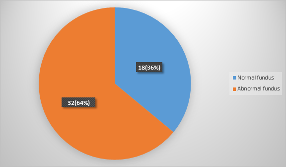
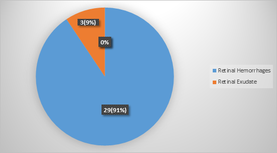
Among 50 patients,32 patients(64%) patients were found to have fundoscopic abnormalities(Figure 3). Figure 4 showing among 32(64%) patients with fundoscopic abnormalities, only 3 patients (9%) had exudates while others (29 , 91%) have retinal hemorrhages(Figure 4).
Hemorrhage was found in majority of ALL and AML patients, 48.28% and 51.28% respectively. Hemorrhage was not found in CLL and CML, 38.10% and 28.57% respectively. Hence hemorrhages were frequently seen in acute than chronic leukemia (p<0 p=0.25) p=0.08).>
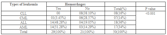
Table 2 shows out of 50 leukemic patients, 8 cases were CLL, 7 were CML, 18 were ALL and 17 were AML. Among them fundal lesion was detected in 32 patients (64%), retinal hemorrhages being the most common lesion (total 29, 90.63%). Hemorrhage was found in majority of ALL and AML patients, 48.28% and 51.28% respectively. Hemorrhage was less frequently found in CLL and CML patients, 38.10% and 28.57% respectively. Hence hemorrhages were frequently seen in acute than chronic leukemia (p<0>
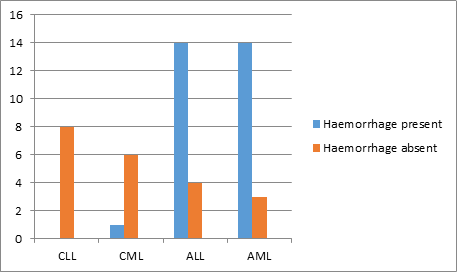
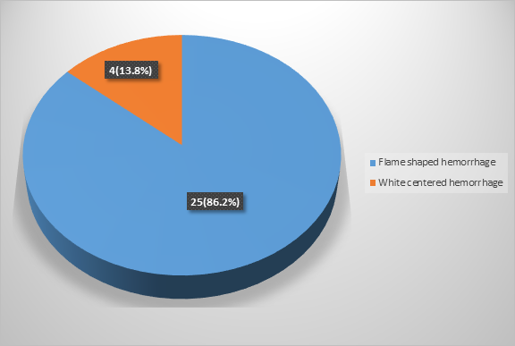
Figure 6 shows majority (25,86.2%) had flame shaped hemorrhage and remaining 4(13.8%) had white centered hemorrhage
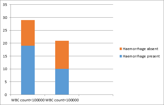
Figure 7 shows 19 out of 29 patients without high leukocyte count have haemorrhages and 11 out of 21 patients with high leukocyte count(white blood cells more than 100000/cmm) have no hemorrhages. P value was >0.05 which was not statistically significant.
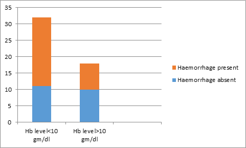
Figure 8 shows 21 out of 32 patients with anemia have retinal haemorrhages and 10 out of 18 patients without anemia (hemoglobin less than 10gm/dl) have no haemorrhages. There were no statistically significant correlation between anemia and retinal Hemorrhages (p value>0.05).
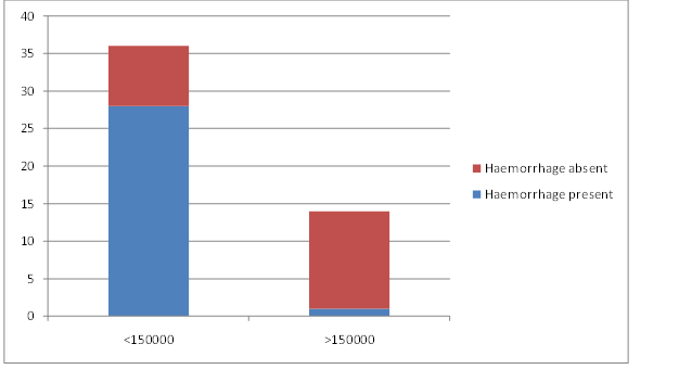
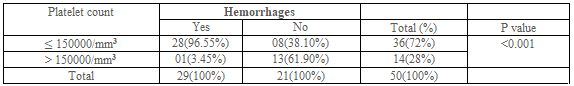
Table 3 shows 28 out of 36 patients with thrombocytopenia (platelet count less than 150000/cmm) have retinal hemorrhages and 13 out of 14 patients without thrombocytopenia have no haemorrhages. Statistically significant relation was found between thrombocytopenia and retinal hemorrhages.
Leukemic retinopathy is the term most used to denote fundus manifestations of anemia, thrombocytopenia and increased blood viscosity seen in patients with leukemia.9In the present study, higher prevalence (64%) of leukemic retinopathy was noted than the prevalence of 42%, 43% and 49% reported by Guyer et al [10], Abu el-Asrar et al [16] and S. C. Reddy and N. Jackson.16 respectively. Intra retinal hemorrhages were seen more often than above three studies. This very high percentage of fundoscopic abnormalities may due to majority of patients with acute leukemia and co-occurrence of thrombocytopenia and anemia in the majority of patients.
In the present study, 28 out of 36 patients with thrombocytopenia have retinal hemorrhages and 13 out of 14 patients without thrombocytopenia have no hemorrhages. Statistically significant relation was found between thrombocytopenia and retinal hemorrhages.
In this study, 21 out of 32 patients with anemia have retinal hemorrhages and 10 out of 18 patients without anemia have no hemorrhages. There were no statistically significant correlation between anemia and retinal Hemorrhages (p value>0.05). However, other researchers have noted an association between anemia and the presence of retinal haemorrhages [14, 16]. This finding may be due to small sample size in this study.
Shivaprasad C and Srinivasan R studied, 120 eyes of 60 consecutive patients with leukemia and lymphoma who presented to the hematology clinic of Jawaharlal Nehru Institute of Postgraduate Medical Education and Research were included in this study. Of the 60 patients examined which included 32 males and 28 females, 36 of them had chronic leukemia, 18 had acute leukemia and 6 patients had lymphomas. Ocular manifestations were present in 42.85% (25 patients). The most common ocular manifestation was conjunctival pallor in 30.95% (18 patients). Retinal changes were seen in 26.19% (16 patients) which included optic disc pallor and retinal vascular changes with hemorrhages and soft exudates. The retinal changes were bilateral and asymmetrical. Patients with optic disc pallor had hemoglobin ranging from 2.0-6 gram%. Of the total 8 patients presenting with retinal hemorrhages and soft exudates, six of them were acute leukemias, one chronic leukemia, another being Non-Hodgkins lymphoma. All of them had reduced platelet count counts ranging from10000 to 59000. The patient with platelet count of 10000 had more retinal hemorrhages which had also involved the macular area. Visual acuity was decreased in 7.1% due to hemorrhage at macula. Correlation between total white cell count, anemia and retinal hemorrhages or cotton wool spots was not observed in their study. No significant association was found between gender and the presence of retinal lesions. Retinal vascular changes with hemorrhages correlated with platelet counts and are commoner in acute leukemia’s [15].
Hemorrhages of the retina are the most striking feature of leukemia. They tend to occur most commonly at the posterior pole, may be at any level of the retina and may extend into the sub-retinal or vitreous spaces. Retinal hemorrhages have been described as dot and blot, or flame-shaped or classically as white-centered hemorrhages. It has been proposed that this white center represent platelet fibrin aggregates or an accumulation of leukemic cells.
In this study, 19 out of 29 patients without high leukocyte count have hemorrhages and 11 out of 21 patients with high leukocyte count have no hemorrhages. P value was >0.05 which was not statistically significant. Similarly, Holt and Gordon-Smith and Mahneke and Videbaek describe that the white blood cell count was not related to the occurrence of White Centered Hemorrhage (WCH) [20] Guyer et al. found a non-significant trend of leukocytosis in patients with WCH [9]. In contrast this study finding is not consistent with the observation of Gibson 1938 and Abu el-Asrar et al. [13].
This prospective, observational case series surveyed adult leukemia patients presenting at Tertiary Hospital population of adult Nigerian Africans departments of Hematology/Immunology and Ophthalmology from July 2003 to August 2008. The demographic profile, clinical data from for each individual in the cohort were statistically collated and analyzed. A P<0>P<0>P< 0>P<0>P = 0.0822) or leukemic ophthalmopathy (P = 0.6624). The prevalence of leukemic ophthalmopathy in Enugu is high. It is often associated with significant ocular co-morbidity and vision loss. These have implications for clinicians involved in leukemia management. Early diagnosis and regular ophthalmic examinations are recommended to optimize treatment outcomes [17].
A prospective study of ocular disorders in adult patients with leukemia at the University of Benin Teaching Hospital, Benin City, Nigeria, between July 2004 and June 2008 was conducted. The patients were interviewed and examined by the authors and the ocular findings were recorded. The means, standard deviation, and the Kruskal-Wallis non parametric test was performed. Forty-seven patients with leukemia were seen. Nineteen patients (40.4%) had CLL, 14(29.8%) had CML, 9(19.1%) had AML and 5(10.6%) had ALL. Seven patients (14.9%) had ocular disorders due to leukemia. The ocular disorders due to the leukemia were proptosis in two patients (4.3%), retinopathy in one patient (2.1%), conjunctival infiltration in one patient (2.1%), periorbital edema in one patient (2.1%), retinal detachment in one patient (2.1%), and subconjunctival hemorrhage in one patient (2.1%). There was no significant difference in rate of the ocular disorders in the various types of leukemia (Kruskal-Wallis KW= 4.019; corrected for ties. P=0.2595). One patient (2.1%) was blind from bilateral exudative retinal detachment while 1 patient (2.1%) had monocular blindness from mature cataract. Ophthalmic disorders that are potentially blinding occur in leukemia. Ophthalmic evaluation is needed in these patients for early identification and treatment of blinding conditions [18].
Chronic myeloid leukemia (CML) is a well-studied entity and advances made in diagnosis and treatment has improved the disease outcome. Patients with ophthalmic manifestation of CML have been reported to have lower 5-year survival rates. Hence, recognizing the early fundus changes may improve outcome by allowing earlier diagnosis and treatment. They report a case of a previously healthy 30-year-old Myanmarese male, who presented with a minor visual disturbance, complaining of seeing a ‘black dot’ in his left visual field for the past 1 week. Fundoscopic examination revealed bilateral retinal blot hemorrhages, white-centered hemorrhage, and pre-retinal hemorrhage over the left fovea. The full blood count and peripheral blood film were abnormal, and bone marrow biopsy confirmed the diagnosis of CML. Cytoreduction therapy was promptly commenced and his symptoms resolved, with improvement in visual acuity. No complications were recorded at 1-year follow-up [21].
The autopsy study by Leonardy et al. [20]. Showed a highly significant correlation between ocular leukemic cell infiltration and white blood cell counts; in a number of cases, leukemic cells were seen migrating in choroidal vessels or infiltrating perivascularly. It is probable that this mechanism may explain the role of a white blood cell count in the pathogenesis of retinal hemorrhages in these patients.
In this study, 35 out of 36 patients with thrombocytopenia have no retinal exudates and 2 out of 14 patients without thrombocytopenia have exudates. Statistically significant relation was not found between thrombocytopenia and retinal exudates (p value>0.05).
Similar to the observation of Guyer et al. [10]. I didn’t find any significant association between the presence of cotton-wool spots and hemoglobin level. However, Abu el-Asrar et al. [13]. reported that ALL patients with cotton-wool spots had significantly lower hemoglobin levels than patients without such lesions. This study showed no statistically significant correlation between Hemoglobin levels with exudates.
It is observed that the mean white cell counts in patients with cotton wool spot (CWS) in AML and ALL groups was higher than in those without CWS, but this was not statistically significant. Abu el-Asrar et al. [16] found a significantly lower leukocyte count in patients with AML with CWS, and higher leukocyte count in patients with ALL with CWS, while Guyer et al[9]. In the present study, there is no statistically significant association between leukocytosis and the presence of CWS.
Cotton-wool spot are often present in patients with leukemia. They are caused by occlusion of precapillary arterioles resulting in retinal ischaemia [23]. This focal ischemia leads to a blockage of exoplasmic flow within the nerve fibre layer of the retina, with the subsequent deposition of intraaxonal organelles [36]. The reason for this occlusion in leukemia is poorly understood. They may be related to ischemia secondary to anemia, direct occlusion by leukemic cells and occlusion by platelet fibrin aggregates or sludging resulting from hyperviscosity [24, 25].
In this study, mean age of the population was 47.00 (±19.72), lowest being 18 and highest being 82 years, 24(48%) were male and 26(52%) were female.
Reddy and Jackson study reported, a total of 127 patients with acute leukemia were examined for retinal changes, of whom 78 (61%) were male and 49 (39%) were female; 83 (65%) had myeloid leukemia and 44 (35%) had lymphoid leukemia. The mean age of the patients was 34.6 years (range 13-77 years) [16].
This study has identified retinopathy as a significant health problem among patients with leukemia and highlighted some of the factors associated with funduscopic abnormalities among them. A high percentage of patients (64%) with leukemia have fundal lesion and most of them (58%) had retinal hemorrhages and were frequently seen in acute than chronic leukemia. Among hemorrhages, flame shaped hemorrhage was the commonest finding. So, recognition of retinopathy is important to improve care of leukemia patients because it has impact on the prognosis and overall management of these patients. Therefore an adequate attention should be paid to funduscopic evaluation while treating leukemic patients.
The present study did not represent the actual scenario of leukemic retinopathy in Bangladesh because the study was conducted in one tertiary level hospital (Bangladesh Medical College and Hospital (BMCH) in Dhaka city only. Sample size and duration of the study was short. Treatment outcomes were also not assessed because of lack of follow up.
There were no conflicts of interests
Appropriately taken
Self-funded
Dear Editorial Team, Clinical Medical Reviews and Reports. My experience with the journal was highly positive. The peer-review process was rigorous, constructive, and completed in a timely manner. The reviewers provided valuable comments that helped improve the quality and clarity of our manuscript. The editorial office was professional, responsive, and supportive throughout all stages of the publication process. Communication was clear and efficient, and any questions were addressed promptly. Overall, I found the journal to maintain high scientific standards and an excellent publication workflow. I would be pleased to consider submitting future work to this journal. Best wishes from, Elena Popa.

It was my pleasure to submit my testimonial concerning the Reviewer Board of our Scientific Journal “Brain and Neurological Disorders”. The Reviewers focused on some modifications and their contribution was helpful. The ladies of our Editorial Office were also supported my efforts. It was my honor to have such a co-operation and I am looking forward for more collaboration.

Dear Grace Pierce, Editorial Coordinator of Journal of Clinical Research and Reports, Thank you for the speedy and efficient peer review process. I appreciate the fact that your peer reviewers do not take months to respond like with some other journals. I would also like to thank the editorial office for responding quickly to my questions. It is an excellent journal. I plan to submit more manuscripts in the future. Best wishes from, Robert W. McGee

Dear Grace Pierce, Editorial Coordinator of Journal of Clinical Research and Reports, Working with you and your team on our recent publication in JCRR has been a truly wonderful and enjoyable experience. The responses were prompt, and the reviewers were patient, constructive, and highly professional. One reviewer in particular gave me the feeling that a professor was carefully reading and commenting on my coursework, which was deeply touching. The entire process was straightforward and hassle‑free, with no tedious online forms to complete. I highly recommend this journal. Best wishes from, DR Aibing Rao, Head of R&D

I Appreciate the Opportunity to Share my Experience with the Journal of Clinical Research and Reports. The peer review process was timely and constructive, and the feedback provided helped improve the quality of our manuscript. The editorial office was professional, responsive, and supportive throughout the process, ensuring smooth communication and efficient handling of the submission. Overall, it was a positive experience collaborating with your team.

Dear Mercy Grace, Editorial Coordinator of Obstetrics Gynecology and Reproductive Sciences, We would like to express our gratitude for your help at all stages of publishing and editing the article. The editors of the magazine answer all the necessary questions and help at every stage. We will definitely continue to cooperate and publish other works in the Obstetrics Gynecology and Reproductive Sciences! Best wishes from, Alla Konstantinovna Politova,
