
Research Article | DOI: https://doi.org/10.31579/CIC-2021/007
1 Department of Cardiology, Istanbul University Cerrahpasa, Institute of Cardiology, Istanbul, Turkey.
2 Laboratory of Clinical Microbiology, Istanbul University Cerrahpasa, Institute of Cardiology, Istanbul, Turkey.
*Corresponding Author: Nazmi Gultekin, Istanbul Universty-Cerrahpasa, Institute of Cardiology, Istanbul, Turkey. Tel: +90 212 4592000; Fax: +90 2124592069.
Citation: Anil Tanki, Nazmi Gultekin, and Emine Kucukates (2021). Evaluation of autophagy and microtubules inhibition through blood beclin-1 levels in subgroups with Heart Failure reduced Ejection Fraction. Clinical and Interventional Cardiology. 1(1); DOI: 10.31579/CIC-2021/007
Copyright: © 2021, Nazmi Gultekin. This is an open access article distributed under the Creative Commons Attribution License, which permits unrestricted use, distribution, and reproduction in any medium, provided the original work is properly cited.
Received: 02 October 2021 | Accepted: 25 October 2021 | Published: 06 November 2021
Keywords: heart failure; dilated cardiomyophathy; cell death; autophagy; beclin-1; microtubules
Background and Aim: In this study, we aimed to compare beclin-1, one of the factors and moderators of autophagy activity, in the serum of heart failure patients with low ejection fraction (HFrEF) with those in the serum of healthy individuals. Also, we investigated serum beclin-1 levels according to etiological classifications (ischemic/non-ischemic subgroups). Additionally, the subset of patients using colchicine as a microtubule inhibitor for at least three months due to HFrEF was included.
Methods: This study included 50 patients with HFrEF (25 with ischemic etiology, 25 with non-ischemic etiology) and 30 healthy subjects between January 2018 and December 2019 in Istanbul University Cardiology Institute. Serum beclin-1 levels were determined by using the ELISA method by the ELISA Kit.
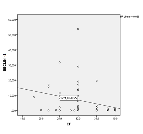
Results: Although serum beclin-1 levels of all HFrEF group compared to the control group did not reach statistical significance, increased serum beclin-1 levels were found (p:0.64). However, NT-proBNP levels were found significantly higher (p:0.01) .Serum beclin-1 levels correlated with ejection fraction in 50 patient with HFrEF (p: 0.018, R²: 0.088). In the non-ischemic etiology subgroup with HFrEF especially had higher serum beclin-1 levels (p:0.01). There was also no significant correlation between creatinine and eGFR levels and autophagic activity (p:0.482). Also, we found lower levels of NT-proBNP that did not reach statistical significance and higher beclin-1 levels to reach statistical significance (p: 0.015 ) in the colchicine using patient subset.
Conclusıons: Beclin-1 levels especially increased in the HFrEF with non-ischemic etiology group. Low dose colchicine affects autophagy, microtubules inhibition, and vesicle trafficking in HFrEF.
The autophagic response has been described in various pathophysiological situations, including neurobiology, cancer, cardiovascular disease, and infectious diseases [1-3]. Membrane trafficking, i.e. endocytic/exocytic, endo-lysosomal and autophagic pathways are well studied. “New era of membrane trafficking diseases” as emerged and de-regulation of vesicle trafficking pathways were shown to be related to disease syndromes. However, their roles in diseases were not fully understood as they can act in different conditions to work towards cell survival or induce cell death [1,4].
As the mammalian ortholog of the yeast Atg6 gene, beclin-1 is an essential mediator of autophagy [5-10]. Beclin-1 forms a multimeric complex with vacuolar protein sorting 34 (Vps34) and class 3 phosphatidylinositol 3-kinase (PI3k), which is necessary for the formation of autophagosome. Component of various PI(3)K complexes beclin-1 interacts with PIK3C3/VPS34 to signal the onset of autophagy. Atg6/beclin-1 Inhibited by binding to Bcl2 [5-10] (Figure 1).
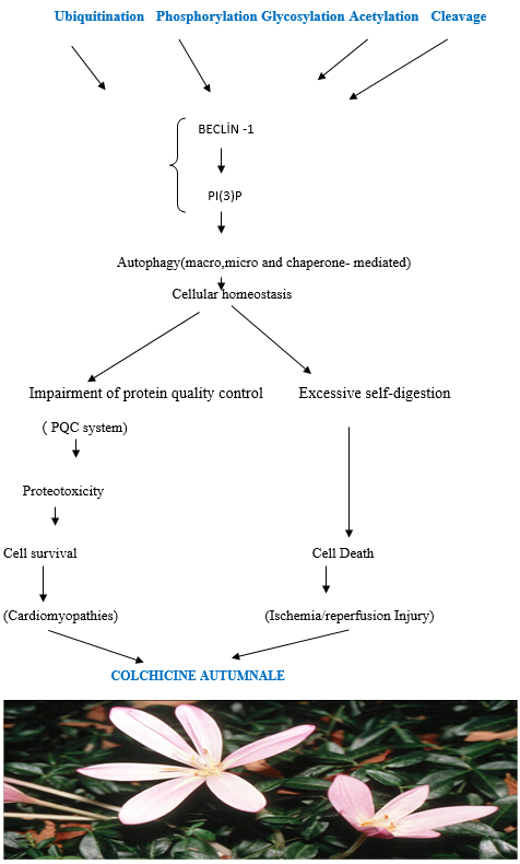
•Post-translational modifications of Beclin 1 affect protein stability, confirmation, activity, and its interactome and can be used as a molecular rheostat to fine-tune autophagic activity.
•Targeting Beclin 1 modifiers to regulate Beclin 1 post-translational modifications could provide a possible therapeutic intervention for upregulatin autophagy.
•Low dose colchicine accelerates the physiological clearance of misfolded proteins from cells through the protein quality control (PQC) system and cytoprotective autophagy and inhibits NLRP3) inflammasome, antigen presentation to T lymphocyte and the lysozymes of cardiomyocytes with acquired autoimmunity.
The mechanism of action of colchicine is through the inhibition of tubulin polymerization and potentially also through effects on cellular adhesion molecules and inflammatory chemokines. Colchicine may also have direct anti-inflammatory effects by inhibiting key inflammatory signaling networks known as the inflammasome and pro-inflammatory cytokines. Through the disruption of the cytoskeleton, colchicine is believed to suppress the secretion of cytokines and chemokines as well as in vitro platelet aggregation. Colchicine interferes with neutrophil adhesion and recruitment to inflamed tissue and antigen presentation to T lymphocyte and the lysozymes of cardiomyocytes with acquired autoimmunity.. Also, when misfolded proteins cannot be efficiently removed by degradative pathways. Misfolded proteins may accumulate and block the ubiquitin-proteasome system (UPS) and autophagy. Colchicine modulates regulation of the protein quality control (PQC) system via chaperone-mediated autophagy and post-translational modification and also by effective mitochondrial dynamics and homeostasis. These modifications have a role in multiple cellular functions, ranging from cell motility, cell cycle progression or cell differentiation to intracellular trafficking and signaling [1,10-12,] (Figure1).
Colchicine is a medication with a relatively low therapeutic index [13]. Because high doses, especially in muscle cells can lead to vacuolization and myopathy should be avoided [11,13]. Considerable researches have highlighted the potential of colchicine in the treatment of cardiovascular diseases mediated by pro-inflammatory processes [13-15]. The sustained benefit of short-term colchicine treatment on survival, cardiac function, and ventricular remodeling after myocardial infarction(MI )seems to be beneficial [13-15]. Whereas in previous studies, the colchicine treatment of Heart Failure with Reduced Ejection Fraction (HrEF) has been reported to be conflicting [16,17].
In this study, it was aimed to compare the serum beclin-1 levels which is one of the markers of autophagic activity in the patients with heart failure reduced ejection fraction (HFrEF), and those healthy subjects. Besides, in the study, variations of serum beclin-1 levels will be investigated in patients who were followed-up due to HFrEF according to etiological classification (ischemic/non-ischemic subgroup), and also in the colchicine-using subset.
Patient and public involvement statement
The patients and public were not involved in designing of, recruitment to, or conduct of the present study.
The sample size was calculated by the formula: n=Z121 α/2S2÷d2(n=80=1.962×(1-0.55/2)×0.502÷0.052)(N= Population Size; n=Sample Size; S= Standard Error;0.05 (5%) (Confidence Interval [CI] 95%); Confidence Interval: upper=95%; Lower: 5%; The Z-values for confidence levels were: 1.645=90 percent confidence level; 1.96= 95 percent confidence level; 2.576 = 99 percent confidence level (Z for p=0.05, 0.01, 0.001 are1.96, 2.58 and 3.28 Z values respectively); d= RelativeStandard Error) and it was confirmed by automatic sample size calculator [18].
Design and definitions
Our study was designed as a prospective cross-sectional observational. Based on ESC 2016 heart failure guidelines, symptoms and findings, biochemical and echocardiographic parameters, and etiology of the disease were evaluated. A total of 80 patients with 50 HFrEF (25 with ischemic etiology and 25 with non-ischemic etiology) and 30 healthy subjects were included in the study. Half of the HFrEF patients included in the study were selected from ischemic dilated cardiomyopathy patients with ischemic etiology, while the other half were selected from non-ischemic dilated cardiomyopathy patients without ischemic etiology [19].
For the control group, healthy individuals without heart failure with similar demographic characteristics were included in the study. In addition, low-dose colchicine (0.5-1 mg) was added to the treatment for at least three months in our 13 patients HFrEF with non-ischemic etiology for various reasons such as pericardial effusion accompanying HFrEF, and/or who developed hyperuricemia as a result of intensive diuretic therapy.
Exclusion criteria
Patients with the following characteristics were excluded from the study: under 18 old, being older than 80, having a history of hospitalization due to acute coronary syndrome in the last 30 days, end-stage renal disease patients, To have an active infection, having a life expectancy of less than 1 year (such as malignancies) due to noncardiac reasons, to have advanced valve disease.
Study population characteristics
The demographic characteristics of all patients included in the study and all individuals included in the control group; Age, gender was recorded in appropriate forms in the study forms, and symptom status was classified according to the NYHA symptom evaluation system. Comorbid diseases; hypertension, Diabetes Mellitus, hyperlipidemia, ischemic heart disease history were recorded in the study form. Heart rhythms (sinus rhythm / atrial fibrillation/battery rhythm), presence of the device (Pacemaker/ICD/CRT-D), and habits of the patients and control group; cigarettes, alcohol, and medications they used were questioned and written to the appropriate places in the study forms. In the study group and control group, the diagnosis of DM was based on at least one history of antidiabetic drug use or HbA1c level> 6.5%.
Recent biochemical tests of all patients and control subjects included total cholesterol, LDL cholesterol, HDL cholesterol, triglycerides, glucose, HbA1c, creatine, eGFR, total protein, albumin, ALT, AST, NT-ProBNP levels and the hemogram parameters (hemoglobin, leukocyte, neutrophil, monocyte, lymphocyte, basophil, platelet, MPV) were written in the appropriate places. No additional tests were performed to determine the necessary blood results of the patients. Glomerular filtration rates (GFR) of the patients were calculated using the MDRD formula. Also, echocardiography parameters of all patients and control groups were examined in detail. EF, left atrium (LA), left ventricular end-diastolic diameter (LVD), right ventricular diameter (RVD), septum thickness (IVS), segmenter or global where the study forms were recorded in the appropriate places (Table1-3).


Parameters B) NT-proBNP and Beclin-1 values of patients with heart failure according to ischemic (N: 25) and non-ischemic (N: 25) etiology included in the study(mean ± SD).,
Parameters C) Differences in NT-ProBNP and Beclin-1 Levels and echocardiographic parameters of colchicine treated(N:13) and non-colchicine treated patients in the HFrEF patient group (N: 37) included in the study(mean ± SD).
There was no significant correlation between creatinine and eGFR levels and beclin-1 (ATG6 analog in yeast cells), an autophagy marker in mammals (p: 0.482) ( Table 3 A). Additionally, in the subgroup analysis of the heart failure group, statistically significant serum beclin-1 and low NT-proBNP levels were found in the non-ischemic etiology group according to the ischemic etiology group (respectively p:0.01:p:0.01) (Table 3 B ). Also, we found lower levels of NT-proBNP that did not reach statistical significance and higher beclin-1 levels to reach statistical significance (p: 0.015 ) in colchicine- using patient group (Table 3 C). Moreover, EFs increased, LVDD decreased.
* Continuous variables are presented as mean ± SD and dichotomous variables as percentages. A two-tailed t-test was used for comparison of means, and an x2-test for percentages in statistical analysis at table1,2,3..
Plasma beclin-1 levels determination
The consent form was obtained from the patients and the control group at the time of admission. Blood samples were taken into 8-ml K-EDTA-tube with Monovette brand vacutainer and centrifuged at 3000 rpm for 10 minutes. Serum samples obtained were stored at -80 ° C for further study. After all the samples were completed, they were studied in the microbiology laboratory of Haseki Training and Research Hospital.
Plasma beclin-1 levels were determined using the human beclin-1 ELISA Kit (Bioassay Technology Laboratory). Each well was previously prepared according to the kit package insert. All reagents, standard solutions, and samples were prepared as instructed. 50μl Standard solution was added to a standard well, 40 μl sample to sample wells and then 10μl anti-BECN1 antibody to sample wells, subsequently 50 μl streptavidin-HRP to sample wells and Standard wells. The plate with a sealer was covered and incubated 60 minutes at 37°C. The plate was washed 5 times with wash buffer. For automated washing, someone was by aspirated all wells and wash by 5 times with wash buffer, overfilling wells with wash buffer. The plates onto paper towels or other absorbent material were blotted. 50 μl substrate solution A to each well and then add 50μl substrate solution B was added to each well and incubated by plate covered with a new sealer for 10 minutes at 37°C in the dark. Subsequently, 50 μl stop solution was added to each well, the blue color will change into yellow immediately. The optical density (OD value) of each well immediately using a microplate reader set to 450 nm within 10 minutes after adding the stop solution. The blank value (incubation buffer + stop solution), was subtracted from all other values obtained.
If the summarized; in serum samples, beclin-1 was measured immunoturbidimetrically by turbidity caused by the antigen-antibody complex formed by binding of monoclonal anti-BECN1 coated latex microparticles with beclin-1 in serum. The curve of the optical densities formed 10 minutes after the addition of the stopping solution was calculated with a compatible computer-based software system.
Statistical analysis was performed by using SPSS (Statistical Package for Social Science) for Windows 23. 0 program. The student's t-test was used when the parameters providing normal distribution conditions were compared according to two independent groups. In the comparison of categorical values, independence control was performed by using Chi-Square (χ2) analysis. Beclin-1 levels of healthy individuals, ischemic dilated cardiomyopathy and non-ischemic dilated cardiomyopathy patients were compared with the Mann-Whitney U test. Statistical significance was accepted as p≤0.05.
All data and statistical comparisons are shown in the legends of (Tables 1, 2, 3) and (Figure 1, 2).
Beclin-1, the mammalian orthologue of yeast Atg6, has a central role in autophagy, a process of programmed cell survival, which is increased during periods of cell stress and extinguished during the cell cycle. It interacts with several cofactors (Atg14L, UVRAG, Bif-1and survivin) to regulate the lipid kinase Vps-34 protein and promote the formation of beclin 1-Vps34-Vps15 core complexes, thereby inducing autophagy [10,20]. In contrast, the BH3 domain of belcin-1 is bound to and inhibited by Bcl-2 or Bcl-XL.This interaction can be disrupted by phosphorylation of Bcl -2 and belcin-1 or ubiquitination of belcin-1. Interestingly, caspase-mediated cleavage of belcin-1 promotes crosstalk between apoptosis and autophagy.Beclin-1 dysfunction has been implicated in many disorders, including cancer and neurodegeneration. IL-6 could inhibit starvation-induced autophagy by the STAT3/Bcl2/beclin-1 pathway in cells [1-8,10,20]. Beclin-1 and PIK3C3 are involved in the complex which signals the onset of autophagy and could be used to monitor autophagy [1,6-10](Figure 1).
In our study, beclin-1 levels were higher in the HFrEF patient group compared to the control group, but not statistically significant (p:0.64); however, NT-proBNP levels were found significantly higher (p:0.01). When the relationship between ejection fraction (EF) and beclin-1 levels of HFrEF group in the study was examined, we found that patients with lower EF had correlated higher beclin-1 levels (p: 0.018, R²: 0.088) (Figure 2; Table 3 A).
In the observational and experimental studies to determine the relationship between heart failure and autophagy, increased autophagic activity was found in individuals with heart failure [21-25]. Although there are many molecular studies, biopsy and autopsy studies and animal experiments to determine the relationship between cardiovascular diseases and autophagy [4,21-25]. We could not find any study in the literature based on the determination of the level of autophagy markers in serum in patients with Heart Failure (HF).
Furthermore, in our study, higher creatinine and decreased eGFR levels were found in the heart failure group with low EF compared to the control group (p:0002;0,05). When HFrEF groups with ischemic and non-ischemic etiology were compared, renal functions were similar. There was also no significant correlation between creatinine, eGFR values and beclin-1levels (ATG6 analog in yeast cells), an autophagy marker in mammals in the patient groups (p: 0.482) (Table 3 B). This proves that other mechanisms such as dehydration, electrolyte imbalance, and hypotensive condition are responsible for the deterioration of renal function in patients with HFrEF rather than autophagy [26].
Also, in the subgroups analysis of the HFrEF group, serum beclin-1 levels were statistically significantly increased in non-ischemic etiologysubgroup but not ischemic subgroup. But, NT-proBNP levels were found low in the non-ischemic etiology subgroup according to the ischemic etiology subgroup (respectively, p:0.01:p:0.01) (Table 2,3 B ). We consider that serum beclin-1 levels were significantly found low in the patients with HFrEF subgroup with ischemic etiology, because of the ischemia /reperfusion process was completed and autophagic activity decreased in the scar tissue. Changes in autophagic flux are seen in essentially all forms of heart disease and all cell types. It can either be beneficial or aid to disease progression. Ischemic heart disease is characterized by lack of nutrients and oxygen to the myocardium [14,15]. Studies looking at models of ischemia have shown that autophagy functions as a pro-survival mechanism by adapting to changing metabolic needs and eliminate damaged mitochondria, which can release ROS and initiate apoptosis to prevent any tissue damage [27,28 ].Inhibition of autophagy can exacerbate cardiac dysfunction and remodeling [1,2,4,20]. Once oxygen and nutrients are restored, autophagy can either be adaptive or detrimental to the tissue. Some studies have shown that reperfusion following an ischemic attack leads to an increase in autophagy but an impairment of autophagosome clearance leading to cell death [13-15.] Also, recent studies have demonstrated that nucleotide-binding oligomerization domain-like receptors, pyrin domain -containing 3 (NLRP3) inflammasome, is associated with endogenous sterile inflammation after Myocardial infarction. NLRP3 is a cytosolic multiprotein complex activated by tissue danger signals and associated with the production of activated interleukin (IL)-1β and IL-18 [14,15]. Colchicine was reported to reduce inflammatory cytokines (including IL-1β and IL-18) and infarction size in MI [13,21]. Therefore, the sustained benefit of short-term colchicine treatment on survival, cardiac function, and ventricular remodeling after MI is to be beneficial [13].
Non-ischemic etiology with heart failure may also be associated with an increased vacuolization [14,15,25]. Because, high dose colchicine can lead to vacuolization and myopathy especially in muscle cells should be avoided [26,29]. Therefore, in our study low-dose colchicine (0.5-1 mg) exclusively was added to the treatment for at least three months in 13 patients with non-ischemic etiology with HFrEF for various reasons. In this subset, we found lower levels of NT-proBNP that did not reach statistical significance (p:0.69) However, statistical significance was reached in the beclin-1 levels of this subset (p:0.015) (Table 3 C). At baseline, low-dose colchicine was used in 13 patients with terminal non-ischemic cardiomyopathy with severe left ventricular dysfunction (p = 0.009) and an enlarged left ventricle (p = 0.002). After 3 months, these patients were detected low NT-proBNP levels and higher beclin-1 levels. Moreover, EFs increased, LVDD decreased. Actually, we did not expect these paradoxical results. Low levels of NT-proBNP and higher beclin-1 levels discordances suggests another mechanism that low dose colchicine probably demonstrates cytoprotective autophagic clearance and regeneration activities via chaperone-mediated autophagy [27,28]. Thus; colchicine effects regulation of the protein quality control (PQC), chaperone-mediated autophagy and post-translational modification and also by effective mitochondrial dynamics and homeostasis [1,11,27,28]. Therefore,increased autophagy in our cases subgroup using colchicine did not correspond to cell death, but to selective cytoprotective autophagic clearance and regeneration activities that enabled cell survival, and reduced autoimmunity of cardiomyocytes according to our opinion.
Frankly; colchicine accelerates misfolded protein are physiologically cleared from the cells by the protein quality control (PQC) system. The PQC system is composed of two main arms: Firstly the molecular chaperone, mainly represented by the heat shock proteins (HSPs), and secondly the degradative pathways, including the proteasome, the autophagic response and the unfolded protein response (UPR). A specific degradative pathway for a given misfolded protein is selected by a defined class of chaperones, with the assistance of co-chaperones. These pathways may determine whether misfolded proteins have to be refolded or degraded. So, the PQC system controls dynamic selective autophagy [1,11].
As is known, autophagic vacuoles have been described in the heart tissue of patients with idiopathic dilated cardiomyopathy [25]. Cardiac hypertrophy is characterized by an increase in cardiomyocyte size, disarrangement of sarcomeric structure, enhanced protein synthesis and re-expression of fetal genes [24]. Cardiac hypertrophy is accomplished by the dysfunction of numerous signaling pathways that are involved in autophagy. PI3K/AKT, GSK3, MAPKs, and mTOR pathways have all been associated with the regulation of cardiomyocyte autophagy [2-10,20]. The deficiency of the autophagy gene LAMP-2 was shown to cause Danon disease [25]. Autophagy acts as a defense mechanism against the of cardiomyocytes. Furthermore, the heart develops cardiac hypertrophy and diastolic dysfunction with aging when autophagy is inhibited [22]. Recent studies have shown that miRNAs play an important role in regulating cardiac autophagy and there is a growing number of studies targeting autophagy regulating miRNAs as a potential strategy in the treatment of cardiac disorders [23].
Under the light of the literature data by we have explained above, this increase in autophagic activity in our study can also be explained by low-dose colchicine’s induction or upregulation of macro and microautophagy and especially selective chaperone-mediated autophagy for the clearance and regeneration from dysfunctional cardiomyocytes which are thought to contribute to heart failure [1,13,28]. On the other hand, in contrast to the high dose, low dose colchicine probably cleans bad vacuolization in HFrEF cases with non-ischemic etiology [1,25]. According to our opinion, regulation of autophagy , miRNAs and vesicle trafficking pathways in microenvironment can either be a cell– protective mechanism or in fact, an alternative cell–death mechanism, given that in cardiomyocytes in heart failure the apoptosis pathway is commonly altered. In this respect, colchicine or similar drugs treatment needs to be tried in more extensive studies.
Limitations of the study were non-randomized prospective cross-sectional observational design, relatively small number of patients, and whether there was not investigated the correlation between serum beclin-1 levels and beclin-1 levels measured in cardiac biopsy materials.
Our study reveals that HFrEF had higher serum Beclin-1 levels, especially in the non-ischemic etiology subgroup with HFrEF because of higher autophagic activity. Low-dose colchicine (0.5-1 mg) probably induces selective and cytoprotective autophagic clearance and regeneration activities in HFrEF and seems to be manifestly beneficial. On the other hand, in contrast to the high dose, low dose colchicine probably cleans bad vacuolization in HFrEF cases with non-ischemic etiology.There was also no significant correlation between creatinine and eGFR levels and autophagic activity. Further study of the autophagic and vesicle trafficking pathways and miRNAs will lead to an increased understanding of the regulation of this critical homeostatic pathway and identify new therapeutic potentials for cardiac diseases.
Dear Editorial Team, Clinical Medical Reviews and Reports. My experience with the journal was highly positive. The peer-review process was rigorous, constructive, and completed in a timely manner. The reviewers provided valuable comments that helped improve the quality and clarity of our manuscript. The editorial office was professional, responsive, and supportive throughout all stages of the publication process. Communication was clear and efficient, and any questions were addressed promptly. Overall, I found the journal to maintain high scientific standards and an excellent publication workflow. I would be pleased to consider submitting future work to this journal. Best wishes from, Elena Popa.

It was my pleasure to submit my testimonial concerning the Reviewer Board of our Scientific Journal “Brain and Neurological Disorders”. The Reviewers focused on some modifications and their contribution was helpful. The ladies of our Editorial Office were also supported my efforts. It was my honor to have such a co-operation and I am looking forward for more collaboration.

Dear Grace Pierce, Editorial Coordinator of Journal of Clinical Research and Reports, Thank you for the speedy and efficient peer review process. I appreciate the fact that your peer reviewers do not take months to respond like with some other journals. I would also like to thank the editorial office for responding quickly to my questions. It is an excellent journal. I plan to submit more manuscripts in the future. Best wishes from, Robert W. McGee

Dear Grace Pierce, Editorial Coordinator of Journal of Clinical Research and Reports, Working with you and your team on our recent publication in JCRR has been a truly wonderful and enjoyable experience. The responses were prompt, and the reviewers were patient, constructive, and highly professional. One reviewer in particular gave me the feeling that a professor was carefully reading and commenting on my coursework, which was deeply touching. The entire process was straightforward and hassle‑free, with no tedious online forms to complete. I highly recommend this journal. Best wishes from, DR Aibing Rao, Head of R&D

I Appreciate the Opportunity to Share my Experience with the Journal of Clinical Research and Reports. The peer review process was timely and constructive, and the feedback provided helped improve the quality of our manuscript. The editorial office was professional, responsive, and supportive throughout the process, ensuring smooth communication and efficient handling of the submission. Overall, it was a positive experience collaborating with your team.

Dear Mercy Grace, Editorial Coordinator of Obstetrics Gynecology and Reproductive Sciences, We would like to express our gratitude for your help at all stages of publishing and editing the article. The editors of the magazine answer all the necessary questions and help at every stage. We will definitely continue to cooperate and publish other works in the Obstetrics Gynecology and Reproductive Sciences! Best wishes from, Alla Konstantinovna Politova,
