AUCTORES
Globalize your Research

Case Report | DOI: https://doi.org/10.31579/2578-8949/064
Department of Dermatology, University Hospital Hassan II of Fez Morocco.
*Corresponding Author: Mounia Bennani, Department of Dermatology, University Hospital Hassan II of Fez Morocco.
Citation: Bennani M, Ziani J, Elloudi S, Douhi Z, Baybay H, Fatima Z Mernissi (2020). Yellowish plaque on the scalp of a nursling, What’s your Diagnosis?. Journal of Dermatology and Dermatitis.5 (1); Doi:10.31579/2578-8949/064
Copyright: © 2020 Sara Oukarfi, This is an open-access article distributed under the terms of The Creative Commons. Attribution License, which permits unrestricted use, distribution, and reproduction in any medium, provided the original author and source are credited.
Received: 01 March 2020 | Accepted: 19 March 2020 | Published: 23 March 2020
Keywords: Juvenile xanthogranuloma; xanthogranulomatous dermal infiltrate; dermoscopy
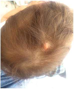
A 6-month-old girl presented with a 2- month history of a solitary slowly enlarging soft yellow plaque located at the vertex, with no other associated sign
General examination had found a baby in good general condition, toned and reactive, with on dermatological examination a yellow plaque of 3 cm of large roughly oval, with hairy surface (Figure 1)
The dermoscopy had objectified: an orange-yellow background coloring with clouds of xanthomatous deposits (Figure 2).

The rest of the somatic examination was unremarkable
Given the age of the infant, the clinical history, the appearance of the lesion, and the dermoscopy strongly suggestive of the diagnosis, a conservative management was adopted, with the beginning of regression of the lesion in the following year, without the appearance of new injury with a hindsight of four years
Juvenile xanthogranuloma (JXG) is a common form of non-Langerhans cell histiocytosis, mild, with good prognosis , First described by Adamson in 1905[1], its current nomenclature was adopted in 1954.[2] and its manifests as asymptomatic yellow-red papulonodules that usually occur in childhood [3] usually in the first months of life [4] and spontaneously regress within a year of formation.[3] Lesions may be solitary or multiple, and although they are most often found in the skin, they can also develop within other organs [5]
In early stages it is pink to red with a yellow tinge , but with time it acquires a yellow-brown hue and may develop occasional telangiectases on the surface[6]
Because of its benignity and transitory character, it is estimated that it is an underdiagnosed entity and, therefore, of unknown incidence [7]
Extracutaneous involvement is described, and is estimated to occur in 4% of cases, affecting any organ or tissue [8] Risk factors for extracutaneous involvement are age under two years and the presence of multiple lesions [5].
Diagnostic is fundamentally clinical , but dermoscopy can improve diagnostic sensitivity by showing a characteristic orange-yellow background colouration, with ‘clouds’ of paler yellow deposits consistent with a xanthogranulomatous dermal infiltrate. The ‘clouds’ of paler yellow deposits are similar to those seen in sebaceous hyperplasia [3] as in our patient.
The prognosis of patients with exclusively cutaneous involvement is excellent, with spontaneous remission in months or a few years, and relapses are rare.[7]In some cases, a small residual hyperpigmented scar may remain.Surgical removal may be considered only for cosmetic reasons, especially in cases of giant JXG[9]
Clearly Auctoresonline and particularly Psychology and Mental Health Care Journal is dedicated to improving health care services for individuals and populations. The editorial boards' ability to efficiently recognize and share the global importance of health literacy with a variety of stakeholders. Auctoresonline publishing platform can be used to facilitate of optimal client-based services and should be added to health care professionals' repertoire of evidence-based health care resources.


