AUCTORES
Globalize your Research

Case Report | DOI: https://doi.org/10.31579/2578-8949/061
1Departement of Dermatology, CHU Hassan II FEZ Morocco.
*Corresponding Author: Elharrouni A Aicha, Departement of Dermatology, CHU Hassan II FEZ Morocco
Citation: Elharrouni A Aicha, Dahhouki S,Douhi Z, Elloudi S, Baybay H et al (2019). Giant Fibrous Histiocytoma : Clinical and Dermoscopic Features 4(4); Doi:10.31579/2578-8949/061
Copyright: © 2019 Elharrouni A Aicha, This is an open-access article distributed under the terms of The Creative Commons. Attribution License, which permits unrestricted use, distribution, and reproduction in any medium, provided the original author and source are credited.
Received: 06 November 2019 | Accepted: 23 November 2019 | Published: 26 December 2019
Keywords: giant fibrous histiocytoma; benign tumor, dermoscopy
Benign fibrous histiocytoma is one of the most frequent benign neoplasms ,the diagnosis of cutaneous benign fibrous histiocytoma is generally easy ,however the atypical fibrous histiocytoma is an uncommon, poorly documented variant of cutaneous fibrous histiocytoma,may be difficult to identify, and the diagnosis only confirmed after exhaustive histopathological examination. Recently the dermoscopy has revealed a new dimension of dermoscopic features giant Fibrous Histiocytoma . Our purpose was to report the clinical and dermoscopic characteristics of giant Fibrous Histiocytoma and review the few cases of this variant of Fibrous Histiocytoma reported in the literature.
Fibrous histiocytoma are harmless growths within the skin that usually have a small diameter. They can vary in color, and the color may change over the years.They are firm to the touch,many people say they feel like a small stone underneath or raised above the skin. Several clinical variants of Fibrous histiocytoma have been described. Giant fibrous histiocytoma have been reported,often they have a pedunculated appearance. Because of their large size, the correct diagnosis is not suspected clinically; a diagnosis of malignancy is often made. We describe the clinical and dermoscopic features of giant fibrous histiocytoma , an unusual variant of dermatofibroma.
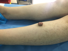
We report a case of 35 years-old patient, with no pathological history. She presented to our institution with a slowly growing mass on her left leg back for seven years. The mass would recur and progressively enlarge each time. Clinical examination showed a brown exophytic mass mesuring 4cm, that was extremely firm on palpation and not freely movable(Figure1).
Dermoscopically exhibiting a central white area combined with globular-like structures associated locally with Dotted and hairpin vessels(Figure2).

The mass was surgically removed under via wide local excision. The histopathological examination showed a epidermis pseudoepitheliomatous hyperplasia and hyperpigmentation of basal cell layer,Mixture of fibroblastic, myofibroblastic-like and histiocytic cells,Spindle cells have scant cytoplasm, thin elongated nuclei with pointed ends , Arranged in a cartwheel or vague storiform pattern,and Mitotic figures are uncommon. The diagnosis of Fibrous Histiocytoma was considered. The patient was reassured of the benign nature.
Fibrous histiocytomas or dermatofibroma are benign fibrohistiocytic (BFH) tumors ,they are a common cutaneous lesion that usually appears as a slowgrowing firm dermal nodule ,and made up of a mixture of fibroblastic and histiocytic cells, collagen and blood vessels[1]. Several clinical variants of Fibrous histiocytomas have been described, Over the years sothers clinical variants have been delineated incuding giant Fibrous histiocytomas[2], The latter variant is uncommon, and it is usually not suspected because of its large size.It can appear as solitary slow-growing, finn, circumscribed dermal nodule or tumor, they often impart a light brown to dark brown, purple-red, or yellow , the most frequent it is located at the legs.[3-4].
Although the clinical diagnosis of Fibrous histiocytomas is rather easy, in some instances the differentiation from other tumors. Dermoscopy is an invaluable technique that has revealed a new dimension of clinical morphologic features in pigmented and nonpigmented tumors.The dermoscopy of a classical Fibrous histiocytomas, revealing a central white patch surrounded by a thin brown network with linear vessels in a radial arrangement and dotted vessels,but of variant presentation is characterized by a peripheral homogeneous area and central white scarlike patch, crystalline structures, combined with globular-like structures,[5] as the case of our patient. However, BFH often deviates the latter atypical pattern and mimics other benign and malignant skin tumors. Moreover, because similar structures,the authors’ findings suggesting and highlighting the importance of histopathologic examination for an accurate diagnosis. However, BFH are characterized histologically by a proliferation of mononuclear, spindle-shaped, or histiocytoid cells and/or multinucleated cells, usually admixed with inflammatory cells[1.2].
In conclusion This rare clinical variant of Fibrous histiocytomas, whose large size is its only distinctive characteristic, invariably has a benign course and simple excision is curative.
Clearly Auctoresonline and particularly Psychology and Mental Health Care Journal is dedicated to improving health care services for individuals and populations. The editorial boards' ability to efficiently recognize and share the global importance of health literacy with a variety of stakeholders. Auctoresonline publishing platform can be used to facilitate of optimal client-based services and should be added to health care professionals' repertoire of evidence-based health care resources.


