AUCTORES
Globalize your Research

Case Report | DOI: https://doi.org/10.31579/2578-8949/055
*Corresponding Author: Rahul Pillai, Kkris skin care center & laser clinic, Kerala.
Citation: Rahul Pillai (2019) Squamous Cell Carcinoma under the Veil of a Trophic Ulcer in a Case of Hansen’s disease 4(3); DOI:10.31579/2578-8949/055
Copyright: © 2019. Rahul Pillai This is an open-access article distributed under the terms of the Creative Commons Attribution License, which permits unrestricted use, distribution, and reproduction in any medium, provided the original author and source are credited.
Received: 30 July 2019 | Accepted: 12 August 2019 | Published: 19 August 2019
Keywords: squamous cell carcinoma; marjolin’s ulcer; trophic ulcer; plantar ulcer; Hansen’s disease; leprosy
Hansen’s disease is seldom associated with trophic ulcers, which over a period of time when neglected has the potential for malignant transformation. This is a case report of a 44 year old man previously treated for Hansen’s disease presenting with non-healing plantar ulcer developing squamous cell carcinoma with lymph node metastasis. We highlight the importance of having a high degree of suspicion in each case as our patient was asymptomatic and repeated biopsies and lymph node FNAC were required to diagnosis.
The resultant neurological deficit in Hansen’s disease often results in chronic non-healing trophic ulcers which is a well-known entity. These ulcers when neglected or not properly managed have the predisposition for malignant transformation over a long period of time.
A 44 year old male was referred to our outpatient clinic with a non-healing chronic plantar ulcer involving the right sole of foot of 2 years duration. He was diagnosed as multi-bacillary leprosy at the age of 15 years and gives history of regular treatment from the leprosy hospital. He had no past history of diabetes or hypertension and was not on any other medications.
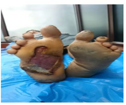
Local examination revealed a large plantar ulcer involving almost the entire sole of right foot extending from the forefoot up to the heel measuring 10 x 6 cms. The ulcer had everted hyperkeratotic edges in the lower lateral border with induration. The floor showed unhealthy granulation tissue with slough in areas with the fascia of the muscle visible. Sensory examination revealed glove and stocking examination. Peripheral nerve examination revealed bilateral ulnar and radial cutaneous nerve enlargement. On general examination the patient had partial clawing of both hands with difficulty walking. There was bilateral inguinal lymphadenopathy largest measuring 2x2 cms on the right horizontal group which was firm and non-tender.
Wedge biopsy done from the infero-lateral quadrant of the ulcer showed cytomorphological changes suggestive of micro invasive well-differentiated squamous cell carcinoma. Ultrasound guided FNAC of the right inguinal lymph node revealed metastasis.
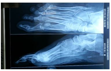
Other investigations included X-ray of the right foot which showed multiple osteophytes in the 5th metatarsal with vascular calcifications. Ultrasonogram of abdomen and pelvis confirmed multiple bilateral lymph nodes more in the right side. Chest X-ray and routine blood investigations were unremarkable.
After due consultation with Plastic surgeons and Oncosurgeons, the patient was taken up for wide local excision of the ulcer with a margin of 2 cms and right sided inguinal lymph node dissection. Biopsies were done from the margins to ensure the complete excision of the tumor. The resultant defect was covered using split thickness graft harvested from the thigh. On subsequent follow-up the patient should good recovery with the graft being taken up well.
French surgeon Jean Nicholas Marjolin in 1828 coined the term ‘Ulcus Marjolini’ or Marjolin’s ulcer to describe chronic leg ulcers which showed warty changes.1 Later in 1903 Da Costa first used the term in describing tumors arising from chronic leg ulcers (2).
Marjolin’s ulcers reflect malignant degeneration arising within pre-existing scar tissue or chronic inflammatory skin lesions.3Marjolin’s ulcers are mostly found in the lower extremity,commonest site being the plantar foot.4 They exhibit slow growth and are painless with a latent period between the primary pathology and malignant transformation. A time lag of up to 70years has been reported in some case series and the shortest reported time is 4 weeks, by Mcleod and Stauffer (5,6,7).
Treves and pack described two clinical types of Marjolin’s ulcer, the flat indurated, infiltrative, ulcerative type and the less frequent exophytic papillary form with the former having poorer prognosis. The malignant change often begins at the edge which may show some warty change with elevation. Occasionally there is the appearance of a mass within the scar or ulcer. The base of the ulcer becomes increasingly indurated with a granular, often necrotic floor with areas sloughing and discharge. Lymphatic spread may occur in the later stages when the tumor exceeds beyond the margins and regional lymph node metastasis is considered the most important prognostic factor (8.9).
The association between leprosy and malignant changes has been well documented in the literature. The reduced cell-mediated immunity in leprosy increases the risk for malignant changes. Cellular mutations are responsible for neoplastic transformations with infections serving as co-carcinogens(10).
The mainstay for treatment for Marjolin’s ulcer remains to be surgery. Wide local excision with a minimum margin of 2cms of healthy tissue have been recommended by many authors (9,11).Recently Moh’s micrographic surgery has ensured complete local excision with safe margins.
Defects following resection are usually covered with a skin graft, except when the bed may not be suitable, then a flap may be used(12). Amputations are indicated when deep invasion makes local excision difficult.
Regional metastases occur in 2 to 6% of cases, therefore the regional nodes should be thoroughly assessed in a patient presenting with Marjolin’s ulcer. Lymph node dissection is generally advised in the presence of clinically palpable nodes and some investigators suggest it as a prophylactic measure against regional node metastases (PND, prophylactic node dissections(9, 13).


We report this case due to the fairly benign nature of the tumor with hardly any symptomatic discomfort to the patient or suspicion of malignancy. Repeated biopsies and FNAC’s were required to substantiate the evidence of the tumor and nodal metastases. Malignant changes in non-healing trophic ulcers of Hansen’s disease may not be common, but can be missed if not vigilant. A high degree of suspicion and early specialist referral can be very vital as in our case.
Clearly Auctoresonline and particularly Psychology and Mental Health Care Journal is dedicated to improving health care services for individuals and populations. The editorial boards' ability to efficiently recognize and share the global importance of health literacy with a variety of stakeholders. Auctoresonline publishing platform can be used to facilitate of optimal client-based services and should be added to health care professionals' repertoire of evidence-based health care resources.


