AUCTORES
Globalize your Research

Case Report | DOI: https://doi.org/10.31579/2578-8949/049
*Corresponding Author: Rahul Pillai, Department of Dermatology, Venereology and Leprology,Mahatma Gandhi Medical College & Research Institute, Pondicherry, Kerala
Citation: Rahul Pillai (2019) A Child’s Search for a Diagnosis-A Case Report of Subcutaneous Zygomycosis.J Dermatol Dermatit 4(2) DOI: 10.31579/2578-8949/049
Copyright: © 2019. Rahul Pillai This is an open-access article distributed under the terms of the Creative Commons Attribution License, which permits unrestricted use, distribution, and reproduction in any medium, provided the original author and source are credited.
Received: 25 July 2019 | Accepted: 29 July 2019 | Published: 09 August 2019
Keywords: subcutaneous zygomycosis ,filamentous fungi
Basidiobolus species are filamentous fungi belonging to the order Entomophthorales. Unlike other zygomycetes, Basidiobolus species can cause subcutaneous zygomycosis in healthy individuals.Basidiobolus ranarum was first described as an isolate from frogs in 1886 and was later cultured from the intestinal contents and the excreta of frogs[2]. It is commonly found in soil, decaying vegetable matter, and the gastrointestinal tracts of amphibians, reptiles, fish and bats.3 Basidiobolus is endemic in Uganda and certain areas of Africa, India and other parts of Asia[2].
Basidiobolus species are filamentous fungi belonging to the order Entomophthorales. Unlike other zygomycetes, Basidiobolus species can cause subcutaneous zygomycosis in healthy individuals.Basidiobolus ranarum was first described as an isolate from frogs in 1886 and was later cultured from the intestinal contents and the excreta of frogs[2]. It is commonly found in soil, decaying vegetable matter, and the gastrointestinal tracts of amphibians, reptiles, fish and bats.3 Basidiobolus is endemic in Uganda and certain areas of Africa, India and other parts of Asia[2].
In the past,the clinical isolates of Basidiobolus were classified as B. ranarum, B. meristosporus and B. haptosporus . But recent taxonomic studies based on antigenic analysis, isoenzyme banding and restriction enzyme analysis of rDNA indicate that all human pathogens belong to B. ranarum[4].
Basidiobolus ranarum was commonly isolated from South India [4,5]. It was mainly isolated from the extremities, trunk, intestinal tract and rarely other parts of the body [2,3,4,5].The disease usually occurs in children, less often in adolescents and rarely in adults. 3 Males are more frequently affected than females [3].
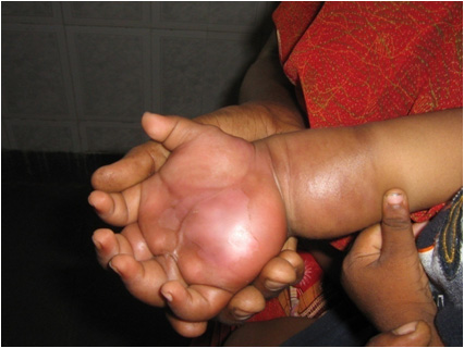
Here we report a case of subcutaneous zygomycosis caused by Basidiobolus ranarum in a 2 year old boy.
A 2 year old boy presented with a firm, painless , gradually progressive swelling of his right hand of 6 months duration.During this time he was subjected to repeated I&Ds suspecting that it was an abscess and antibiotic therapy by various doctors and even ATT for 2 past months.We made a provisional diagnose of deep fungal infection,probably Subcutaneous Zygomycosis.Skin biopsy was done under G.A for histopathological analysis and for tissue culture,while a therapeutic trial of Fluconazole was started at the dose of 50mg daily .The skin biopsy was reported as dense subcutaneous inflammation composed predominantly of eosnophils and plasma cells along with a few non-septate fungal hyphae which were identified with an eosinophilic sheath (Splendore-Hoeppli phenomenon) characteristic of Subcutaneous Zygomycosis but tissue growth was negative for fungal growth after 7 days.This could be probably due to the fact that zygomycetous fungi have primitive coenocytic hyphae that will often be damaged and become non-viable during the biopsy procedure or by the chopping up or tissue grinding process in the laboratory.When the boy presented in our OPD after 2weeks the lesions had subsided by almost 30% and by 70 to80% in another 2 more weeks of treatment and completely resolved in about 2 months treatment. We present this case because investigations support,corroborate and document but may not substitute a clinical diagnosis.Also it’s a rare case as only 4 cases have been reported in IJDVL so far and also to suggest that Fluconazole could be tried in deep fungal infections though books are skeptical about it.
Subcutaneous zygomycosis, is the commonest clinical form of basidiobolomycosis, is endemic in South India.4,5 There was no predisposing factor identified in this case, though trauma could be a probably the mode of entry. It is mainly isolated from the extremities, trunk and intestinal tract, [2,3]whereas the present case it had involved the right hand.
The aggressive nature of disease, as shown by the extradural involvement of the mass, is an unusual presentation of basidiobolomycosis. Diagnosis is mainly based on histopathological examination and fungal culture of biopsy specimen. The typical histopathological feature is the presence of thin-walled, broad, often aseptate hyphae or hyphal fragments with an eosinophilic sheath (Splendore-Hoeppli phenomenon), frequently phagocytised within giant cells[2,3].On fungal culture, thick-walled zygospore with a pair of conical projection (tubular protuberances) is visualised which is the characteristic feature of Basidiobolus ranarum[2].An immunodiffusion test has also been developed for specific diagnosis of the disease, 2,3but it is not routinely available.
Most patients with Basidiobolomycosis respond very well to oral potassium iodide therapy as also to azoles particularly itraconazole. Treatment with amphotericin B gave unsatisfactory results, with some strains and even showed in vitro resistance to this drug[6,7].The infection could be fatal or cause disabilities if not treated properly or treated very late, however our patient got completely cured after 2 months of treatment with fluconazole[8].

Clearly Auctoresonline and particularly Psychology and Mental Health Care Journal is dedicated to improving health care services for individuals and populations. The editorial boards' ability to efficiently recognize and share the global importance of health literacy with a variety of stakeholders. Auctoresonline publishing platform can be used to facilitate of optimal client-based services and should be added to health care professionals' repertoire of evidence-based health care resources.


