AUCTORES
Globalize your Research

Research Article | DOI: https://doi.org/10.31579/2578-8949/037
*Corresponding Author: Collin Jared, Department of Dermatology, Manchester, UK.
Citation: Collin Jared, Diego Luis, Jonathan Noah, and Benjamin Dylan, Transdermal diffusion of xenon from lipophilic solution of capric triglyceride and from water. J .Dermatology and Dermatitis. Doi: 10.31579/2578-8949/037
Copyright: © 2018 Collin Jared. This is an open-access article distributed under the terms of the Creative Commons Attribution License, which permits unrestricted use, distribution, and reproduction in any medium, provided the original author and source are credited.
Received: 10 May 2018 | Accepted: 02 June 2018 | Published: 10 June 2018
Keywords: xenon; rat skin; transdermal delivery; franz diffusion cell
Aim: The purpose of the study was to characterize transdermal delivery of xenon through rat skin from a lipophilic solution and from water.
Methods: Sections of skin were obtained from adult rats (n=12) and were placed into static Franz diffusion cells for 24 h. Xenon diffusion coefficients were determined for diffusion from a lipophilic solution (n=6) and from water (n=6) to phosphate buffered saline (PBS) through skin and for diffusion from a lipophilic solution to PBS through a phase boundary in the absence of skin (n=6).
Results: Xenon flux (JXe) through skin from the lipophilic solution was 0.036 mg/hour×cm2 and permeability coefficient (Kp) was 0.003 cm/h; JXe through skin from water was 0.029 mg/h×cm2 and the Kp was 0.002 cm/h. Total time for xenon diffusion through skin from lipophilic solution and from water was ~2 h.
Conclusion: The study presents the first characterization of xenon diffusion through rat skin from multiphase solutions to PBS. These data may be useful for the development of xenon-rich pharmaceutical products for external use.
Xenon has been considered an ideal anaesthetic agent due to rapid induction and emergence of anaesthesia, efficient analgesia, and low toxicity [1,2]. Moreover, xenon is known as a medication for treatment of pain, insomnia, depression, and drug addiction [3,4]. Also, scientific literature presents evidence of the neuroprotective effects of xenon [5] in treatment of traumatic [6] and ischemic [7] brain injury. Data suggest that blockade of NMDA receptors in the nervous system by xenon plays a key role in neuroprotective effects of this agent [5,8-10]. Recently, NMDA receptors have been found in the axons of human skin [15] and in the keratinocytes of the epidermis [16,17]. These receptors are involved in skin barrier function [18], calcium regulation [19], and dermatitis development [20]. We expect that xenon will exert similar blocking action when interacting with NMDA receptors in skin providing that this gas penetrates skin.
Earlier studies demonstrated xenon solubility in blood with solubility coefficients of 0.14 [11] and 0.115 [12]. These values are significantly higher than those for water: 0.085 [11] or 0.096 [12], but smaller than for oil: 1.7 [11]. Xenon is amphiphilic and able to dissolve in multiphase liquids, lipophilic, hydrophilic, and emulsified solutions and so is promising for research, development, and implementation of intravenous and oral formulations delivering xenon to target receptors [13,14].
The purpose of this work was to quantify xenon diffusion through rat skin from lipophilic solution and water.
Chemicals
High purity xenon (>99.999%, Tianjin Dongchuangrixing Technology Co. Ltd, CHINA) was used in the study. A universal cosmetic component, capryl/capric triglyceride (trade name: Tegosoft CT, Evonik Goldschmidt GmbH, Germany), was used as lipophilic solvent for xenon. Xenon was dissolved in capryl/capric triglyceride by using homogenizer (3000 rpm) with the airtight tank. Peristaltic pump was used to deliver exact amounts of xenon. Detailed protocol for the process of dissolving xenon in liquids is described in the patent [21]. Phosphate buffered saline (PBS, рН=7.4) was prepared by using high purity chemical reagents (Seebio Biotech, Inc., China). Urethane (Shanhai Jinsul Bio-Technology Co., Ltd, China) was used for anaesthesia.
Full-thickness skin preparations (epidermis and derma) were obtained from 3-month-old male Wistar rats (220 to 290 g). All operations with laboratory animals were performed according to international standards (Ethics Committee of Siberian State Medical University, Resolution # 3886). Animal skin samples were obtained under general anaesthesia induced by infusion of 20% urethane solution (1 mL/100 g of body weight). When rats were under anaesthesia, abdominal skin surface was depilated with scissors and electric shaver avoiding skin damage. Dissected patch of skin was rinsed with 0.9%-NaCl saline solution and carefully cleaned of residual connective and adipose tissue so that only derma remained. Round preparations with diameter of 3.2 to 3.4 сm were dissected from the skin patch. Prepared skin samples were immediately mounted in the diffusion cells between the receptor and donor chambers. The receptor chamber was filled with PBS solution (8 mL); air bubbles were extracted from the solution;then the PBS solution was stabilized at the receptor chamber temperature of 37°C for 1 h. This methodology assured that the skin surface facing the donor chamber was maintained at 32°±0.1°C during the entire experiment.
All experiments were divided into three groups. In Group 1, we quantified xenon diffusion from lipophilic capryl/capric triglyceride to PBS solution. Group 1 results are essential to assess skin barrier function in xenon diffusion. In Group 2, we used lipophilic capryl/capric triglyceride to create a high xenon concentration in contact with the skin. Capryl/capric triglyceride and the stratum corneum of the skin are both lipophilic. It is good for xenon mass transport. In Group 3, we eliminated the close contact between lipophilic capryl/ capric triglyceride and the surface of the skin by adding a layer of water between the skin and the triglyceride.The results of this group show the diffusion of xenon through the skin from the water.
All experiments were performed in static Franz diffusion cells (PermeGear Inc., USA); a standard for studying dermal permeability in vitro [22,23]. Sets of three Franz diffusion cells were used in all experiments. The orifice area between the donor and receptor chambers was 1 сm2, the donor chamber volume was 2 mL and the receptor chamber volume was 8 mL. Receptor chamber content was continuously mixed with a magnetic stirrer (600 rpm). Receptor chamber temperature was maintained at 37°C by using an ultra-thermostat SC-15 (NingBo Scientz Biotechnology Co., Ltd, China).
Control parameters of xenon diffusion were obtained without skin when xenon diffused directly through the interphase boundary from capryl/capric triglyceride to PBS solution (control group, Group 1).
Receptor chambers were filled with 8 mL of PBS and allowed to come to the equilibrium temperature, 37°C, for 1 hour. Donor chambers were then top-filled with 1 mL of capryl/capric triglyceride saturated with xenon and sealed. An interphase boundary between capryl/capric triglyceride and PBS demarcated the boundary between donor and receptor chambers so that the diffusion system was formed as follows: xenon-rich capryl/capric triglyceride-PBS (Figure 1, Group 1).

In Group 2, 1 mL of xenon-rich capryl/capric triglyceride solution (Figure 1, Group 2) was applied to the skin, mounted between the donor and receptor chambers, to study xenon diffusion through skin from a lipophilic media. This diffusion system configuration is: xenon-rich capryl/capric triglycerideskin- PBS.
In Group 3, 0.5 mL of water was first applied to the skin surface. Then, 1 mL of xenon-rich capryl/capric triglyceride solution was carefully applied over the water layer (Figure 1, Group 3). Contact of capryl/capric triglyceride with skin was thus avoided. In this setup, the xenon-rich capryl/capric triglyceride solution was a source of xenon for the water layer that was adjacent to skin surface. This approach enabled us to achieve high xenon content in the water layer to establish the diffusion system of xenon-rich water-skin-PBS. In preliminary experiments, we tested xenon diffusion from capryl/ capric triglyceride solution into water through the interphase boundary similarly to control Group 1. After maintaining the system for 2 hours, xenon content in water in the receptor chamber was 1.453±0.043 mg (n=3).
After addition of 1 mL of xenon-rich capryl/capric triglyceride, the donor chambers were sealed. Samples (1 mL) were taken from the receptor chambers every 2 hours for 24 hours for xenon analysis. Precautions were taken to prevent the entry of air into the system. After each sampling event, 1 mL of PBS was immediately added back into the receptor chamber to maintain constant volume. The extracted samples were immediately placed into the custom 20-mL glass containers and sealed. Xenon content in the samples was determined by gas chromatography.
Xenon content in the samples was determined by using a gas chromatograph Agilent 7697A, 7890A (Agilent Technologies, USA). Headspace analysis in the presence of thermodynamic equilibrium was used to measure xenon content in the liquids. Calibration was carried out at 20°C by injecting pure xenon in 0.1 mL (0.549 mg) increments up tо 2.5 mL (13.725 mg) into the 20-mL airtight containers that were empty or had 1 mL of the test liquid with and without xenon. Sample volume was 1 mL and the container volume was 20 mL. Conditions of xenon content measurements were as follows: temperature of 250°C, pressure of 5.9204 psi, and purge flow rate of 2 mL/ min. Helium was used as carrier gas. Steady-state flow rate in chromatographic column (length of 30 m; inner diameter of 0.53 mm) was 4 mL/min at initial temperatures of 50°C to 120°C. Temperature of termodesorber`s detector was 300°C, flow rate was 10 mL/min and the signal was 5 HZ/0.04 min. Sample testing lasted for 0.5 min at gas chromatography cycle of 15 min.
Xenon diffusion was studied for 24 consecutive hours; xenon content in the samples was determinedevery two hours. Based on the xenon concentration in 1 mL of PBS, we calculated total amount of xenon (Q) in the receptor chamber as a function of time. The flux (J) was calculated as amount of xenon (mg) crossing 1 сm2 of interphase boundary per 1 h. Under steady-state conditions with constant xenon flow rate, the permeability coefficient (Kp) was calculated by formula:
Kp = J/Cd
where Cd is xenon concentration in capryl/capric triglyceride in the donor chamber [24].
Each series consisted of six experiments. Data are presented as the mean±SD calculated in Microsoft Excel. For comparison of Group 2 and Group 3, values were considered statistically significant when P was <0.05 (Student's t-test).
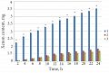
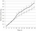
Initial concentration of xenon in capryl/capric triglyceride solution was 12.225±0.074 mg/mL (n=18). Figure 2 shows total amount of xenon (mg) in 8 mL and is the amount of xenon that has diffused through the interphase boundary (Group 1), through the skin from lipophilic media (Group 2) and through the lipophilic-hydrophilic media (Group 3, respectively). Figure 3 shows increasing xenon concentrations at different time points for the systems of diffusion through skin from the lipophilic (Group 2: solid line) and hydrophilic media (Group 3: dashed line). The figure shows that the difference between xenon diffusion in Group 2 and Group 3 occurs after hour 10. Therefore, the nature of the solvent does not significantly affect the diffusion during the first eight hours.


Transdermal xenon transport obeys Fick's first law of diffusion where in the diffusive flux J and is characterized by the amount of matter transferred through a unit of skin surface area per a unit of time. Kp can be calculated from the relationship Kp=Q/[Ad–Cr)] where Q is the total amount of xenon traversing the membrane in time t, and A is the area of exposed membrane in cm2, Cd is the concentration of the xenon on the donor chamber and Cr is the concentrations of the xenon on the receptor chamber. Usually Cd can be simplified as the donor concentration and Cras 0. The units of Kp are cm/h. The permeability coefficient Kp describes the rate of penetration.
We present results on transdermal delivery of xenon. Transdermal delivery is typically studied by using skin preparations from humans, pigs, and other laboratory animals including rodents [25,26]. It is important to note that comparative results for penetration of different compounds through the skin of different species, including hairless mice and humans, depend on the physical-chemical properties of the substances [27]. A lot of work has been done on rat skin and and rat skin is a relevant model [28,29]. In this study we present data on transdermal delivery for xenon for the first time. In Group 2 and Group 3, we detected xenon in the receptor chamber after 2 hours which represents the time for diffusion through skin. In Group 3, we identified xenon in only 3 out of 6 cases.
Water is an additional pathway for diffusion. During the period from 4 to 8 hours, xenon diffusion rates did not differ between Group 2 and Group 3. Data on xenon diffusion through skin from 10 h to 24 h in Group 3 can be explained by the fact that the presence of water adjacent to the skin surface increased time of xenon diffusion as the mean xenon diffusion rate decreased (by 17.5%). During this period, the decrease in diffusion could be caused by hydration of the epidermis. Hydration can induced skin swelling, thickening, and alteration of horny layer architectonics [30-33]. When the distance of xenon diffusion through the skin increases, diffusion rate decreases.
Comparison of diffusion rates for xenon diffusion from capryl/capric triglyceride to receptor chamber through the interphase boundary with no skin (Group 1) and through the skin (Group 2) demonstrated that the presence of skin slows down diffusion by ~31 times compared to the 2 h time point. Complete penetration of xenon through skin, as evidenced by detection of this gas in the receptor chamber, took ~2 h. After 2 h, the difference in diffusion rates gradually decreased15 fold at 4 hand 7.7 fold at 8 h. Starting from 10 h and throughout the rest of the experiment, this difference continued to decrease from 5.7 to 4.2 times. These data are important because they demonstrate the time of saturation of the skin with xenon. High xenon solubility in lipophilic liquids enabled us to achieve high topical xenon concentration on the skin surface ensuring the possibility of transdermal diffusion through all skin layers and water layer. In our study, xenon concentration in capryl/caprictriglyceride corresponded to its solubility coefficient of 1.2-1.3. At such concentrations, xenon-rich lipophilic solution remains stable and can be used for several months if stored in an airtight container.
Our data on transdermal diffusion of xenon through skin from multiphase solutions offer exciting possibilities for the development of xenon-rich medications designed for external use. The proposed mechanism of action of these medications is blockade of NMDA receptors by xenon. There is evidence that efficacy of many agents designed for treatment of dermal diseases associated with pain syndromes depends on their ability to suppress the activity of NMDA receptors (NMDA receptors antagonists) [34]. Continuing research and development of xenon-rich compounds for medicinal use requires further experiments done on different skin types including human preparations. Next steps will involve studying the effects of xenon on the activity of NMDA receptors in the skin and comparing with other known antagonists of NMDA receptors. In summary: xenon is a promising agent for anesthesia and therapy. It could be used separately or in a combination with other biologically active substances or enhancers. Mechanisms of xenon effects on skin and modalities of delivery require further studies.
In vitro transdermal diffusion of xenon from lipophilic solution of capryl/capric triglyceride and from water was characterized. Diffusion parameters flow (J) and permeability coefficient (Kp) were identified.
Diffusion rate for xenon diffusion through skin from water was significantly lower (17.5%) than t diffusion from capryl/ capric triglyceride solution for the time points from 10 h to 24 h.
Time for xenon penetration from capryl/capric triglyceride solution and from water through rat skin was 2 h.
Clearly Auctoresonline and particularly Psychology and Mental Health Care Journal is dedicated to improving health care services for individuals and populations. The editorial boards' ability to efficiently recognize and share the global importance of health literacy with a variety of stakeholders. Auctoresonline publishing platform can be used to facilitate of optimal client-based services and should be added to health care professionals' repertoire of evidence-based health care resources.


