AUCTORES
Globalize your Research

Editorial Note | DOI: https://doi.org/10.31579/2578-8949/032
*Corresponding Author: Hayden Wyatt, Department of Dermatology, Division of Immunology and Rheumatology, USA.
Citation: Hayden Wyatt, The Clinical Manifestation of Childhood Systemic Lupus Erythematous. J .Dermatology and Dermatitis. Doi: 10.31579/2578-8949/032
Copyright: © 2018 Hayden Wyatt. This is an open-access article distributed under the terms of the Creative Commons Attribution License, which permits unrestricted use, distribution, and reproduction in any medium, provided the original author and source are credited.
Received: 06 April 2018 | Accepted: 30 April 2018 | Published: 05 May 2018
Keywords: childhood; complement; c-reactive protein; malar rash; systemic lupus erythematosus
The clinical manifestation of childhood systemic lupus erythematosus (SLE) is similar to that of adult SLE; however, the former tends to be more severe and more aggressive than the latter. The prevalence of malar rash in SLE patients was reported to be 50% to 60%, and the rash may sometimes mimic sunburn erythema, as in our patient; thus, the underlying disease may be misdiagnosed in certain cases.
The clinical manifestation of childhood systemic lupus erythematosus (SLE) is similar to that of adult SLE; however, the former tends to be more severe and more aggressive than the latter. The malar rash in SLE patients may sometimes mimic sunburn erythema, as in our patient; thus, the underlying disease may be misdiagnosed in certain cases.
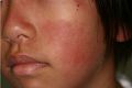
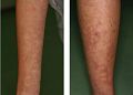
A previously healthy 12-year-old Japanese girl presented to our outpatient clinic with a rash in early September. She had been well except for easy fatigability and not had an excessive sun exposure. Her physician administered a topical ointment (hydrocortisone butyrate) as treatment for sunburn erythema on the face and an antihistaminergic drug (10mg/ day of bepotastine besilate ) for urticaria on the trunk and extremities for two weeks. Physical examination revealed a flat, brownish rash on the cheeks and nasal bridge (Figure 1), and an itchy erythematous rash on the trunk and extensor surfaces of the extremities (Figure 2); however, no lymphadenopathy, hepatosplenomegaly, or arthritis was noted.
In addition, chest auscultation revealed normal breath sounds. The laboratory findings were as follows: white blood cell count, 3,200/μL with 34% lymphocytes; hemoglobin level, 11.8 g/dL; platelet count, 17.2 × 10⁴/μL; aspartate aminotransferase level, 67 IU/L; alanine aminotransferase level, 69 IU/L; lactate dehydrogenase level, 400 IU/L; C-reactive protein (CRP) level, 0.02 mg/dL; ferritin level, 117.8 ng/mL; immunoglobulin G level, 3,120 mg/dL; erythrocyte sedimentation rate (ESR), 120 mm/h; complement (C) 3 level, 34 mg/dL (reference range, 77–195 mg/dL) ; C4 level, 1 mg/dL (reference range, 7–40 mg/dL); anti-nuclear antibody titer, 320× with a speckled pattern; and anti-dsDNA antibody level, 124.5 IU/mL (reference range, <10 IU/mL). On urinalysis, protein (2+) with cellular casts was found. Based on these results, we diagnosed the patient with systemic lupus erythematosus (SLE), because of the presence of 5 of the 11 criteria of the American College of Rheumatology (ACR) 1997 revised classification criteria for SLE [1]. Histopathological examination by a renal biopsy revealed class IV nephritis according to the World Health Organization classification (diffuse proliferative glomerulonephritis affecting >50% of glomeruli) [2]. After the diagnosis, she was treated with 40 mg/day of prednisolone and 150 mg/day of mizoribine and the disease had been controlled well.
Childhood-onset SLE is rare, with an estimated incidence rate of <1 case per 100,000 children. The clinical manifestation of childhood SLE is similar to that of adult SLE; however, the former tends to be more severe and more aggressive than the latter [3]. The prevalence of malar rash in SLE patients was reported to be 50% to 60%. In the literature, malar rash is typically described as a slightly raised, bright, erythematous rash on the light-exposed areas of the face (the "butterfly" area). However, the rash may sometimes mimic sunburn erythema [4], as in our patient; thus, the underlying disease may be misdiagnosed in certain cases. In addition to the ACR criteria, low serum complement levels and elevated ESRs with normal CRP levels could be helpful in the diagnosis of SLE [5].
Clearly Auctoresonline and particularly Psychology and Mental Health Care Journal is dedicated to improving health care services for individuals and populations. The editorial boards' ability to efficiently recognize and share the global importance of health literacy with a variety of stakeholders. Auctoresonline publishing platform can be used to facilitate of optimal client-based services and should be added to health care professionals' repertoire of evidence-based health care resources.


