AUCTORES
Globalize your Research

Case Report
*Corresponding Author: Smail Ammari, General Surgery Department, Ain Taya Hospital, 16029, Algiers, Algeria.
Citation: S. Ammari, N. Nait S, Y. Benhocine, N. Bounab, A. Bouzid, et al., (2023), Psoas Muscle, An Exceptional Location for Cystic Echinococcosis: About A Case, Journal of Clinical Surgery and Research, 4(5); DOI:10.31579/2768-2757/093
Copyright: © 2023, Smail Ammari. This is an open access article distributed under the Creative Commons Attribution License, which permits unrestricted use, distribution, and reproduction in any medium, provided the original work is properly cited.
Received: 01 September 2023 | Accepted: 15 September 2023 | Published: 22 September 2023
Keywords: cystic echinococcosis; hydatic cyst; psoas muscle
Introduction:
Primary extra-hepatic and extra-pulmonary locations of cystic echinococcosis are rare. Psoas muscle, is one of the unusual locations of cystic echinococcosis, few series of cases have been reported.
Purpose: report a new case of psoas muscle cystic echinococcosis, and review the literature to discuss diagnostic circumstances and therapeutic modalities
Observation: A 29-year-old man, consulted for painful swelling, extending from the right flank of the abdomen to the root of the right thigh. Preoperative diagnosis is facilitated by ultrasound, CT, and serology. The patient is operated by a median laparotomy. The intimate relationship of the cyst with the adjascente structures including the nerve structures prevented the realization of a complete kystectomy. Post-operative evolution is simple. An Albendazole-based medical treatment was associated in post-operative for one year.
Conclusion: Psoas muscle is one of the rare locations of cystic echinococcosis. Know how to think about it before any lumbar or iliac cystic formation. Surgical treatment remains the first line of therapy.
Cystic echinococcosis, a name now replacing that of hydatic cyst. It’s a cosmopolitan anthropozoonose, widespread throughout the world [1]. Liver is the most frequently affected organ (50percentage to to 70percentage to), followed by the lung (20percentage toto 30percentage to) [2]. Primary muscle location is exceptional, and uncommon, accounting for 0.4percentage to 1percentage to of all locations [2]. The pathophysiology of psoas muscle involvement remains hypothetical and imprecise [3]. The symptomatology is poor, and variable. Preoperative diagnosis is based on ultrasound, CT and serology. Treatment, can be radical, by a complete cystectomy, or conservative, by a partial resection of the cyst, with treatment of the parasite, and prevention of recurrence.
Our aim is reporting a new case of psoas muscle cystic echinococcosis, and to review the literature to discuss diagnostic circumstances and therapeutic modalities.
A 29-year-old patient, with no history of disease, consulted for right iliac fossa (RIF) and right thigh root pain, evolving for two weeks. The clinical examination finds a patient in good general condition, apyretic, body mass index (BMI) at 25. Visible and palpable oblong mass occupying right flank, the RIF, and the root of the right thigh; it was firm, painless, adherent to the deep plane, extended on about 20 cm of long axis. There’s a limitation in the extension of the right thigh in relation to psoitis. Digital rectal exam was without anomalies, hernial orifices were free.
Abdominal ultrasound showed a multicystic mass of the right iliac psoas muscle, compressive, evoking cystic echinococcosis.
Abdominopelvian computed tomography (CT-scann) found a multi-loculate formation, with a bi-sac wall, of the right pelvic retroperitoneum, contained a central calcification, measuring 22.2 cm high by 9.9 cm x 9.5 cm of transverse axis, extending from the retrocecal region to the root of the right thigh through the crural orifice.
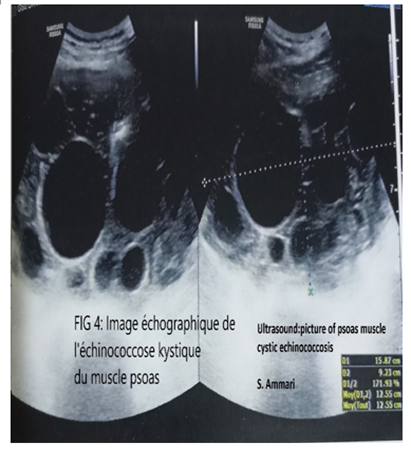
Figure 1: Psoas muscle cystic echinococcosis ultrasound image.
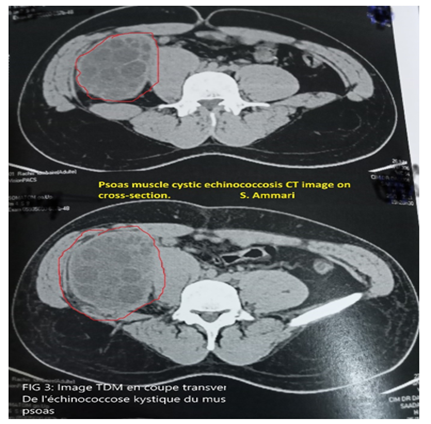
Figure 2: Psoas muscle cystic echinococcosis CT image on cross-section.
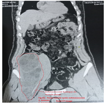
Figure 3: Psoas muscle cystic echinococcosis Front CT image.
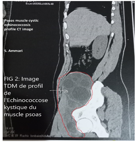
Figure 4: Psoas muscle cystic echinococcosis profile CT image.
Hydatic serology using the ELISA technique was positive (3.28 IU/L). Intradermal reaction (IDR) to tuberculin was negative. Other biological tests were normal.
We operated on the patient. Laparotomy under umbilical. Intraoperative exploration found a large retro peritoneal cystic mass, protruding under the right psoas muscle aponeurosis. This collection extended from the root of the psoas muscle at the height of the 3rd and 4th lumbar vertebra to the right coxo-femoral joint about 20 cm tall and 10 cm wide, pushing the right colon forward and towards the midline.
The attempt to perform a closed complete cystectomy was unsuccessful due to the cyst’s intimate contact with adjacent structures, particularly the nerves of the right lower limb.
After protecting the abdominal cavity with compresses soaked with hydrogen peroxide, we proceeded to a complete aspiration of the cyst, treatment of the parasite and sterilization of the residual cavity with hydrogen peroxide, with partial resection of the cystic cavity.
A drainage irrigation system left in the residual cavity. In postoperative, it allowed us to do iterative washings of this cavity.
Postoperative recoveries were simple and the patient was released on the 7th post-operative day.
Medical treatment, based on Albendazole at a rate of 10 mg/kg/day for one year (Started one month before the intervention and continued for one year), no recurrence noted.
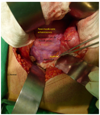
Figure 6: Intraoperative photo of the hydatic cyst of the psoas, pushing back the right colon.
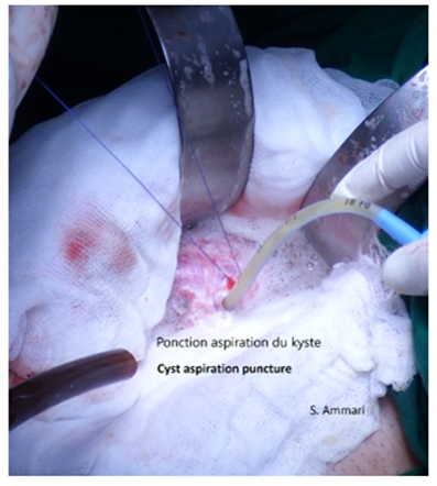
Figure 7: Intra-operative photo of the puncture and aspiration of the cyst.
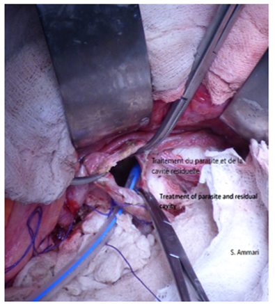
Figure 8: Intra-operative photo of parasite sterilization and residual cavity treatment.
The liver, then the lung, are the first filters that limit the passage of echinococcus granulosis to the general circulation. Rare to have primary involvement to other organs without liver and/or lung involvement. The psoas muscle, is one of the exceptional and uncommon locations of cystic echinococcosis.
The scarcity of muscular location is due to several factors, including the difficulty of local implantation of the embryo, caused by continuous muscle contractions and the production of lactic acid hindering the nesting of the embryo [3, 4, 5], the efficacy of liver and pulmonary filters that oppose the easy migration of the hexacanthe embryo into the systemic circulation [6. 7], the alternating muscle contraction-relaxation does not allow uniform vascularization and
exerts a compression preventing the parasite fixation on the muscle [6, 8,9, 10]. Finally, the absence of particular tropism of echinococcosis strains for muscle is the last possibility reported by the authors [6, 11].
The pathophysiology of psoas muscle hydatic cyst involvement remains hypothetical. Several contamination pathways are possible. First, the haematogenic pathway; after passing the liver and lung filters, the larva of the echinococcus granulosis is carried by the great circulation and is located in the most richly vascularized organs including the spleen, and muscles [3]. The second reported, is lymphatic contamination pathway or shunt from the gastrointestinal tract [2,3]. For our patient, these two contamination pathways are possible, and could explain this primitive localization in the psoas muscle. Other contamination pathways, have been reported, such as contiguous contamination from vertebrospinal hydatidosis is possible [3, 12]. This last-mentioned contamination pathway is not consistent with the case of our patient, who does not have a spinal injury.
Clinical manifestations of cystic echinococcosis of psoas muscle are variable. It’s generally a banal swelling, of slow evolution, long well supported, and therefore unknown [2]. Some cysts may be revealed by complications such as nerve compression, urinary, vascular, or haematogenic superinfection that can lead to sometimes severe sepsis [4, 13]. For our patient, the revelation of this disease was made by a painful swelling occupying the right flank, and the right iliac fossa (FID) of the abdomen.
Imaging allows visualization of the hydatic cyst and its constituent parts. His techniques are efficient and allow to establish a diagnosis, to judge complications, to carry out mass tests, and to perform instrumental treatments [2]. Ultrasound is a reliable examination and can clarify the hydatic nature of the cyst in more than 95% of cases [3, 6, 14,15,16]. Ultrasound is superior to CT for identification of the hydatic nature of the cyst, but the latter is more effective in the accuracy of its topography and ratios [3]. Ultrasound can also be used to specify the type according to Gharbi classification (16) and vascular and urinary ratios [3, 17]. The use of high-frequency probes refines the ultrasound study of the cystic wall [5]. Computed tomography (CT) shows morphological aspects similar to those shown by ultrasound [3, 5, 9, 18]. Cystic echinococcosis is characterized by the absence of contrast enhancement after injection [3]. The additional contribution of CT, compared to ultrasound, lies in accurate topographical diagnosis [3, 5, 18]. Moreover, CT is very useful in the precision of the hydatic nature of a mass (In case of type IV ultrasound) [3]. For our patient, ultrasound and CT have greatly contributed and facilitated positive diagnosis, with accurate topographical diagnosis to CT.
Magnetic resonance imaging (MRI) is reserved for cases where the diagnosis remains doubtful [3, 17]. The cyst image appears as a multi-vesicular lesion with or without peripheral hypo-signal on the T1 and T2-weighted Rim-sign sequences. There is often a parietal enhancement after gadolinium injection [6, 19, 20]. MRI, allows a detailed study of the wall, and cystic content [21]. It is useful in assessing cyst vitality by showing a hyper-signal in the daughter vesicles on T2-weighted sequences [6, 19]. In our case, MRI was not necessary.
Arteriography and cavography are currently abandoned. These two invasive scans were previously performed to assess the vascular impact of a retro peritoneal cyst [3].
Biology is essentially hydatic serology. It’s of great diagnostic value when it is positive [4]. Its main role lies in post-operative surveillance, looking for a possible recurrence, when it shows an ascent in antibody levels [3]. The qualitative (Immunoelectrophoresis, electrosyneresis) and quantitative methods (Indirect Haemagglutination, Immunofluorescence, ELISA) are difficult to interpret, however, the Western Blot and Immunoimprint are more sensitive and specific [5, 13]. In order to improve the sensitivity/specificity ratio, most authors prefer to combine two serological techniques, one quantitative: indirect haeaglutination, immunofluorescence, ELISA and the other qualitative: immunoelectrophoresis, electrosyneresis [4, 22]. In the case of our patient, ELISA is the technique that was used.
Eosinophilia is inconstant [3, 23]. It is concomitant with the invasion phase fades rapidly, sometimes persisting (in 7-15% of cases) at a moderate level. It may reappear during cyst cracking but fails in case of bacterial superinfection [2].
Surgery remains the first-line treatment for cystic echinococcosis of psoas. Medical treatment with Albendazole or Mebendazole remains an alternative for inoperable cases or in case of massive recurrence in addition to surgery [24]. In our patient, Albendazole has been systematically associated with surgical treatment. More recently, laparoscopy has been used, and its indications are being evaluated, it is a pathway that is not yet validated [2, 5, 25, 26].
Classical surgery remains the gold standard for cystic echinococcosis. The extraperitoneal surgical pathway is preferable to avoid any risk of intraperitoneal hydatic dissemination [24, 27, 28]. The transperitoneal pathway through a median, may be useful to treat at the same time other associated intraperitoneal hydatic lesions and especially hepatic [3]. Sometimes the recommended median incision, due to the large volume of the cyst [3]. In our case, we used the transperitoneal medial pathway because of the large volume of the cyst.
To avoid the spread of hydatic fluid and especially scolex in the abdominal cavity, it is essential to protect the surgical field by compresses soaked with parasiticide solutions [2].
Radical treatment is based on total kystectomy. However, adhesions to vasculonerveux elements can make this complete resection difficult or even dangerous [24]. Association with vertebral involvement is another contraindication of total cyst ectomy since, in this case, the parasite behaves maliciously by developing between bone trabeccles without forming a clean cystic wall [24, 29].
In some situations, radical treatment is impossible to achieve; it is then necessary to limit to a partial cyst, leaving a pericyst cap against the vascular and nervous elements to avoid their trauma during dissection [3]. In our case, we tried in vain to achieve a total kystectomy; the volume of the cyst and its
intimate relations with the vasculonerous structures intended for the lower limbs, forced us to limit ourselves to a partial kystectomy. The depth of the residual cavity, prompted us to leave in place an irrigation-drainage system, in order to carry out repeated washings postoperatively; thus, avoiding complications of the residual cavity, especially infectious complications.
Based on the literature, mortality is estimated at 4% and morbidity at 8.6% [3]. Paresthesia, in the territory of the crural nerve, may persist in postoperative stages due to intra-operative microtrauma of this nerve and which are often self-limiting [22, 30]. In our patient, mobility and mortality are zero, no recurrence is noted after one year of follow-up.
Psoas muscle is one of the uncommon locations of cystic echinococcosis. The rarity of this muscular localization is due to several factors, including difficulty of local implantation of the embryo, caused by continuous muscle contractions and the effectiveness of liver and lung filters. Clinical symptomatology is not specific. The diagnosis must be evoked before any lumbar or iliac fluid mass. Confirmation is based on imaging (Ultrasound, and CT), and hydatic serology. Radical total kystectomy surgery should be preferred whenever possible. Medical treatment remains an alternative for inoperable cases or in case of massive recurrence in addition to surgery. Prevention remains the best treatment.
Ethics approval
The results of this work come from a thesis work carried out by the main author (S. Ammari), and supervised by Professor M. Taieb at general surgery department of Ain Taya University Hospital.
Before starting this thesis work. A project was submitted to 03 experts at Algiers Faculty of Medicine who gave their approval to begin this research work. Thus, we had the authorization of the scientific council of Algiers faculty of medicine.
All patients are consenting for their inclusion in this work and for the publication of the results.
The author and co-authors declare that they have no conflicts of interest.
All authors contributed to this work
Funding will be provided by the lead author, with no funding from any other source.
The data (Patient records, information sheets for each patient) are available and entered in Excel and Word format
Clearly Auctoresonline and particularly Psychology and Mental Health Care Journal is dedicated to improving health care services for individuals and populations. The editorial boards' ability to efficiently recognize and share the global importance of health literacy with a variety of stakeholders. Auctoresonline publishing platform can be used to facilitate of optimal client-based services and should be added to health care professionals' repertoire of evidence-based health care resources.

Journal of Clinical Cardiology and Cardiovascular Intervention The submission and review process was adequate. However I think that the publication total value should have been enlightened in early fases. Thank you for all.

Journal of Women Health Care and Issues By the present mail, I want to say thank to you and tour colleagues for facilitating my published article. Specially thank you for the peer review process, support from the editorial office. I appreciate positively the quality of your journal.

Journal of Clinical Research and Reports I would be very delighted to submit my testimonial regarding the reviewer board and the editorial office. The reviewer board were accurate and helpful regarding any modifications for my manuscript. And the editorial office were very helpful and supportive in contacting and monitoring with any update and offering help. It was my pleasure to contribute with your promising Journal and I am looking forward for more collaboration.

We would like to thank the Journal of Thoracic Disease and Cardiothoracic Surgery because of the services they provided us for our articles. The peer-review process was done in a very excellent time manner, and the opinions of the reviewers helped us to improve our manuscript further. The editorial office had an outstanding correspondence with us and guided us in many ways. During a hard time of the pandemic that is affecting every one of us tremendously, the editorial office helped us make everything easier for publishing scientific work. Hope for a more scientific relationship with your Journal.

The peer-review process which consisted high quality queries on the paper. I did answer six reviewers’ questions and comments before the paper was accepted. The support from the editorial office is excellent.

Journal of Neuroscience and Neurological Surgery. I had the experience of publishing a research article recently. The whole process was simple from submission to publication. The reviewers made specific and valuable recommendations and corrections that improved the quality of my publication. I strongly recommend this Journal.

Dr. Katarzyna Byczkowska My testimonial covering: "The peer review process is quick and effective. The support from the editorial office is very professional and friendly. Quality of the Clinical Cardiology and Cardiovascular Interventions is scientific and publishes ground-breaking research on cardiology that is useful for other professionals in the field.

Thank you most sincerely, with regard to the support you have given in relation to the reviewing process and the processing of my article entitled "Large Cell Neuroendocrine Carcinoma of The Prostate Gland: A Review and Update" for publication in your esteemed Journal, Journal of Cancer Research and Cellular Therapeutics". The editorial team has been very supportive.

Testimony of Journal of Clinical Otorhinolaryngology: work with your Reviews has been a educational and constructive experience. The editorial office were very helpful and supportive. It was a pleasure to contribute to your Journal.

Dr. Bernard Terkimbi Utoo, I am happy to publish my scientific work in Journal of Women Health Care and Issues (JWHCI). The manuscript submission was seamless and peer review process was top notch. I was amazed that 4 reviewers worked on the manuscript which made it a highly technical, standard and excellent quality paper. I appreciate the format and consideration for the APC as well as the speed of publication. It is my pleasure to continue with this scientific relationship with the esteem JWHCI.

This is an acknowledgment for peer reviewers, editorial board of Journal of Clinical Research and Reports. They show a lot of consideration for us as publishers for our research article “Evaluation of the different factors associated with side effects of COVID-19 vaccination on medical students, Mutah university, Al-Karak, Jordan”, in a very professional and easy way. This journal is one of outstanding medical journal.

Dear Hao Jiang, to Journal of Nutrition and Food Processing We greatly appreciate the efficient, professional and rapid processing of our paper by your team. If there is anything else we should do, please do not hesitate to let us know. On behalf of my co-authors, we would like to express our great appreciation to editor and reviewers.

As an author who has recently published in the journal "Brain and Neurological Disorders". I am delighted to provide a testimonial on the peer review process, editorial office support, and the overall quality of the journal. The peer review process at Brain and Neurological Disorders is rigorous and meticulous, ensuring that only high-quality, evidence-based research is published. The reviewers are experts in their fields, and their comments and suggestions were constructive and helped improve the quality of my manuscript. The review process was timely and efficient, with clear communication from the editorial office at each stage. The support from the editorial office was exceptional throughout the entire process. The editorial staff was responsive, professional, and always willing to help. They provided valuable guidance on formatting, structure, and ethical considerations, making the submission process seamless. Moreover, they kept me informed about the status of my manuscript and provided timely updates, which made the process less stressful. The journal Brain and Neurological Disorders is of the highest quality, with a strong focus on publishing cutting-edge research in the field of neurology. The articles published in this journal are well-researched, rigorously peer-reviewed, and written by experts in the field. The journal maintains high standards, ensuring that readers are provided with the most up-to-date and reliable information on brain and neurological disorders. In conclusion, I had a wonderful experience publishing in Brain and Neurological Disorders. The peer review process was thorough, the editorial office provided exceptional support, and the journal's quality is second to none. I would highly recommend this journal to any researcher working in the field of neurology and brain disorders.
Dear Agrippa Hilda, Journal of Neuroscience and Neurological Surgery, Editorial Coordinator, I trust this message finds you well. I want to extend my appreciation for considering my article for publication in your esteemed journal. I am pleased to provide a testimonial regarding the peer review process and the support received from your editorial office. The peer review process for my paper was carried out in a highly professional and thorough manner. The feedback and comments provided by the authors were constructive and very useful in improving the quality of the manuscript. This rigorous assessment process undoubtedly contributes to the high standards maintained by your journal.

International Journal of Clinical Case Reports and Reviews. I strongly recommend to consider submitting your work to this high-quality journal. The support and availability of the Editorial staff is outstanding and the review process was both efficient and rigorous.

Thank you very much for publishing my Research Article titled “Comparing Treatment Outcome Of Allergic Rhinitis Patients After Using Fluticasone Nasal Spray And Nasal Douching" in the Journal of Clinical Otorhinolaryngology. As Medical Professionals we are immensely benefited from study of various informative Articles and Papers published in this high quality Journal. I look forward to enriching my knowledge by regular study of the Journal and contribute my future work in the field of ENT through the Journal for use by the medical fraternity. The support from the Editorial office was excellent and very prompt. I also welcome the comments received from the readers of my Research Article.

Dear Erica Kelsey, Editorial Coordinator of Cancer Research and Cellular Therapeutics Our team is very satisfied with the processing of our paper by your journal. That was fast, efficient, rigorous, but without unnecessary complications. We appreciated the very short time between the submission of the paper and its publication on line on your site.

I am very glad to say that the peer review process is very successful and fast and support from the Editorial Office. Therefore, I would like to continue our scientific relationship for a long time. And I especially thank you for your kindly attention towards my article. Have a good day!

"We recently published an article entitled “Influence of beta-Cyclodextrins upon the Degradation of Carbofuran Derivatives under Alkaline Conditions" in the Journal of “Pesticides and Biofertilizers” to show that the cyclodextrins protect the carbamates increasing their half-life time in the presence of basic conditions This will be very helpful to understand carbofuran behaviour in the analytical, agro-environmental and food areas. We greatly appreciated the interaction with the editor and the editorial team; we were particularly well accompanied during the course of the revision process, since all various steps towards publication were short and without delay".

I would like to express my gratitude towards you process of article review and submission. I found this to be very fair and expedient. Your follow up has been excellent. I have many publications in national and international journal and your process has been one of the best so far. Keep up the great work.

We are grateful for this opportunity to provide a glowing recommendation to the Journal of Psychiatry and Psychotherapy. We found that the editorial team were very supportive, helpful, kept us abreast of timelines and over all very professional in nature. The peer review process was rigorous, efficient and constructive that really enhanced our article submission. The experience with this journal remains one of our best ever and we look forward to providing future submissions in the near future.

I am very pleased to serve as EBM of the journal, I hope many years of my experience in stem cells can help the journal from one way or another. As we know, stem cells hold great potential for regenerative medicine, which are mostly used to promote the repair response of diseased, dysfunctional or injured tissue using stem cells or their derivatives. I think Stem Cell Research and Therapeutics International is a great platform to publish and share the understanding towards the biology and translational or clinical application of stem cells.

I would like to give my testimony in the support I have got by the peer review process and to support the editorial office where they were of asset to support young author like me to be encouraged to publish their work in your respected journal and globalize and share knowledge across the globe. I really give my great gratitude to your journal and the peer review including the editorial office.

I am delighted to publish our manuscript entitled "A Perspective on Cocaine Induced Stroke - Its Mechanisms and Management" in the Journal of Neuroscience and Neurological Surgery. The peer review process, support from the editorial office, and quality of the journal are excellent. The manuscripts published are of high quality and of excellent scientific value. I recommend this journal very much to colleagues.

Dr.Tania Muñoz, My experience as researcher and author of a review article in The Journal Clinical Cardiology and Interventions has been very enriching and stimulating. The editorial team is excellent, performs its work with absolute responsibility and delivery. They are proactive, dynamic and receptive to all proposals. Supporting at all times the vast universe of authors who choose them as an option for publication. The team of review specialists, members of the editorial board, are brilliant professionals, with remarkable performance in medical research and scientific methodology. Together they form a frontline team that consolidates the JCCI as a magnificent option for the publication and review of high-level medical articles and broad collective interest. I am honored to be able to share my review article and open to receive all your comments.

“The peer review process of JPMHC is quick and effective. Authors are benefited by good and professional reviewers with huge experience in the field of psychology and mental health. The support from the editorial office is very professional. People to contact to are friendly and happy to help and assist any query authors might have. Quality of the Journal is scientific and publishes ground-breaking research on mental health that is useful for other professionals in the field”.

Dear editorial department: On behalf of our team, I hereby certify the reliability and superiority of the International Journal of Clinical Case Reports and Reviews in the peer review process, editorial support, and journal quality. Firstly, the peer review process of the International Journal of Clinical Case Reports and Reviews is rigorous, fair, transparent, fast, and of high quality. The editorial department invites experts from relevant fields as anonymous reviewers to review all submitted manuscripts. These experts have rich academic backgrounds and experience, and can accurately evaluate the academic quality, originality, and suitability of manuscripts. The editorial department is committed to ensuring the rigor of the peer review process, while also making every effort to ensure a fast review cycle to meet the needs of authors and the academic community. Secondly, the editorial team of the International Journal of Clinical Case Reports and Reviews is composed of a group of senior scholars and professionals with rich experience and professional knowledge in related fields. The editorial department is committed to assisting authors in improving their manuscripts, ensuring their academic accuracy, clarity, and completeness. Editors actively collaborate with authors, providing useful suggestions and feedback to promote the improvement and development of the manuscript. We believe that the support of the editorial department is one of the key factors in ensuring the quality of the journal. Finally, the International Journal of Clinical Case Reports and Reviews is renowned for its high- quality articles and strict academic standards. The editorial department is committed to publishing innovative and academically valuable research results to promote the development and progress of related fields. The International Journal of Clinical Case Reports and Reviews is reasonably priced and ensures excellent service and quality ratio, allowing authors to obtain high-level academic publishing opportunities in an affordable manner. I hereby solemnly declare that the International Journal of Clinical Case Reports and Reviews has a high level of credibility and superiority in terms of peer review process, editorial support, reasonable fees, and journal quality. Sincerely, Rui Tao.

Clinical Cardiology and Cardiovascular Interventions I testity the covering of the peer review process, support from the editorial office, and quality of the journal.

Clinical Cardiology and Cardiovascular Interventions, we deeply appreciate the interest shown in our work and its publication. It has been a true pleasure to collaborate with you. The peer review process, as well as the support provided by the editorial office, have been exceptional, and the quality of the journal is very high, which was a determining factor in our decision to publish with you.

The peer reviewers process is quick and effective, the supports from editorial office is excellent, the quality of journal is high. I would like to collabroate with Internatioanl journal of Clinical Case Reports and Reviews journal clinically in the future time.

Clinical Cardiology and Cardiovascular Interventions, I would like to express my sincerest gratitude for the trust placed in our team for the publication in your journal. It has been a true pleasure to collaborate with you on this project. I am pleased to inform you that both the peer review process and the attention from the editorial coordination have been excellent. Your team has worked with dedication and professionalism to ensure that your publication meets the highest standards of quality. We are confident that this collaboration will result in mutual success, and we are eager to see the fruits of this shared effort.

Dear Dr. Jessica Magne, Editorial Coordinator 0f Clinical Cardiology and Cardiovascular Interventions, I hope this message finds you well. I want to express my utmost gratitude for your excellent work and for the dedication and speed in the publication process of my article titled "Navigating Innovation: Qualitative Insights on Using Technology for Health Education in Acute Coronary Syndrome Patients." I am very satisfied with the peer review process, the support from the editorial office, and the quality of the journal. I hope we can maintain our scientific relationship in the long term.

Dear Monica Gissare, - Editorial Coordinator of Nutrition and Food Processing. ¨My testimony with you is truly professional, with a positive response regarding the follow-up of the article and its review, you took into account my qualities and the importance of the topic¨.

Dear Dr. Jessica Magne, Editorial Coordinator 0f Clinical Cardiology and Cardiovascular Interventions, The review process for the article “The Handling of Anti-aggregants and Anticoagulants in the Oncologic Heart Patient Submitted to Surgery” was extremely rigorous and detailed. From the initial submission to the final acceptance, the editorial team at the “Journal of Clinical Cardiology and Cardiovascular Interventions” demonstrated a high level of professionalism and dedication. The reviewers provided constructive and detailed feedback, which was essential for improving the quality of our work. Communication was always clear and efficient, ensuring that all our questions were promptly addressed. The quality of the “Journal of Clinical Cardiology and Cardiovascular Interventions” is undeniable. It is a peer-reviewed, open-access publication dedicated exclusively to disseminating high-quality research in the field of clinical cardiology and cardiovascular interventions. The journal's impact factor is currently under evaluation, and it is indexed in reputable databases, which further reinforces its credibility and relevance in the scientific field. I highly recommend this journal to researchers looking for a reputable platform to publish their studies.

Dear Editorial Coordinator of the Journal of Nutrition and Food Processing! "I would like to thank the Journal of Nutrition and Food Processing for including and publishing my article. The peer review process was very quick, movement and precise. The Editorial Board has done an extremely conscientious job with much help, valuable comments and advices. I find the journal very valuable from a professional point of view, thank you very much for allowing me to be part of it and I would like to participate in the future!”

Dealing with The Journal of Neurology and Neurological Surgery was very smooth and comprehensive. The office staff took time to address my needs and the response from editors and the office was prompt and fair. I certainly hope to publish with this journal again.Their professionalism is apparent and more than satisfactory. Susan Weiner

My Testimonial Covering as fellowing: Lin-Show Chin. The peer reviewers process is quick and effective, the supports from editorial office is excellent, the quality of journal is high. I would like to collabroate with Internatioanl journal of Clinical Case Reports and Reviews.

My experience publishing in Psychology and Mental Health Care was exceptional. The peer review process was rigorous and constructive, with reviewers providing valuable insights that helped enhance the quality of our work. The editorial team was highly supportive and responsive, making the submission process smooth and efficient. The journal's commitment to high standards and academic rigor makes it a respected platform for quality research. I am grateful for the opportunity to publish in such a reputable journal.

My experience publishing in International Journal of Clinical Case Reports and Reviews was exceptional. I Come forth to Provide a Testimonial Covering the Peer Review Process and the editorial office for the Professional and Impartial Evaluation of the Manuscript.
