AUCTORES
Globalize your Research
Research Article
*Corresponding Author: Aubrey C. Galloway, MD Seymour Cohn Professor Department of Cardiothoracic Surgery NYU Grossman School of Medicine 530 1st Avenue, Suite 9V, New York, NY 10016.
Citation: Michael P. Dorsey, Navneet Narula, Harvey I. Pass, Les James, Valeria Mezzano, et al, (2024), post-COVID-19 Aortitis with Acute Thoracic Aortic Aneurysms: A Gene Expression and Molecular Pathway Analysis, J, Surgical Case Reports and Images, 7(7); DOI:10.31579/2690-1897/199
Copyright: © 2024, Aubrey C. Galloway. This is an open access article distributed under the Creative Commons Attribution License, which permits unrestricted use, distribution, and reproduction in any medium, provided the original work is properly cited.
Received: 14 June 2024 | Accepted: 03 July 2024 | Published: 14 August 2024
Keywords: digital spatial profiling, covid19, inflammatory thoracic aortic aneurysms
Post-COVID-19 aortitis (AoC19) with aneurysm formation has been described, but little is known about the molecular pathophysiology. This study utilized Digital Spatial Profiling (DSP) to evaluate differentially expressed genes (DEG) and molecular pathway activation in the aorta of AoC19 compared to Giant Cell aortitis (AoGC).
Methods: Histopathology analysis was performed on AoC19 and AoGC and regions of interest (ROI) based on varying degrees of inflammation were chosen and encircled. ROIs were stained with fluorescent anti-cytokeratin, anti-leukocyte, anti-nuclear antibodies and profiling agents for 1,850 RNA targets using the COVID-19 Immune Response Atlas. DSP was performed to compare DEG in AoC19 compared to AoGC, and across zones of AoC19 itself. Molecular pathway activation and gene primary ontology functions were analyzed.
Results: 103 DGEs were demonstrated when comparing AoC19 to AoGC. In AoC19, the pathways activated were associated with viral protein interaction with cytokines and cytokine receptors, cytokine-cytokine receptor interactions, and COVID-19; while in AoGC, pathways activated were associated with human leukocyte antigens HLA-DRB1, HLA-DRB4, and CD4 T cells. 42 DGEs were demonstrated when comparing ROIs across AoC19 itself, activating molecular pathways associated with cytokine-cytokine receptor interactions, complement and coagulation cascades, signaling pathways for TNF, IL-17 and nuclear factor-kappa ß, and rheumatoid arthritis. The primary molecular ontology functions of the DGEs across AoC19 were for COVID-19, Kawasaki Disease/Takayasu arteritis, and aneurysm formation.
Conclusions: This study demonstrated differential gene expression in the aortic wall of AoC19 with the activation of molecular pathways shared between COVID-19, Kawasaki Disease/Takayasu arteritis and aneurysm formation.
AoC19 aortic aneurysm specimen from patient with post-COVID-19 aortitis
AoGC aortic aneurysm specimen from patient with Giant Cell aortitis
COVID-19 coronavirus 2019 disease
DEG differentially expressed genes
DSP digital spatial profiling
FDR false discovery rate
KEGG Kyoto Encyclopedia of Genes and Genomes
ROI regions of interest
Patients with COVID-19 can develop serious cardiovascular complications resulting in increased morbidity and mortality, including acute myocardial infarction, myocarditis, vasculitis, vascular thrombosis and thromboembolic events.[1,2] The SARS-CoV-2 viral spike (S) protein enters cells through angiotensin-converting enzyme 2 (ACE2) receptors, which are highly expressed in the heart, vascular system, respiratory tract and gastrointestinal tract. RNA sequencing data from multiple vascular beds, however, has demonstrated a low level of ACE2 expression in vascular endothelial cells compared to epithelial cells in the respiratory track and enterocytes in the gastrointestinal tract.[3-6] Therefore, much of the vascular endothelial cell damage in COVID-19 may be mediated by the host inflammatory immune response through the activation of inflammatory cytokines, macrophage and lymphocyte infiltration, and platelet and complement activation. The SARS-CoV-2 viral spike (S) protein also binds directly to toll-like receptors and nucleotide-binding and oligomerization domain (NOD)-like receptors on host leukocytes to activate the host immune system directly. In the most severely ill COVID-19 patients the host immune response was found to be dysregulated and maladaptive, which contributed to multisystem organ damage and the high mortality in these patients.[7] The host immune transcriptomic response signature to COVID-19 can persist for 6 weeks to 8 months after the initial infection and, result in late post-acute COVID-19 sequelae in certain patients.8,9 Late sequelae most commonly occurs 2-3 months after the initial infection, and the etiology is thought to be due to a persistent, maladaptive host immune response pattern rather than ongoing infection.[9] The development post COVID-19 aortic aneurysms has been reported as a rare late complication, although the molecular mechanism is unknown. [10,11,12]
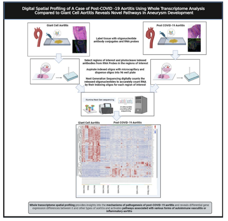
This study was generated after we treated a patient who presented with large vessel aortitis and acute thoracic aortic aneurysms six weeks after a mild COVID-19 respiratory illness. Based on the temporal relationship to the initial COVID-19 infection, and the unusual operative findings of acute inflammatory thoracic aortic aneurysms, with areas localized dissection, acute thrombus, and multiple pseudoaneurysms, our hypothesis was that this likely represented a late post-acute COVID-19 sequelae of large vessel inflammatory aortitis (AoC19). This study was designed to evaluate gene expressing and the molecular pathway activating patterns in the aortic wall of this patient (AoC19) using Digital Spatial Profiling (DSP) and to compare this to patient with Giant Cell aortitis (AoGC).
Patient Population
A 59-year-old female with a history of hypertension, diabetes, obesity, and metabolic syndrome presented with mild upper respiratory symptoms, normal oxygen saturation scattered mild infiltrates on Chest X-ray, a normal mediastinum, and tested positive for COVID-19 on nasopharyngeal swab. She did not require oxygen or hospital admission and was discharged. At home she remained clinically well, without further symptom, and apparently recovered, but developed left chest pain and back pain 6 weeks later and represented to the emergency room. The follow-up chest x-ray revealed clearing of the prior pulmonary infiltrates, but demonstrated a newly enlarged mediastinum. The nasopharyngeal swab for COVID-19 was negative, while blood work demonstrated anti-COVID-19 immunoglobulins (IgG.3.4, IgG 16.9, and IgM 3.9) consistent with a prior COVID-19 infection, and elevated inflammatory markers (D-dimer 1258 ng/mL, Ferritin 1035 ng/mL, and C-reactive protein 220 mg/L). Work up for infection (blood cultures, human immunodeficiency virus) and autoimmune disease (antinuclear antibodies, rapid plasma reagin, and antiphospholipid antibodies) was enegative. A computed tomographic angiography (CTA) of the chest demonstrated diffuse thickening of the thoracic aorta consistent with aortitis, and saccular pseudo-aneurysms in both the ascending and descending thoracic aorta. (Figure 1) Due to ongoing left lower back pain she underwent urgent endovascular aortic repair (TEVAR) of the descending thoracic aorta. Her pain resolved and she was discharged home on a course of steroids for presumed inflammatory aortitis, with plans for follow-up elective open repair of the ascending aorta and aortic arch in 2-3 weeks. However, she developed recurrent chest pain in 2 weeks and was readmitted for urgent surgery for ascending aorta and zone 2 aortic arch replacement. The operative findings included extensive inflammation and thickening of the aorta, with multiple areas of localized dissection, thrombus, and pseudoaneurysms. (Figure 2, B, C) She recovered uneventfully and was discharged home on postoperative day 5. The postoperative CT scan demonstrating the ascending aorta and zone II aortic arch replacement is shown in Figure 3.
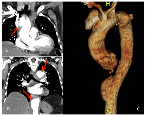
Figure 1. CTA coronal view with arrow demonstrating 4.5cm saccular pseudoaneurysm of the ascending aorta CTA coronal view with arrows demonstrating two descending thoracic aortic pseudoaneurysm (arrows) CTA three-dimensional reconstruction demonstrating multiple thoracic aortic pseudoaneurysms.
CTA, computed tomographic angiography

Figure 2. A. Completion aortogram after initial endovascular repair of the descending thoracic aorta. B. Intraoperative image demonstrating the whitish, inflamed and thickened ascending aorta. C. Intraoperative image demonstrating intimal thickening and multiple penetrating defects in the inner curvature of the open aortic arch.
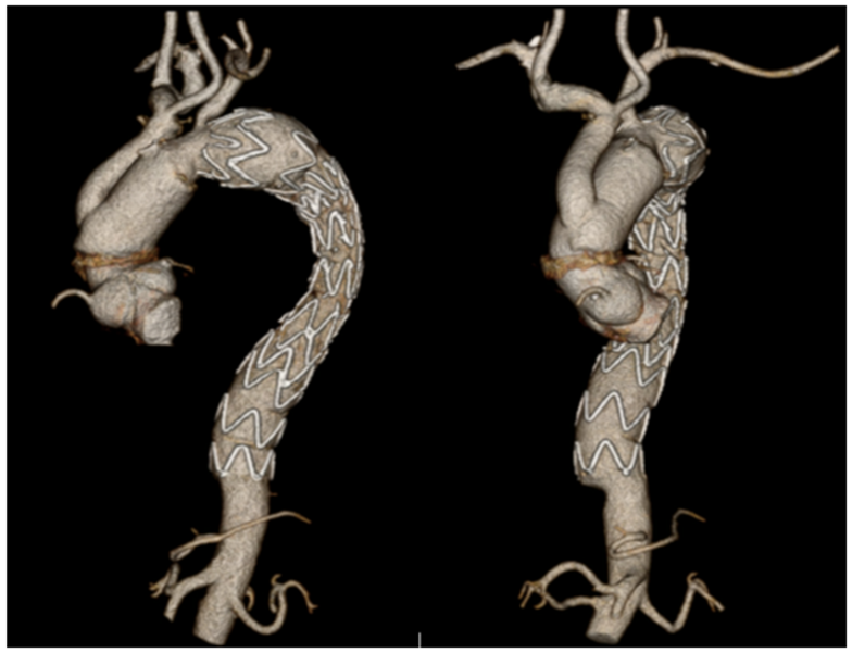
Figure 3. Postoperative CT scan demonstrating the ascending aorta and zone II aortic arch repair with debranching, and prior descending thoracic aorta stent graft. CT (computed tomographic).
This study was conducted under the NYU Grossman School of Medicine Lung Cancer Biomarker Center (#I8896) and underwent expedited IRB review and approval in 2021. Histopathologic analysis, along with special bacterial and fungal stains, was performed on the AoC19 specimen, and on a specimen from a patient with Giant Cell aortitis (AoGC) who had previously undergone ascending aneurysm repair. Electron microscopy to test for viral particles was also performed on the AoC19 specimen. Regions of interest (ROI) in different zones of the aortic, based on varying degrees of inflammation or fibrosis, and were marked and encircled for subsequent analysis. Formalin-fixed, paraffin-embedded sections of the specimens were subjected to a standard antigen retrieval protocol and stained with fluorescent anti-cytokeratin (i.e. pan-cytokeratin), anti-leukocyte (i.e. CD45) antibodies, and DNA binding SYTO-13 for nuclear recognition. The barcodes for each fluorescent DNA target in the ROIs were then selected for analysis and decoded for sequencing. (Figure 4) DSP) was then performed to evaluate DEGs, using the COVID-19 Immune Response Atlas (NanoString, WA, USA), which included a combination of the GeoMx Cancer Transcriptome Atlas of 1850 immune genes with an additional 30 genes from a specially designed COVID-19 panel. DEG analyses were performed by comparing ROI’s in AoC19 to AoGC, and by comparing ROIs across various zones across
AoC19 itself, representing normal areas, and zones with varying degrees of inflammation or fibrosis. Log fold changes with a false discovery rate (FDR) of <0>
Histopathology
Histopathologic analysis of AoC19 demonstrated patchy necrosis of the aortic wall, with localized areas of dissection, thrombus, and multiple sterile abscesses. Stains for fungal and bacterial organisms were negative, and no viral particles were identified in the AoC19 specimen by electron microscopy. Immunofluorescence microscopy of AoC19 demonstrated acute thrombus with infiltrating neutrophils, with surrounding areas of lymphoplasmacytic inflammation, and areas of fibrosis in the adventitia and in AoGC demonstrated histiocytic inflammation, with granulomatous aortitis and giant cells, as is typically seen in AoGC. ROIs of each specimen, based on normal appearing areas and areas with varying degrees of inflammation or fibrosis, were encircled for subsequent analysis by DSP. (Figure 5, sidebars)
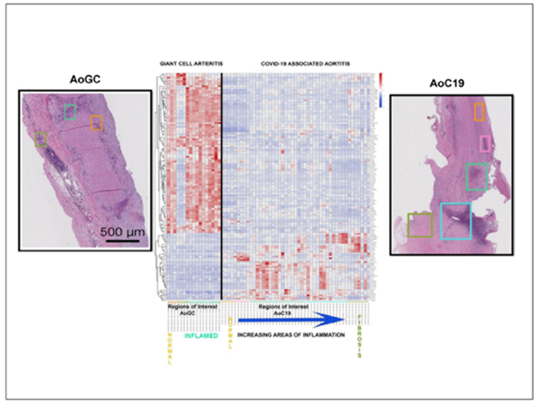
Figure 5. Digital Spatial transcriptomic heat-map identifying differentially expressed genes when comparing post-COVID-19 aortitis (AoC19) to giant cell arteritis (AoGC). Histologic sections of each =specimen are demonstrated on the side panels, with the colored squares in the heat map corresponding to the regions of interest (ROI)). The black line separates the AoGC clustes (left) from the AoC19 clusters (right). At the bottom of the heat map the ROI squares corresponding to the varying degrees of progressive inflammation across AoC19 itself are arranged from left to right.
Digital Spatial Profiling and KEGG Pathway Analysis
In comparing AoC19 to AoGC, 103 DGEs were demonstrated. The DEGs in AoC19 activated KEGG pathways associated with viral protein interaction with cytokines and cytokine receptors, cytokine-cytokine receptor interactions, and COVID-19; while in AoGC the KEGG pathways activated were associated with human leukocyte antigens HLA-DRB1, HLA-DRB4, and CD4 T cells. 42 DGEs were demonstrated when comparing ROIs across various inflammatory zones of AoC19 itself, (Table 1) activating KEGG pathways associated with cytokine-cytokine receptor interactions (q value=0.002); complement and coagulation cascades (q value=0.0009); signaling pathways for TNF (q value=0.004), IL-17 (q value=0.009), and NF-kß (q value=0.013); and rheumatoid arthritis (q value=0.009); Citrullinated H3-5, CLEC5A and CXCL2 are associated with neutrophil extracellular trap formation, neutrophil and macrophage migration, Kawasaki Disease and COVID-19.[15,16], The primary ontology functions of the DEGs identified across the zones of AoC19 were for COVID-19 infection (purple font), Kawasaki Disease/Takayasu arteritis (green font), and aneurysm formation (red font). (Figure 6)
| Gene | Log Fold Change | FDR |
| ITGA8 | -1.372 | 0.012 |
| CES3 | -0.939 | 0.013 |
| CFI | -0.611 | 0.022 |
| ACTA2 | -1.541 | 0.034 |
| IL22 | -0.646 | 0.035 |
| USP9Y | -0.899 | 0.035 |
| TNFRSF11B | -1.675 | 0.036 |
| ARG2 | -0.760 | 0.040 |
| ITGAX | 1.724 | 0.000 |
| H3-3A | 1.702 | 0.001 |
| AQP9 | 1.130 | 0.002 |
| CXCR4 | 1.714 | 0.006 |
| KRT14 | 0.872 | 0.008 |
| CD55 | 0.879 | 0.010 |
| RPS27A | 1.505 | 0.012 |
| CSF3R | 1.689 | 0.012 |
| VEGFA | 2.389 | 0.012 |
| SIRPA | 0.942 | 0.012 |
| IER3 | 1.898 | 0.012 |
| RPL7A | 1.374 | 0.013 |
| TREM1 | 1.163 | 0.015 |
| CXCL2 | 0.988 | 0.015 |
| SGK1 | 0.984 | 0.015 |
| ALDOA | 1.601 | 0.015 |
| TNFAIP6 | 0.857 | 0.015 |
| TNFAIP3 | 1.200 | 0.018 |
| ICAM1 | 1.017 | 0.022 |
| SERPINA1 | 2.346 | 0.023 |
| IL1R2 | 1.018 | 0.030 |
| TNFRSF1B | 0.730 | 0.030 |
| PTPN11 | 0.694 | 0.030 |
| MMP1 | 2.677 | 0.032 |
| BMP2 | 1.421 | 0.032 |
| CD44 | 1.734 | 0.033 |
| CEBPB | 0.908 | 0.035 |
| NFKB2 | 0.639 | 0.035 |
| DDIT4 | 1.050 | 0.035 |
| CLEC5A | 1.053 | 0.038 |
| CD53 | 1.035 | 0.040 |
| H3-5 | 1.075 | 0.040 |
| FPR1 | 0.835 | 0.043 |
| VSIR | 0.623 | 0.050 |
Table 1. The significant differentially expressed genes across zones of AoC19 itself when comparing normal zones to zones with varying degrees of inflammation or fibrosis.
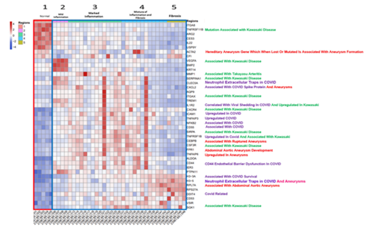
Figure 6: The digital spatial transcriptomic heat-map that displays the 42 significant differentially expressed genes (DEGs) across the various inflammatory zones of AoC19 along the vertical y-axis, and displays the zones of AoC19 with increasing degrees of inflammation or fibrosis from left to right along the horizontal x-axis. The primary ontology functions of the DEGs were for COVID-19 (purple font), Kawasaki Disease/Takayasu arteritis (green font), and aneurysm formation (red font).
The host inflammatory process plays a major role in the development of thoracic aortic aneurysms, producing endothelial cell dysfunction, vascular smooth muscle cell stress with a change in phenotype, and the activation of matrix metalloproteases in the extracellular matrix. (13) An analysis of bulk transcriptomome and single-cell RNA sequencing data identified 1525 DEGs in thoracic aortic aneurysms compared to normal aortic controls, activating multiple immune cell types, including macrophages, neutrophils, and T cells, and molecular pathways involved with leukocyte trans-endothelial migration, and the activation of complement and coagulation cascades. Thirty-nine (39) DEGs were identified in the macrophage population of and 30 DEGs in the T cell population. These were associated with increased infiltration of M2 macrophages and CD8+ T cells into the aortic wall, suggesting a major role of these cell types in development of thoracic aneurysms. (13)
Large vessel vasculitis with inflammatory thoracic aneurysms are most commonly due to Giant Cell aortitis (AoGC), Takayasu’s arteritis, and isolated aortitis. (14,15) While the exact etiology is unknown, the molecular pathophysiology is thought to be antigen related, with the activation of inflammatory cytokines, macrophages, and T cells (13) Using global transcriptomic profiling and bulk RNA sequencing, Hur and associates compared DEG signatures between inflammatory and non-inflammatory aneurysms, and demonstrated 159 upregulated DEGs and 93 downregulated DEGs in the inflammatory group. (16) Gene ontology enrichment demonstrated that the top 3 gene ontology terms were for immune response, host defense response, and inflammatory response; and pathways associated with increased cytokine and chemokine activity, and autoimmune disease. (16). Patients with Giant Cell aortitis have been shown to have DEGs in the aortic wall and circulating leukocytes that activate CD4 T cells and macrophages, and molecular pathways associated with a Type I interferon signature.[17]
Multisystem inflammatory syndrome in children (MIS-C) is a systemic medium vessel vasculitis thought to be triggered by SARS-CoV-2. It typically develops 3-4 weeks after the initial infection, (18,19,20) MIS-C) and is distinct from Kawasaki Disease, but with overlapping transcriptomic pathways. (18). An artificial intelligence-guided review of host immune signatures in the COVID-19 pandemic revealed shared host immune response patterns between COVID-19 (SARS-Co-V-2), MIS-C, and Kawasaki disease. (20).
The patient in this study presented 6 weeks after a mild COVID-19 infection with acute large vessel vasculitis and thoracic aortic aneurysms. There were no viral particles in the aortic specimen and the patient did not have an active infection. Our findings, however, demonstrated DEGs in the aortic wall that activated KEGG pathways associated with viral protein interaction with cytokines and cytokine receptors, cytokine-cytokine receptor interactions, and COVID-19 with primary ontology functions for COVID-19, Kawasaki Disease/Takayasu arteritis, and aneurysm formation. These findings suggest a shared and overlapping expression of host immune transcriptomic pathways in large vessel inflammatory vasculitis, inflammatory thoracic aneurysms, MIS-C, Kawasaki disease and autoimmune aortitis, The study is unique in the use of multiplexed DSP to analyze DEGs and molecular pathway activation patterns in specific areas of the aortic wall. This approach is considerably different than the more widely used bulk transcriptomic and single cell RNA methods, which measure DEGs in the entire aortic specimen. Based on this pilot study it is possible that DSP might be useful in analyzing molecular mechanisms in specific zones of aortic wall in various types of aneurysms, which could lead to a better understanding of the molecular pathophysiology involved.
This was a pilot study with only one patient and one control, and has many limitations. Based on the small sample size, the statistical significance of the findings should be considered marginal and the findings need to be validated across a larger number of patients. A major limitation is that the study does not have a normal aorta as a control. These findings only represent DEG between specific areas within the aortic wall across the AoC19 specimen, and DEGs in comparison to Giant Cell aortitis, and do not identify DEGs compared to a normal aorta. While DSP may have certain advantages in allowing the spatial comparison of gene expression in the microenvironment of tissue specimens, that is also a limitation, and the use of bulk transcriptomics and single cell RNA may be more robust in identifying major tissue wide transcriptomic patterns across a large number of specimens or patients.
Pass, Mezzano, Zhou, Tsirigos, Ramaswami, James, Peterson, and Smith, have nothing to disclose. Dr. Galloway has intellectual property with Medtronic and receives royalties for valve repair devices.
This study was supported by NYU Grossman School of Medicine, Department of Cardiothoracic Surgery Research Fund. The Experimental Pathology and Genome Technology Center core laboratories are a part of the NYU Grossman School of Medicine, Division of Advanced Research Technologies, and are funded in part by the National Institutes of Health/National Cancer Institute [5 P30CA16087].
Abstract presented at 2022 AATS Aortic Symposium: “Post-COVID-19 Aortitis with Aneurysm Formation.”
The study was conducted under the NYU Grossman School of Medicine Lung Cancer Biomarker Center (#I8896) and underwent expedited IRB review.
The GeoMx gene tag sequencing data sets generated in this study will be available at GEO, and Code will be available at GitHub. Microscopy image data is stored in public OMERO Plus and will be accessible through the NYU Data Catalog upon publication.
Clearly Auctoresonline and particularly Psychology and Mental Health Care Journal is dedicated to improving health care services for individuals and populations. The editorial boards' ability to efficiently recognize and share the global importance of health literacy with a variety of stakeholders. Auctoresonline publishing platform can be used to facilitate of optimal client-based services and should be added to health care professionals' repertoire of evidence-based health care resources.

Journal of Clinical Cardiology and Cardiovascular Intervention The submission and review process was adequate. However I think that the publication total value should have been enlightened in early fases. Thank you for all.

Journal of Women Health Care and Issues By the present mail, I want to say thank to you and tour colleagues for facilitating my published article. Specially thank you for the peer review process, support from the editorial office. I appreciate positively the quality of your journal.
Journal of Clinical Research and Reports I would be very delighted to submit my testimonial regarding the reviewer board and the editorial office. The reviewer board were accurate and helpful regarding any modifications for my manuscript. And the editorial office were very helpful and supportive in contacting and monitoring with any update and offering help. It was my pleasure to contribute with your promising Journal and I am looking forward for more collaboration.

We would like to thank the Journal of Thoracic Disease and Cardiothoracic Surgery because of the services they provided us for our articles. The peer-review process was done in a very excellent time manner, and the opinions of the reviewers helped us to improve our manuscript further. The editorial office had an outstanding correspondence with us and guided us in many ways. During a hard time of the pandemic that is affecting every one of us tremendously, the editorial office helped us make everything easier for publishing scientific work. Hope for a more scientific relationship with your Journal.

The peer-review process which consisted high quality queries on the paper. I did answer six reviewers’ questions and comments before the paper was accepted. The support from the editorial office is excellent.

Journal of Neuroscience and Neurological Surgery. I had the experience of publishing a research article recently. The whole process was simple from submission to publication. The reviewers made specific and valuable recommendations and corrections that improved the quality of my publication. I strongly recommend this Journal.

Dr. Katarzyna Byczkowska My testimonial covering: "The peer review process is quick and effective. The support from the editorial office is very professional and friendly. Quality of the Clinical Cardiology and Cardiovascular Interventions is scientific and publishes ground-breaking research on cardiology that is useful for other professionals in the field.

Thank you most sincerely, with regard to the support you have given in relation to the reviewing process and the processing of my article entitled "Large Cell Neuroendocrine Carcinoma of The Prostate Gland: A Review and Update" for publication in your esteemed Journal, Journal of Cancer Research and Cellular Therapeutics". The editorial team has been very supportive.

Testimony of Journal of Clinical Otorhinolaryngology: work with your Reviews has been a educational and constructive experience. The editorial office were very helpful and supportive. It was a pleasure to contribute to your Journal.

Dr. Bernard Terkimbi Utoo, I am happy to publish my scientific work in Journal of Women Health Care and Issues (JWHCI). The manuscript submission was seamless and peer review process was top notch. I was amazed that 4 reviewers worked on the manuscript which made it a highly technical, standard and excellent quality paper. I appreciate the format and consideration for the APC as well as the speed of publication. It is my pleasure to continue with this scientific relationship with the esteem JWHCI.

This is an acknowledgment for peer reviewers, editorial board of Journal of Clinical Research and Reports. They show a lot of consideration for us as publishers for our research article “Evaluation of the different factors associated with side effects of COVID-19 vaccination on medical students, Mutah university, Al-Karak, Jordan”, in a very professional and easy way. This journal is one of outstanding medical journal.
Dear Hao Jiang, to Journal of Nutrition and Food Processing We greatly appreciate the efficient, professional and rapid processing of our paper by your team. If there is anything else we should do, please do not hesitate to let us know. On behalf of my co-authors, we would like to express our great appreciation to editor and reviewers.

As an author who has recently published in the journal "Brain and Neurological Disorders". I am delighted to provide a testimonial on the peer review process, editorial office support, and the overall quality of the journal. The peer review process at Brain and Neurological Disorders is rigorous and meticulous, ensuring that only high-quality, evidence-based research is published. The reviewers are experts in their fields, and their comments and suggestions were constructive and helped improve the quality of my manuscript. The review process was timely and efficient, with clear communication from the editorial office at each stage. The support from the editorial office was exceptional throughout the entire process. The editorial staff was responsive, professional, and always willing to help. They provided valuable guidance on formatting, structure, and ethical considerations, making the submission process seamless. Moreover, they kept me informed about the status of my manuscript and provided timely updates, which made the process less stressful. The journal Brain and Neurological Disorders is of the highest quality, with a strong focus on publishing cutting-edge research in the field of neurology. The articles published in this journal are well-researched, rigorously peer-reviewed, and written by experts in the field. The journal maintains high standards, ensuring that readers are provided with the most up-to-date and reliable information on brain and neurological disorders. In conclusion, I had a wonderful experience publishing in Brain and Neurological Disorders. The peer review process was thorough, the editorial office provided exceptional support, and the journal's quality is second to none. I would highly recommend this journal to any researcher working in the field of neurology and brain disorders.

Dear Agrippa Hilda, Journal of Neuroscience and Neurological Surgery, Editorial Coordinator, I trust this message finds you well. I want to extend my appreciation for considering my article for publication in your esteemed journal. I am pleased to provide a testimonial regarding the peer review process and the support received from your editorial office. The peer review process for my paper was carried out in a highly professional and thorough manner. The feedback and comments provided by the authors were constructive and very useful in improving the quality of the manuscript. This rigorous assessment process undoubtedly contributes to the high standards maintained by your journal.

International Journal of Clinical Case Reports and Reviews. I strongly recommend to consider submitting your work to this high-quality journal. The support and availability of the Editorial staff is outstanding and the review process was both efficient and rigorous.

Thank you very much for publishing my Research Article titled “Comparing Treatment Outcome Of Allergic Rhinitis Patients After Using Fluticasone Nasal Spray And Nasal Douching" in the Journal of Clinical Otorhinolaryngology. As Medical Professionals we are immensely benefited from study of various informative Articles and Papers published in this high quality Journal. I look forward to enriching my knowledge by regular study of the Journal and contribute my future work in the field of ENT through the Journal for use by the medical fraternity. The support from the Editorial office was excellent and very prompt. I also welcome the comments received from the readers of my Research Article.

Dear Erica Kelsey, Editorial Coordinator of Cancer Research and Cellular Therapeutics Our team is very satisfied with the processing of our paper by your journal. That was fast, efficient, rigorous, but without unnecessary complications. We appreciated the very short time between the submission of the paper and its publication on line on your site.

I am very glad to say that the peer review process is very successful and fast and support from the Editorial Office. Therefore, I would like to continue our scientific relationship for a long time. And I especially thank you for your kindly attention towards my article. Have a good day!

"We recently published an article entitled “Influence of beta-Cyclodextrins upon the Degradation of Carbofuran Derivatives under Alkaline Conditions" in the Journal of “Pesticides and Biofertilizers” to show that the cyclodextrins protect the carbamates increasing their half-life time in the presence of basic conditions This will be very helpful to understand carbofuran behaviour in the analytical, agro-environmental and food areas. We greatly appreciated the interaction with the editor and the editorial team; we were particularly well accompanied during the course of the revision process, since all various steps towards publication were short and without delay".

I would like to express my gratitude towards you process of article review and submission. I found this to be very fair and expedient. Your follow up has been excellent. I have many publications in national and international journal and your process has been one of the best so far. Keep up the great work.

We are grateful for this opportunity to provide a glowing recommendation to the Journal of Psychiatry and Psychotherapy. We found that the editorial team were very supportive, helpful, kept us abreast of timelines and over all very professional in nature. The peer review process was rigorous, efficient and constructive that really enhanced our article submission. The experience with this journal remains one of our best ever and we look forward to providing future submissions in the near future.

I am very pleased to serve as EBM of the journal, I hope many years of my experience in stem cells can help the journal from one way or another. As we know, stem cells hold great potential for regenerative medicine, which are mostly used to promote the repair response of diseased, dysfunctional or injured tissue using stem cells or their derivatives. I think Stem Cell Research and Therapeutics International is a great platform to publish and share the understanding towards the biology and translational or clinical application of stem cells.

I would like to give my testimony in the support I have got by the peer review process and to support the editorial office where they were of asset to support young author like me to be encouraged to publish their work in your respected journal and globalize and share knowledge across the globe. I really give my great gratitude to your journal and the peer review including the editorial office.

I am delighted to publish our manuscript entitled "A Perspective on Cocaine Induced Stroke - Its Mechanisms and Management" in the Journal of Neuroscience and Neurological Surgery. The peer review process, support from the editorial office, and quality of the journal are excellent. The manuscripts published are of high quality and of excellent scientific value. I recommend this journal very much to colleagues.

Dr.Tania Muñoz, My experience as researcher and author of a review article in The Journal Clinical Cardiology and Interventions has been very enriching and stimulating. The editorial team is excellent, performs its work with absolute responsibility and delivery. They are proactive, dynamic and receptive to all proposals. Supporting at all times the vast universe of authors who choose them as an option for publication. The team of review specialists, members of the editorial board, are brilliant professionals, with remarkable performance in medical research and scientific methodology. Together they form a frontline team that consolidates the JCCI as a magnificent option for the publication and review of high-level medical articles and broad collective interest. I am honored to be able to share my review article and open to receive all your comments.

“The peer review process of JPMHC is quick and effective. Authors are benefited by good and professional reviewers with huge experience in the field of psychology and mental health. The support from the editorial office is very professional. People to contact to are friendly and happy to help and assist any query authors might have. Quality of the Journal is scientific and publishes ground-breaking research on mental health that is useful for other professionals in the field”.

Dear editorial department: On behalf of our team, I hereby certify the reliability and superiority of the International Journal of Clinical Case Reports and Reviews in the peer review process, editorial support, and journal quality. Firstly, the peer review process of the International Journal of Clinical Case Reports and Reviews is rigorous, fair, transparent, fast, and of high quality. The editorial department invites experts from relevant fields as anonymous reviewers to review all submitted manuscripts. These experts have rich academic backgrounds and experience, and can accurately evaluate the academic quality, originality, and suitability of manuscripts. The editorial department is committed to ensuring the rigor of the peer review process, while also making every effort to ensure a fast review cycle to meet the needs of authors and the academic community. Secondly, the editorial team of the International Journal of Clinical Case Reports and Reviews is composed of a group of senior scholars and professionals with rich experience and professional knowledge in related fields. The editorial department is committed to assisting authors in improving their manuscripts, ensuring their academic accuracy, clarity, and completeness. Editors actively collaborate with authors, providing useful suggestions and feedback to promote the improvement and development of the manuscript. We believe that the support of the editorial department is one of the key factors in ensuring the quality of the journal. Finally, the International Journal of Clinical Case Reports and Reviews is renowned for its high- quality articles and strict academic standards. The editorial department is committed to publishing innovative and academically valuable research results to promote the development and progress of related fields. The International Journal of Clinical Case Reports and Reviews is reasonably priced and ensures excellent service and quality ratio, allowing authors to obtain high-level academic publishing opportunities in an affordable manner. I hereby solemnly declare that the International Journal of Clinical Case Reports and Reviews has a high level of credibility and superiority in terms of peer review process, editorial support, reasonable fees, and journal quality. Sincerely, Rui Tao.

Clinical Cardiology and Cardiovascular Interventions I testity the covering of the peer review process, support from the editorial office, and quality of the journal.

Clinical Cardiology and Cardiovascular Interventions, we deeply appreciate the interest shown in our work and its publication. It has been a true pleasure to collaborate with you. The peer review process, as well as the support provided by the editorial office, have been exceptional, and the quality of the journal is very high, which was a determining factor in our decision to publish with you.
The peer reviewers process is quick and effective, the supports from editorial office is excellent, the quality of journal is high. I would like to collabroate with Internatioanl journal of Clinical Case Reports and Reviews journal clinically in the future time.

Clinical Cardiology and Cardiovascular Interventions, I would like to express my sincerest gratitude for the trust placed in our team for the publication in your journal. It has been a true pleasure to collaborate with you on this project. I am pleased to inform you that both the peer review process and the attention from the editorial coordination have been excellent. Your team has worked with dedication and professionalism to ensure that your publication meets the highest standards of quality. We are confident that this collaboration will result in mutual success, and we are eager to see the fruits of this shared effort.

Dear Dr. Jessica Magne, Editorial Coordinator 0f Clinical Cardiology and Cardiovascular Interventions, I hope this message finds you well. I want to express my utmost gratitude for your excellent work and for the dedication and speed in the publication process of my article titled "Navigating Innovation: Qualitative Insights on Using Technology for Health Education in Acute Coronary Syndrome Patients." I am very satisfied with the peer review process, the support from the editorial office, and the quality of the journal. I hope we can maintain our scientific relationship in the long term.
Dear Monica Gissare, - Editorial Coordinator of Nutrition and Food Processing. ¨My testimony with you is truly professional, with a positive response regarding the follow-up of the article and its review, you took into account my qualities and the importance of the topic¨.

Dear Dr. Jessica Magne, Editorial Coordinator 0f Clinical Cardiology and Cardiovascular Interventions, The review process for the article “The Handling of Anti-aggregants and Anticoagulants in the Oncologic Heart Patient Submitted to Surgery” was extremely rigorous and detailed. From the initial submission to the final acceptance, the editorial team at the “Journal of Clinical Cardiology and Cardiovascular Interventions” demonstrated a high level of professionalism and dedication. The reviewers provided constructive and detailed feedback, which was essential for improving the quality of our work. Communication was always clear and efficient, ensuring that all our questions were promptly addressed. The quality of the “Journal of Clinical Cardiology and Cardiovascular Interventions” is undeniable. It is a peer-reviewed, open-access publication dedicated exclusively to disseminating high-quality research in the field of clinical cardiology and cardiovascular interventions. The journal's impact factor is currently under evaluation, and it is indexed in reputable databases, which further reinforces its credibility and relevance in the scientific field. I highly recommend this journal to researchers looking for a reputable platform to publish their studies.

Dear Editorial Coordinator of the Journal of Nutrition and Food Processing! "I would like to thank the Journal of Nutrition and Food Processing for including and publishing my article. The peer review process was very quick, movement and precise. The Editorial Board has done an extremely conscientious job with much help, valuable comments and advices. I find the journal very valuable from a professional point of view, thank you very much for allowing me to be part of it and I would like to participate in the future!”

Dealing with The Journal of Neurology and Neurological Surgery was very smooth and comprehensive. The office staff took time to address my needs and the response from editors and the office was prompt and fair. I certainly hope to publish with this journal again.Their professionalism is apparent and more than satisfactory. Susan Weiner

My Testimonial Covering as fellowing: Lin-Show Chin. The peer reviewers process is quick and effective, the supports from editorial office is excellent, the quality of journal is high. I would like to collabroate with Internatioanl journal of Clinical Case Reports and Reviews.

My experience publishing in Psychology and Mental Health Care was exceptional. The peer review process was rigorous and constructive, with reviewers providing valuable insights that helped enhance the quality of our work. The editorial team was highly supportive and responsive, making the submission process smooth and efficient. The journal's commitment to high standards and academic rigor makes it a respected platform for quality research. I am grateful for the opportunity to publish in such a reputable journal.
My experience publishing in International Journal of Clinical Case Reports and Reviews was exceptional. I Come forth to Provide a Testimonial Covering the Peer Review Process and the editorial office for the Professional and Impartial Evaluation of the Manuscript.

I would like to offer my testimony in the support. I have received through the peer review process and support the editorial office where they are to support young authors like me, encourage them to publish their work in your esteemed journals, and globalize and share knowledge globally. I really appreciate your journal, peer review, and editorial office.
Dear Agrippa Hilda- Editorial Coordinator of Journal of Neuroscience and Neurological Surgery, "The peer review process was very quick and of high quality, which can also be seen in the articles in the journal. The collaboration with the editorial office was very good."

We found the peer review process quick and positive in its input. The support from the editorial officer has been very agile, always with the intention of improving the article and taking into account our subsequent corrections.
