AUCTORES
Globalize your Research

research article
*Corresponding Author: M. Bharathselvi, R.S. Mehta Jain Department of Biochemistry and cell Biology, Vision Research Foundation, Sankara Nethralaya, 41, College Road, Chennai-600 006.
Citation: Muthuve B, Konerirajapuram N. S, Ramanujam S, Jyothirmay B. (2022). Inflammation mediated alterations in the iron homeostasis of Eales’ Disease. J. Clinical Research Notes. 3(5); DOI: 10.31579/2690-8816/075
Copyright: © 2022 M. Bharathselvi, this is an open-access article distributed under the terms of the Creative Commons Attribution License, which permits unrestricted use, distribution, and reproduction in any medium, provided the original author and source are credited.
Received: 27 June 2022 | Accepted: 04 July 2022 | Published: 20 July 2022
Keywords: eales’ disease; idiopathic retinal vascular disease; iron transport; hepcidin
Background: Eales’ Disease is an idiopathic retinal periphlebitis, characterized by recurrent vitreous hemorrhage, neovascularization and inflammation. The disease distresses the retina of adult males between 15 and 45 years. In the present study proteins involved in iron homeostasis were assessed in serum and peripheral blood mononuclear cells.
Methods: Forty male subjects, were recruited for the study. Their blood samples were used to measure the ferritin, transferrin, soluble transferrin receptor, hepcidin, ferroportin and heme. Besides ALAS, HO, HIF-2 and other relevant parameters were also measured.
Results: In the ED group, significantly increased in the levels of heme, heme oxygenase, ferritin and VEGF were observed in serum and monocytes of ED, besides decrease in the levels of transferrin. Interestingly, the expression levels of hepcidin and HIF were increased whereas the ferroportin was found to be decreased.
Conclusions: These results propose evidence for the involvement of altered iron homeostasis.
Eales’ disease (ED), characterized as an idiopathic, inflammatory veno-occlusive disease, which affects, the peripheral retinal veins especially of young males [1,2]. The disease is found to be, prevalent in Indian subcontinent with an incidence rate of 1 in 200-250 ophthalmic patients [3,5]. ED presents with periphlebitis that causes retinal ischemia and persistent ischemia leads to retinal neovascularization and consequential blindness [6]. Although, ED is associated with multiple causes, its etiopathogenesis is not clearly known, till date [7]. Among various causes, notable changes in iron homeostasis were observed in patients with ED viz., increased ferric (Fe3+) to ferrous (Fe2+) ion ratio in the serum and increased levels of iron in vitreous and monocytes, of patients with ED [8,9] . Apart from ED; iron is also, involved in various other ocular diseases, like age related macular degeneration (AMD), cataract, glaucoma conditions which results in intraocular hemorrhage [10]. Moreover, iron is a potent producer of most reactive hydroxyl radicals and can be the reason, for the considerable oxidative stress. Oxidative stress, hydroxyl radicals and decreased antioxidants levels were reported in vitreous, serum and monocytes obtained from ED patients [9-11]. The increased level of advanced glycation end product - carboxy methyl lysine also, confirms the involvement of iron induced glycoxidation in Eales' disease [12]. Further a novel 88 kDa proteins, present in serum and vitreous of ED patients, characterized to be a glycoprotein with iron binding capacity and antioxidant role [12].
Dietary iron; imported by the DMT1 (Divalent metal transporter-1) which is expressed on the apical side of absorptive cells, is reduced from Fe3+ to Fe2+ form by duodenal cytochrome B [10]. The iron thus, imported is either stored in cytosolic iron-storage molecule ferritin or secreted into plasma by the basolateral iron exporter ferroportin [13]. The plasma enzymes ceruloplasmin and hephaestin reconvert Fe2+ to Fe3+ that bind to transferrin and transferred to heme group. In case of iron overload; hepcidin, a regulatory protein of iron absorption, triggers the degradation of ferroportin, thereby, preventing release of excess iron into the circulation [14,15].
It is noteworthy that, hepcidin, a key regulator of iron homeostasis [15]. is induced as a result of inflammation, a key factor involved in the pathogenesis of ED. To understand the relevance of iron homeostasis and its deviation in pathophysiology of ED, there exists a need to decipher the functionality of proteins involved in iron metabolism.Therefore, the study aims to unravel the derangement in iron metabolism in ED patients, by using serum and PBMCs from case and control subjects and this study, is first of its kind to provide insights in the understanding of the iron-mediated etiopathology of the disease.
Patients
Twenty patients with ED, of unknown etiology (as diagnosed after detailed fundus examination by an ophthalmologist), and twenty healthy adult volunteers were recruited for the study. ED was diagnosed based on clinical features such as vasculitis, vitreous hemorrhage, peribhlebitis, vascular sheathing, peripheral nonperfusion, presence of floaters and decrease in visual acuity. The demographic details of the patients are given in table 1. All forty test subjects were males, between the ages of 15 and 45, and were generally healthy. None of them were smokers or alcoholics or taking vitamin supplements. The authors’ Institute’s Research Board and Ethics Committee approved this study. Informed written consent for participating in the study was obtained from all participants. All experiments pertaining to human subjects were performed in adherence to the tenets of the Helsinki declaration.
S. NO | Age/Sex | Clinical Diagnosis | Medications at time of collection |
Patients | |||
1 2 3 4 5 6 7
8 9 10 11
12 13
14 15 16 17 18 19 20 | 25/M 20/M 21/M 26/M 20/M 43/M 18/M
32/M 42/M 33/M 23/M
30/M 42/M
28/M 15/M 23/M 43/M 17/M 30/M 17/M | active vasculitis, cotton wool spot (OD,OS) active vasculitis, vitreous hemorrhage active vasculitis, vitreous hemorrhage OD-perivasculitis,choroiditis active vasculitis,vitreous hemorrhage,ED Vasculitis Active vasculitis, dispersed vitreous hemorrhage active vasculitis Vitreous hemorrhage, healed vasculitis Vasculitis, mild vitreous hemorrhage active vasculitis, neovascularization active vasculitis, vitreous hemorrhage, Bronchial tuberculosis active vasculitis, vitreous hemorrhage peripheralvasculitis,vitreous hemorrhage,phlebitis, healed vasculitis active vasculitis, subhyaloid hemorrhage active vasculitis,vitreous exudates, ED active vasculitis,vitreous hemorrhage, active vasculitis,hemorrhage active vasculitis,vitreous hemorrhage activevasculitis,vitreous hemorrhage, periphlebitis | Nil Nil Nil Nil Nil Nil Nil
Nil Nil Nil Nil
Nil Nil
Nil Nil Nil Nil, prednisone Nil Nil Nil |
Controls | |||
|
| 20 healthy volunteers with the age group of 27.5 ± 0.9 | Nil |
Table 1: Demographic details of the patients
Sample collection
From each participant, 12 ml of blood sample was collected. In the 4 ml blood sample was collected in plain tubes and serum was separated from blood cells by centrifugation at 3000 g at 25ºC for 10 minutes, aliquoted and stored at -80ºC for further analysis. 8 ml of heparinized blood sample was collected and monocytes were isolated by ficoll density gradient centrifugation.
Biochemical parameters analyzed in serum sample were as follows; heme, aminolevulinic acid synthase, heme oxygenase, ferritin, transferrin, soluble transferrin receptor and VEGF by following various methods described below. Parameters analysed in monocytes included ferritin, heme, heme oxygenase, aminolevulinic acid synthase and VEGF. RNA was isolated and mRNA expression of hepcidin, ferroportin, and HIF2α were quantified by Q-PCR.
Determination of Heme, Heme oxygenase activity (HO) and Aminolevulinic acid synthase (ALAS)
Heme content in serum and monocytes was determined using colorimetric determination at 400 nm by using a QuantiChrom Heme assay kit described by Berry et al. [16] (Bio Assay Systems, Hayward, CA, USA). The concentration of heme was expressed as µM in serum and µm / mg of protein in monocytes. HO activity assay in serum and monocytes was performed by using the method described by Bussolati et al., [17]. The specific activity of HO was expressed as pmoles of bilirubin formed / mg of protein / h in serum and monocytes [17]. Aminolevulinic acid synthase activity in serum and monocytes was performed by the method described by Hunter et al [18]. The concentration of ALAS was expressed as U/L in serum and units/mg protein in monocytes.
Determination of Ferritin, Transferrin and soluble transferrin receptor
Ferritin in serum and monocytes were determined by turbidimetry method described by Bernard [19]. The concentration of ferritin in serum and monocytes were expressed as µg/L in serum and in monocytes µg/mg of protein. Transferrin in serum was determined by turbidimetry method described by Kreutzer [20]. and the concentration of transferrin in serum was expressed as mg/dL and soluble transferrin receptor was performed by the method of enzyme linked immunosorbent assay described by Samuelson [21] and the concentration of serum transferrin receptor in serum was expressed as mg/L (ELISA, BioVendor, Canada).
Determination of Vascular endothelial Growth Factor (VEGF)
VEGF in serum and monocytes was determined by using Quantikine VEGF Immunoassay kit (R&D, USA) in accordance with the manufacturer’s instructions. The concentration of VEGF in serum was expressed as pg/mL and in monocytes was expressed as pg/mg of proteins. The optical density of well was determined using a microplate reader at 450 nm.
Isolation of total RNA, reverse transcriptase and real-time PCR assays
RNA was extracted by using TRIZOL reagent (sigma), cDNA quality and concentration were estimated by optical density using Nanodrop1000 (Thermo Scientific, Wilmington, DE). Quantitative real-time PCR assays were developed using RNA Master SYBR Green Kit according to the manufacture’s protocol using Applied Bio systems [22] (Foster City, CA). Levels of hepcidin, ferroportin and hypoxia inducible factor - HIF 2α were determined and the details of primers given in table 2.
S.NO | Gene Name | Accession number | Forward primer | Reverse primer |
1 | Hepcidin | 5′GACACCCACTTCCCCATCTG3′ | 5′ GCAGGGCAGGTAGGTTCTAC3′ | |
2 | Ferroportin | NM_014585.5 | 5′ TTGGGGAGATCGGATGTGGC 3′ | 5′ GTCACCGATGATGGCTCCCA 3′ |
3 | HIF 2 α | NM_001430.4 | 5′AGGTGGAGCTAACAGGACATAG3′ | 5′GCTGACTTGAGGTTGACAGTAC3′ |
4 | GAPDH | NM_002046 | 5′GCCAAGGTCATCCATGACAAC 3′ | 5′GTCCACCACCCTGTTGCTGTA 3′ |
Table 2: RT- PCR primers
Statistics
All values were expressed as mean ± standard error mean. With SPSS software, (version: 16.0) the raw data were analyzed for statistical significance using student “t” test. P value of < 0>
The 2 –ΔΔCt method was used to estimate relative transcript levels with GAPDH as endogenous control. Relative quantification data were obtained using ABI prism 7000 SDS software. The PCR reactions were set in triplicate, the mean values used for calculations.
Results
Increased levels of ALAS, Heme, Heme oxygenase and VEGF
The serum and intracellular levels of ALAS, Heme, Heme oxygenase and VEGF are given in table 3 and figure 1. Since serum haemoglobin was more in ED, may be of excess production or breakdown of haemoglobin. To address this, we measured ALAS. There was a significant increased levels of serum ALAS, Heme, HO and VEGF in ED patients compared to the control subjects (p < 0 xss=removed xss=removed xss=removed xss=removed xss=removed xss=removed>
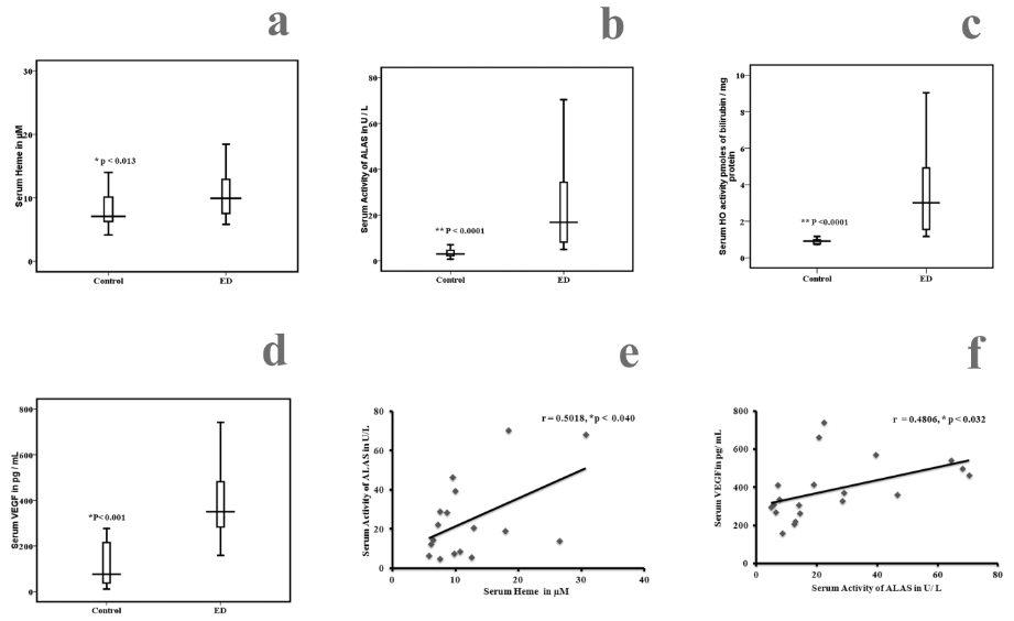
Figure 1: Comparison of serum Heme, aminolevulinic acid synthase, Heme oxygenase (HO) and VEGF among ED and healthy controls. Statistical significance between control (n = 20) and ED
(n = 20), a) Heme p < 0>
Correlation between serum aminolevulinic acid synthase, serum Heme, and serum VEGF in ED and controls, e) Positive correlation between serum Heme and aminolevulinic acid synthase (r = 0.50018, p < 0 xss=removed>
Levels of Ferritin, Transferrin and Soluble transferrin receptor
We measured the levels of serum ferritin, serum transferrin and soluble transferrin receptor levels in study subjects. The results are given in figure 2. There was a significant increase in the levels of serum and intracellular ferritin (table 3) in ED patients compared to the controls (p < 0 xss=removed>
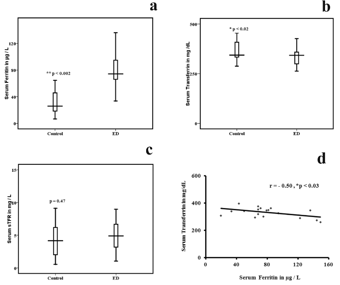
Figure 2: Comparison of serum ferritin, Transferrin and soluble transferrin receptor among ED and healthy controls. Statistical significance between control (n = 20) and ED (n = 20),
a) Serum ferritin p < 0 xss=removed>
d) Negative correlation between serum ferritin and serum Transferrin in ED and controls,
(r = -0.50, p < 0>
Expression of Iron regulators
Why there is an increased intracellular ferritin levels, in ED subjects, provoking interest in knowing the status of iron regulators, their role in the disease and hence to measure the mRNA expression of hepcidin an iron sensor and ferroportin an iron exporter. The mRNA expression of hepcidin was up regulated and ferroportin was down regulated in ED patients when compared to the controls (figure 3). Thus, the result shows a potential role for that hepcidin in ED. This increase in the expression of hepcidin may be due to increased iron, inflammation or infection. It is known that hepcidin can bind to ferroportin, triggering its internalization and degradation resulting in the accumulation of intracellular iron in the form of ferritin.
Over expression of Hypoxia inducible factor (HIF2 α)
Since, both VEGF and HO are elevated in ED; it is of interest to know further on the status of hypoxia inducible proteins. Hence, the expressions of hypoxia (HIF 2) were measured. The mRNA expression of HIF 2 was up regulated in ED compared to the controls as shown (figure 3).
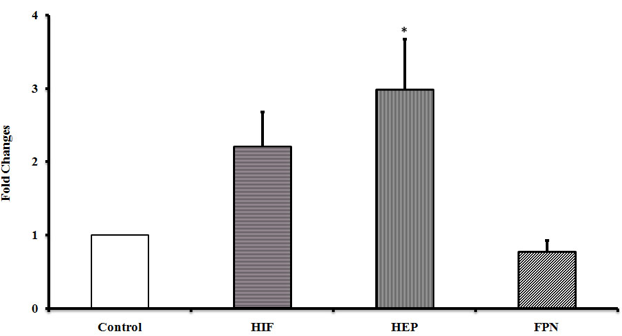
Figure 3: mRNA expressions in human PBMC among ED and healthy controls. Error bars represents SE and significance represents * of the relative expression levels normalized against GAPDH.
The aetiopathogenesis of ED is not clearly known. However, recently literature associated ED with tuberculosis, but still now the active organism was not cultivated. The disease still remains idiopathic, with no diagnostic test available in the lab. Direct evidence of infection is not made. Similar to homocysteine, Iron also acts as an independent risk factor of cardiovascular diseases. Roest [23] observed increased iron stores leads to cardiovascular diseases similar to that of hyperhomocysteine. Sullivan [24] observed that increased iron stores lead to endothelial dysfunction and that elevated homocysteine is dependent on iron overload. This excess iron generates free hydroxyl radical and causes oxidative stress through Fenton reaction. Schiepers [25] observed that changes in both homocysteine and ferritin levels in cardiovascular disease. The serum ferritin is frequently used as a measure of iron stores. The present findings indicate iron storage since; there is an elevated level of ferritin and lowered transferrin in ED.
It is known that, red cell breakdown can occur outside or inside the vascular compartment in ED. Increased serum haemoglobin in ED indicates the vascular damage due to intra vascular and extra vascular hemolysis. Extra vascular hemolysis is occurred due to phagocytosis whereas intravascular hemolysis as a result of mechanical injury and toxic factors. Heme is the prosthetic group of heme proteins such as haemoglobin, is an essential molecule plays a crucial role in cell differentiation and other functions. Free heme namely unbound heme can be toxic to cells, because it results in production of reactive oxygen species and causes cell damage. The absolute levels of Heme are regulated by its biosynthesis and catabolism. Heme biosynthesis is regulated by ALAS and its degradation to Fe, bilirubin and CO is catalyzed by HO, in this study, it is observed that both the enzymes are elevated in ED.
Heme oxygenase plays an important role in regulating the heme level by catalyzing the initial and rate limiting step of heme degradation and resulting in the formation of carbon monoxide, iron and bilirubin. Heme oxygenase exists as three isoforms; HO-1, HO-2 and HO-3. HO-1, the inducible 32-kDa isoform, HO-2, the constitutive 36-kDa isoform, and HO-3, has no activity and is not expressed in humans. The HO-1 is a member of the heat shock proteins, and its expression is influenced by hypoxia [26] heavy metals, ROS such as H2O2 [27], reactive nitrogen oxides [28], TNF α, interleukin β and interferon ϒ [29]. The biological functions of HO-1 are associated against oxidative and cellular stress. HO-1 represents a crucial mediator of antioxidants and possesses anti-inflammatory and anti- apoptotic properties [30]. L’Abbate et al., and Bharathselvi et al.,[31, 32] have shown that induction of HO-1 was associated with a parallel increase in the serum levels of adiponectin, which has a well-documented anti-inflammatory property. The peroxisome proliferator-activated receptor (PPAR-ϒ) regulates the expression of HO in human vascular cells [33].
Over expression of HO-1 contributes in the revascularization of damaged tissue. In term of neovascularization, HO-1 having a pro-angiogenic, anti-inflammatory and anti-apoptotic enzyme in regulation of wound healing [34,35]. Product of HO activity the bilirubin is a powerful antioxidant thereby, protecting the retinal cells. However, during haemorrhage, the iron and bilirubin excessively produced and are neurotoxic, have deleterious consequences. HO-1 is an inducible enzyme whose activity increases in response to iron as well as heme, light, oxidative stress, and inflammation. The main condition for the initiation of neovascularization is hypoxia [36]. Hypoxia-inducible factor activates several genes related to iron metabolism such as HO-1, endothelin-1, transferrin, transferrin receptor and ceruloplasmin [36].
HO, cleavage of the heme ring will release intracellular iron, which in turn increases the stimulation of ferritin. Dulak et.al observed that HO plays an important role in angiogenesis during hypoxia, similarly nitric oxide synthase, by VEGF production [37]. Alternately VEGF can stimulate HO-1 to promote angiogenesis and inhibiting leukocyte adhesion and transmigration [17]. In the present study, it’s observed that both HO and VEGF increased due to inflammation and non-inflammatory responses.
Hepcidin, a circulating peptide hormone is mainly synthesized by the liver hepatocytes, and also in eye plays a major role in regulating iron homeostasis in the body [38]. The mature form is 25 amino acids with four inter subunit disulfide bonds. The massive iron overload found in hepcidin knock-out mice suggests that hepcidin is an iron stores regulator involved in communication of body iron status to the intestine and also in the retinal pigment epithelial cells [38]. The mechanism of hepcidin activity depends on hepcidin interactions with ferroportin. Ferroportin is the only known mammalian cellular iron exporter. Hepcidin regulates post translational ferroportin expression [38]. Hepcidin binds to ferroportin and causes its internalization and degradation in turn blocks the iron transport via ferroportin [38]. Hepcidin is reported to be elevated in chronic inflammation condition, anemia, more specifically wherein iron accumulates inside the cells [39].
Hepcidin synthesized can be induced by inflammatory cytokine IL-6, inflammation, and infection (40)[Prentice, 2012 #102]. IL‐6 acts via its receptor and causes phosphorylation of signal transducer and activator of transcription 3 (STAT 3), STAT3 activation requires the presence of SMAD 4 to affect the HAMP gene expression [39]. Under normal conditions HAMP gene expression is regulated by BMP/SMAD and STAT3 pathways. Another hepatocyte iron sensors activating hepcidin synthesis are hemochromatosis protein (HFE) and transferrin receptor 2 (TfR2) [39].
Due to an intracellular storage of iron, there is an increased production of hepcidin. In the present study, there is an increased storage of ferritin levels and up regulation of hepcidin. Inflammation leads to cellular iron sequestering through IL-6 up regulation of hepcidin. Iron can be export from the cell with the help of iron exporter ferroportin. In the present study, we also observed that hepcidin expression was increased and ferroportin was decreased in ED compared to controls. Interestingly cellular iron accumulation caused diminished ferroportin after hepcidin binding. There is an increased expression of hepcidin may be due to increased iron, inflammation and infection. Importantly, a cellular iron act as a cofactor of HIF, in the present study, it shows that there is an increased expression of HIF2α. A previous report says that hypoxia is a negative regulator of hepcidin expression [39], but in the present study, shows that HIF 2α expression was increased in ED may be due to inflammation and also by IL-6. The interaction between hepcidin and ferroportin binding is a key step to control an iron homeostasis.
From the present study, we conclude that increased serum and intracellular ferritin, heme and HO, hepcidin play an important role in the vasculitis. The present study establishes the role for iron in disease pathogen. In our study thus the increased levels of these proteins may be due to an infection and inflammation conditions. The real significance of these findings needs to be understood in an animal model or an in-vitro cell culture experiment.
Biochemical Parameters | Control subjects (n = 20) | Eales’ disease (n = 20) | P value |
Ferritin (µg / mg of proteins) | 20.9 ± 1.4 | 65.7 ± 2.5 | 0.001 |
Heme (µm / mg of proteins) | 8.9 ± 1.7 | 27.2 ± 5.1 | 0.001 |
Heme oxygenase (pmoles of bilirubin / mg protein) | 1.5 ± 0.2 | 6.4 ± 0.5 | 0.001 |
Aminolevulinic acid synthase (U / mg of proteins) | 2.4 ± 0.2 | 12.5 ± 3.0 | 0.001 |
Vascular endothelial growth factor (pg/mg of proteins) | 29.8 ± 4.0 | 147.5 ± 40.2 | 0.005 |
Table 3: Intracellular Levels of Biochemical parameters measured in control and Eales’ disease subjects.
ED - Eales’ Disease
Hcy - Homocysteine
Hb - Haemoglobin;
HO - Heme oxygenase;
VEGF- Vascular endothelial growth factor;
ALAS - aminolevulinic acid Synthase;
IL - Interleukin;
Hep - Hepcidin;
FPN - Ferroportin.
Ethical approval: Institutional Ethics committee.
Consent to participate: Informed written consent for participating in the study was obtained from all participants. All experiments pertaining to human subjects were performed in adherence to the tenets of the Helsinki declaration.
Availability of data and materials: Data and materials are in the manuscript.
Competing Interests: No conflict of interest.
Funding: Supported by Grant from (ICMR) Indian council of medical research – ICMR/53/18/2009-BMS.
Authors’ Contributions: MB processing the sample analyzed the data and wrote the paper, RS processing the sample; KNS conceived the work, analyzed the data and wrote the paper. JB was the ophthalmic consultant who treated the patients, obtained their consent, recruited and in filling the clinical proforma in this study.
Acknowledgement: We thank the Indian Council of Medical research (ICMR) for their funding.
Authors’ Information (Optional): Not applicable
Clearly Auctoresonline and particularly Psychology and Mental Health Care Journal is dedicated to improving health care services for individuals and populations. The editorial boards' ability to efficiently recognize and share the global importance of health literacy with a variety of stakeholders. Auctoresonline publishing platform can be used to facilitate of optimal client-based services and should be added to health care professionals' repertoire of evidence-based health care resources.

Journal of Clinical Cardiology and Cardiovascular Intervention The submission and review process was adequate. However I think that the publication total value should have been enlightened in early fases. Thank you for all.

Journal of Women Health Care and Issues By the present mail, I want to say thank to you and tour colleagues for facilitating my published article. Specially thank you for the peer review process, support from the editorial office. I appreciate positively the quality of your journal.

Journal of Clinical Research and Reports I would be very delighted to submit my testimonial regarding the reviewer board and the editorial office. The reviewer board were accurate and helpful regarding any modifications for my manuscript. And the editorial office were very helpful and supportive in contacting and monitoring with any update and offering help. It was my pleasure to contribute with your promising Journal and I am looking forward for more collaboration.

We would like to thank the Journal of Thoracic Disease and Cardiothoracic Surgery because of the services they provided us for our articles. The peer-review process was done in a very excellent time manner, and the opinions of the reviewers helped us to improve our manuscript further. The editorial office had an outstanding correspondence with us and guided us in many ways. During a hard time of the pandemic that is affecting every one of us tremendously, the editorial office helped us make everything easier for publishing scientific work. Hope for a more scientific relationship with your Journal.

The peer-review process which consisted high quality queries on the paper. I did answer six reviewers’ questions and comments before the paper was accepted. The support from the editorial office is excellent.

Journal of Neuroscience and Neurological Surgery. I had the experience of publishing a research article recently. The whole process was simple from submission to publication. The reviewers made specific and valuable recommendations and corrections that improved the quality of my publication. I strongly recommend this Journal.

Dr. Katarzyna Byczkowska My testimonial covering: "The peer review process is quick and effective. The support from the editorial office is very professional and friendly. Quality of the Clinical Cardiology and Cardiovascular Interventions is scientific and publishes ground-breaking research on cardiology that is useful for other professionals in the field.

Thank you most sincerely, with regard to the support you have given in relation to the reviewing process and the processing of my article entitled "Large Cell Neuroendocrine Carcinoma of The Prostate Gland: A Review and Update" for publication in your esteemed Journal, Journal of Cancer Research and Cellular Therapeutics". The editorial team has been very supportive.

Testimony of Journal of Clinical Otorhinolaryngology: work with your Reviews has been a educational and constructive experience. The editorial office were very helpful and supportive. It was a pleasure to contribute to your Journal.

Dr. Bernard Terkimbi Utoo, I am happy to publish my scientific work in Journal of Women Health Care and Issues (JWHCI). The manuscript submission was seamless and peer review process was top notch. I was amazed that 4 reviewers worked on the manuscript which made it a highly technical, standard and excellent quality paper. I appreciate the format and consideration for the APC as well as the speed of publication. It is my pleasure to continue with this scientific relationship with the esteem JWHCI.

This is an acknowledgment for peer reviewers, editorial board of Journal of Clinical Research and Reports. They show a lot of consideration for us as publishers for our research article “Evaluation of the different factors associated with side effects of COVID-19 vaccination on medical students, Mutah university, Al-Karak, Jordan”, in a very professional and easy way. This journal is one of outstanding medical journal.

Dear Hao Jiang, to Journal of Nutrition and Food Processing We greatly appreciate the efficient, professional and rapid processing of our paper by your team. If there is anything else we should do, please do not hesitate to let us know. On behalf of my co-authors, we would like to express our great appreciation to editor and reviewers.

As an author who has recently published in the journal "Brain and Neurological Disorders". I am delighted to provide a testimonial on the peer review process, editorial office support, and the overall quality of the journal. The peer review process at Brain and Neurological Disorders is rigorous and meticulous, ensuring that only high-quality, evidence-based research is published. The reviewers are experts in their fields, and their comments and suggestions were constructive and helped improve the quality of my manuscript. The review process was timely and efficient, with clear communication from the editorial office at each stage. The support from the editorial office was exceptional throughout the entire process. The editorial staff was responsive, professional, and always willing to help. They provided valuable guidance on formatting, structure, and ethical considerations, making the submission process seamless. Moreover, they kept me informed about the status of my manuscript and provided timely updates, which made the process less stressful. The journal Brain and Neurological Disorders is of the highest quality, with a strong focus on publishing cutting-edge research in the field of neurology. The articles published in this journal are well-researched, rigorously peer-reviewed, and written by experts in the field. The journal maintains high standards, ensuring that readers are provided with the most up-to-date and reliable information on brain and neurological disorders. In conclusion, I had a wonderful experience publishing in Brain and Neurological Disorders. The peer review process was thorough, the editorial office provided exceptional support, and the journal's quality is second to none. I would highly recommend this journal to any researcher working in the field of neurology and brain disorders.
Dear Agrippa Hilda, Journal of Neuroscience and Neurological Surgery, Editorial Coordinator, I trust this message finds you well. I want to extend my appreciation for considering my article for publication in your esteemed journal. I am pleased to provide a testimonial regarding the peer review process and the support received from your editorial office. The peer review process for my paper was carried out in a highly professional and thorough manner. The feedback and comments provided by the authors were constructive and very useful in improving the quality of the manuscript. This rigorous assessment process undoubtedly contributes to the high standards maintained by your journal.

International Journal of Clinical Case Reports and Reviews. I strongly recommend to consider submitting your work to this high-quality journal. The support and availability of the Editorial staff is outstanding and the review process was both efficient and rigorous.

Thank you very much for publishing my Research Article titled “Comparing Treatment Outcome Of Allergic Rhinitis Patients After Using Fluticasone Nasal Spray And Nasal Douching" in the Journal of Clinical Otorhinolaryngology. As Medical Professionals we are immensely benefited from study of various informative Articles and Papers published in this high quality Journal. I look forward to enriching my knowledge by regular study of the Journal and contribute my future work in the field of ENT through the Journal for use by the medical fraternity. The support from the Editorial office was excellent and very prompt. I also welcome the comments received from the readers of my Research Article.

Dear Erica Kelsey, Editorial Coordinator of Cancer Research and Cellular Therapeutics Our team is very satisfied with the processing of our paper by your journal. That was fast, efficient, rigorous, but without unnecessary complications. We appreciated the very short time between the submission of the paper and its publication on line on your site.

I am very glad to say that the peer review process is very successful and fast and support from the Editorial Office. Therefore, I would like to continue our scientific relationship for a long time. And I especially thank you for your kindly attention towards my article. Have a good day!

"We recently published an article entitled “Influence of beta-Cyclodextrins upon the Degradation of Carbofuran Derivatives under Alkaline Conditions" in the Journal of “Pesticides and Biofertilizers” to show that the cyclodextrins protect the carbamates increasing their half-life time in the presence of basic conditions This will be very helpful to understand carbofuran behaviour in the analytical, agro-environmental and food areas. We greatly appreciated the interaction with the editor and the editorial team; we were particularly well accompanied during the course of the revision process, since all various steps towards publication were short and without delay".

I would like to express my gratitude towards you process of article review and submission. I found this to be very fair and expedient. Your follow up has been excellent. I have many publications in national and international journal and your process has been one of the best so far. Keep up the great work.

We are grateful for this opportunity to provide a glowing recommendation to the Journal of Psychiatry and Psychotherapy. We found that the editorial team were very supportive, helpful, kept us abreast of timelines and over all very professional in nature. The peer review process was rigorous, efficient and constructive that really enhanced our article submission. The experience with this journal remains one of our best ever and we look forward to providing future submissions in the near future.

I am very pleased to serve as EBM of the journal, I hope many years of my experience in stem cells can help the journal from one way or another. As we know, stem cells hold great potential for regenerative medicine, which are mostly used to promote the repair response of diseased, dysfunctional or injured tissue using stem cells or their derivatives. I think Stem Cell Research and Therapeutics International is a great platform to publish and share the understanding towards the biology and translational or clinical application of stem cells.

I would like to give my testimony in the support I have got by the peer review process and to support the editorial office where they were of asset to support young author like me to be encouraged to publish their work in your respected journal and globalize and share knowledge across the globe. I really give my great gratitude to your journal and the peer review including the editorial office.

I am delighted to publish our manuscript entitled "A Perspective on Cocaine Induced Stroke - Its Mechanisms and Management" in the Journal of Neuroscience and Neurological Surgery. The peer review process, support from the editorial office, and quality of the journal are excellent. The manuscripts published are of high quality and of excellent scientific value. I recommend this journal very much to colleagues.

Dr.Tania Muñoz, My experience as researcher and author of a review article in The Journal Clinical Cardiology and Interventions has been very enriching and stimulating. The editorial team is excellent, performs its work with absolute responsibility and delivery. They are proactive, dynamic and receptive to all proposals. Supporting at all times the vast universe of authors who choose them as an option for publication. The team of review specialists, members of the editorial board, are brilliant professionals, with remarkable performance in medical research and scientific methodology. Together they form a frontline team that consolidates the JCCI as a magnificent option for the publication and review of high-level medical articles and broad collective interest. I am honored to be able to share my review article and open to receive all your comments.

“The peer review process of JPMHC is quick and effective. Authors are benefited by good and professional reviewers with huge experience in the field of psychology and mental health. The support from the editorial office is very professional. People to contact to are friendly and happy to help and assist any query authors might have. Quality of the Journal is scientific and publishes ground-breaking research on mental health that is useful for other professionals in the field”.

Dear editorial department: On behalf of our team, I hereby certify the reliability and superiority of the International Journal of Clinical Case Reports and Reviews in the peer review process, editorial support, and journal quality. Firstly, the peer review process of the International Journal of Clinical Case Reports and Reviews is rigorous, fair, transparent, fast, and of high quality. The editorial department invites experts from relevant fields as anonymous reviewers to review all submitted manuscripts. These experts have rich academic backgrounds and experience, and can accurately evaluate the academic quality, originality, and suitability of manuscripts. The editorial department is committed to ensuring the rigor of the peer review process, while also making every effort to ensure a fast review cycle to meet the needs of authors and the academic community. Secondly, the editorial team of the International Journal of Clinical Case Reports and Reviews is composed of a group of senior scholars and professionals with rich experience and professional knowledge in related fields. The editorial department is committed to assisting authors in improving their manuscripts, ensuring their academic accuracy, clarity, and completeness. Editors actively collaborate with authors, providing useful suggestions and feedback to promote the improvement and development of the manuscript. We believe that the support of the editorial department is one of the key factors in ensuring the quality of the journal. Finally, the International Journal of Clinical Case Reports and Reviews is renowned for its high- quality articles and strict academic standards. The editorial department is committed to publishing innovative and academically valuable research results to promote the development and progress of related fields. The International Journal of Clinical Case Reports and Reviews is reasonably priced and ensures excellent service and quality ratio, allowing authors to obtain high-level academic publishing opportunities in an affordable manner. I hereby solemnly declare that the International Journal of Clinical Case Reports and Reviews has a high level of credibility and superiority in terms of peer review process, editorial support, reasonable fees, and journal quality. Sincerely, Rui Tao.

Clinical Cardiology and Cardiovascular Interventions I testity the covering of the peer review process, support from the editorial office, and quality of the journal.

Clinical Cardiology and Cardiovascular Interventions, we deeply appreciate the interest shown in our work and its publication. It has been a true pleasure to collaborate with you. The peer review process, as well as the support provided by the editorial office, have been exceptional, and the quality of the journal is very high, which was a determining factor in our decision to publish with you.

The peer reviewers process is quick and effective, the supports from editorial office is excellent, the quality of journal is high. I would like to collabroate with Internatioanl journal of Clinical Case Reports and Reviews journal clinically in the future time.

Clinical Cardiology and Cardiovascular Interventions, I would like to express my sincerest gratitude for the trust placed in our team for the publication in your journal. It has been a true pleasure to collaborate with you on this project. I am pleased to inform you that both the peer review process and the attention from the editorial coordination have been excellent. Your team has worked with dedication and professionalism to ensure that your publication meets the highest standards of quality. We are confident that this collaboration will result in mutual success, and we are eager to see the fruits of this shared effort.

Dear Dr. Jessica Magne, Editorial Coordinator 0f Clinical Cardiology and Cardiovascular Interventions, I hope this message finds you well. I want to express my utmost gratitude for your excellent work and for the dedication and speed in the publication process of my article titled "Navigating Innovation: Qualitative Insights on Using Technology for Health Education in Acute Coronary Syndrome Patients." I am very satisfied with the peer review process, the support from the editorial office, and the quality of the journal. I hope we can maintain our scientific relationship in the long term.

Dear Monica Gissare, - Editorial Coordinator of Nutrition and Food Processing. ¨My testimony with you is truly professional, with a positive response regarding the follow-up of the article and its review, you took into account my qualities and the importance of the topic¨.

Dear Dr. Jessica Magne, Editorial Coordinator 0f Clinical Cardiology and Cardiovascular Interventions, The review process for the article “The Handling of Anti-aggregants and Anticoagulants in the Oncologic Heart Patient Submitted to Surgery” was extremely rigorous and detailed. From the initial submission to the final acceptance, the editorial team at the “Journal of Clinical Cardiology and Cardiovascular Interventions” demonstrated a high level of professionalism and dedication. The reviewers provided constructive and detailed feedback, which was essential for improving the quality of our work. Communication was always clear and efficient, ensuring that all our questions were promptly addressed. The quality of the “Journal of Clinical Cardiology and Cardiovascular Interventions” is undeniable. It is a peer-reviewed, open-access publication dedicated exclusively to disseminating high-quality research in the field of clinical cardiology and cardiovascular interventions. The journal's impact factor is currently under evaluation, and it is indexed in reputable databases, which further reinforces its credibility and relevance in the scientific field. I highly recommend this journal to researchers looking for a reputable platform to publish their studies.

Dear Editorial Coordinator of the Journal of Nutrition and Food Processing! "I would like to thank the Journal of Nutrition and Food Processing for including and publishing my article. The peer review process was very quick, movement and precise. The Editorial Board has done an extremely conscientious job with much help, valuable comments and advices. I find the journal very valuable from a professional point of view, thank you very much for allowing me to be part of it and I would like to participate in the future!”

Dealing with The Journal of Neurology and Neurological Surgery was very smooth and comprehensive. The office staff took time to address my needs and the response from editors and the office was prompt and fair. I certainly hope to publish with this journal again.Their professionalism is apparent and more than satisfactory. Susan Weiner

My Testimonial Covering as fellowing: Lin-Show Chin. The peer reviewers process is quick and effective, the supports from editorial office is excellent, the quality of journal is high. I would like to collabroate with Internatioanl journal of Clinical Case Reports and Reviews.

My experience publishing in Psychology and Mental Health Care was exceptional. The peer review process was rigorous and constructive, with reviewers providing valuable insights that helped enhance the quality of our work. The editorial team was highly supportive and responsive, making the submission process smooth and efficient. The journal's commitment to high standards and academic rigor makes it a respected platform for quality research. I am grateful for the opportunity to publish in such a reputable journal.

My experience publishing in International Journal of Clinical Case Reports and Reviews was exceptional. I Come forth to Provide a Testimonial Covering the Peer Review Process and the editorial office for the Professional and Impartial Evaluation of the Manuscript.
