AUCTORES
Globalize your Research

Research Article
*Corresponding Author: Yuri Pivovarenko, Research and Training Center ‘Physical and Chemical Materials Science’ Under Kyiv Taras Shevchenko University and NAS of Ukraine, Kiev, Ukraine.
Citation: Yuri Pivovarenko, (2024), Electrical Forces that Build and Recover Neurons, J New Medical Innovations and Research, 5(2); DOI:10.31579/2767-7370/090
Copyright: © 2024, Yuri Pivovarenko. This is an open access article distributed under the Creative Commons Attribution License, which permits unrestricted use, distribution, and reproduction in any medium, provided the original work is properly cited.
Received: 03 February 2024 | Accepted: 12 February 2024 | Published: 19 February 2024
Keywords: nerve; recovery; extracellular filaments; pulse therapy; laser therapy
It was previously shown that it is the electric charge (potential) of water that determines its structuring properties in relation to substances both dissolved in water and in contact with it. In particular, it has been shown that negative electrization of the water used causes the formation of needle-like, thread-like and plant-like crystals and sediments. Taking all this into account, the formation of dendritic structures in aqueous solutions of chlorides exposed to pulsed electromagnetic fields (PEMFs) has been proposed to be explained by the negative electrization of such solutions; accordingly, the regenerative effect of PEMFs on nerve tissue was proposed to be explained in the same way. Over time, it became clear that such explanations are incomplete because they do not take into account the electric fields created by charged surfaces, in particular the outer sides of the cytoplasmic membranes of neurons. To eliminate this incompleteness, a number of additional modeling studies were carried out. In particular, the effect of various charged surfaces on aqueous solutions was studied. As a result, it was shown that in aqueous solutions in contact with charged surfaces, filaments are formed that are collinear to the vectors of electric forces created by such surfaces. It was also shown that these same electrical forces are capable of determining both the integrity of the formed filaments and their mutual separation. Naturally, all the results obtained suggested that it is the electrical forces acting on the outer sides of the cytoplasmic membranes that create all extracellular filaments, including neuronal ones.
It was previously established that positively charged water promotes the formation of compact salt crystals and sediments, and negatively charged water promotes the formation of needle-like or tree-like ones (Figures 1 – 4) [1 – 4].
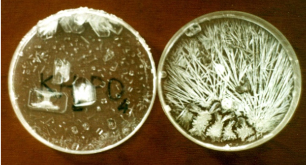
Figure 1: It is the crystals that formed after the drying of solutions of KH2PO4 prepared on the water with potentials of +250 mV (left) and –250 mV (right) [1, 2].
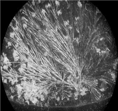
Figure 2: "Bouquets of flowers" are formed in drying NaCl solutions prepared in negatively charged water [1].

Figure 3: Left: these are intense blue prismatic crystals that formed in a copper sulfate solution prepared with positively charged water, which has a high hydrating capacity and high surface tension. Right: these are pale green, grass-like crystals formed in a copper sulfate solution prepared with negatively charged water, which has a low hydrating capacity and low surface tension.
It is worth noting here that fully hydrated copper sulfate, namely CuSO4●5H2O, has an intense blue color (left), while partially hydrated cooper sulfate, for example CuSO4●3H2O, is pale green (right) and completely anhydrous copper sulfate, namely CuSO4, is colorless [6].
All this makes it possible to use this particular salt as a visual indicator of the electrization of water.

Figure 4: It is copper oxide Cu2O dispersed in the water containing a gradient of electric potentials; water with positive potential is above and water with negative potential is below [1].
At the same time, it was found that positive electrization of water increases its surface tension, while negative electrization of water decreases it; in particular, this dependence manifests itself both in the tendency of positively charged water to compress and thereby increase its contact angle (Figure 5, right), and in the tendency of negatively charged water to spread and thereby reduce its contact angle (Figure 5, left)
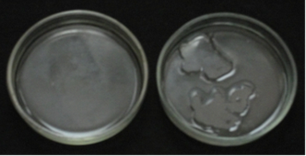
Figure 5: Left: 5 ml of water with an electric potential of –200 mV cover all the bottom of a Petri dish. Right: 5 ml of water with an electric potential of +200 mV do not cover the bottom of a Petri dish; the surface of such water decreases rapidly after mixing [1].
(Apparently, it is worth recalling here that the surface tension of any liquid directly correlates with its contact angle [5].)
Naturally, all this led to the conclusion that the increased surface tension of positively charged water promotes the formation of compact structures, and the reduced surface tension of negatively charged water promotes the formation of needle-like, thread-like and tree-like structures [1].
Apparently, it is worth adding here that the results obtained (Figures 1 – 5) also agree well with the ability of aqueous protons, with which
positively charged water is enriched, to form both oxonium ions, namely H3O+, and very compact symmetrical aggregates, namely H3O+•3H2O [6]. Also consistent with the same results is the ability of aqueous hydroxyl ions, which enrich negatively charged water, to form linear aggregates, namely OH–•3H2O [6].
One way or another, it was the knowledge of the established dependencies (Figures 1 – 5) that made it possible to purposefully create the desired structures, including those characteristic of living organisms (Figure 6).

Figure 6: These crystals, whose shape is similar to plant fragments and bird feathers (top right), formed after CuSO4 solutions prepared in water at various negative potentials dried [7].
One way or another, all the presented results (Figures 1 – 6) ultimately led to the conclusion that it is the electric charge (potential) of water that determines its structuring properties in relation to substances both in contact with water and dissolved in it [1]. All this, in particular, allowed
assuming that the appearance of dendrite-like structures in aqueous solutions of chlorides exposed to pulsed electromagnetic fields (PEMFs) (Figure 7) is also due to the negative electrization of the water contained in these solutions [3].
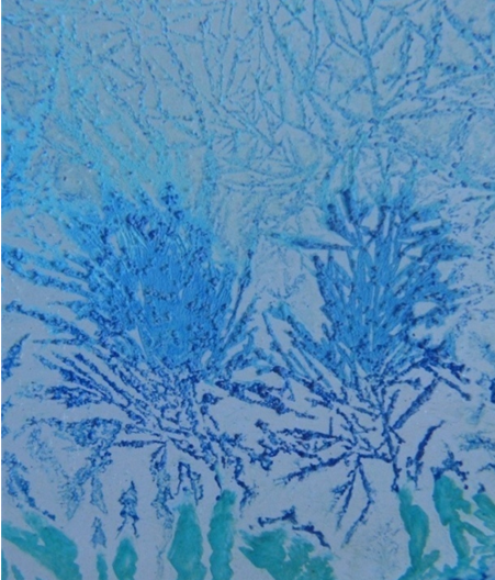
Figure 7: The crystals formed after drying an aqueous solution of CuCl2, which was previously subjected to the action of EMF, pulsing with a frequency of 10 Hz for 10 minutes
For contrast, the crystals formed were treated with ammonia vapors [3].
Considering the obvious similarity of these same salt structures (Figure 7) with nerve dendrites and nerve endings (Figure 8), it was also assumed that the regenerative effect of PEMFs on nervous tissues [8 – 17] is based on their ability to negatively electrify the latter [3]; the predominance of chlorides in nerve tissues [18] was also taken into account.
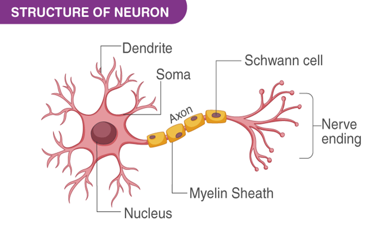
Figure 8: Diagram of a typical neuron.
In fact, it was assumed that the root cause of the regenerative effect of PEMFs and negatively charged electrets on nervous tissues [3] is the same.
Over time, it became clear that all these assumptions do not allow taking into account the contribution of cytoplasmic membranes of neurons, the
outer sides of which form negatively charged surfaces [18], in the formation of nerve dendrites, axons and endings (Figure 8); at the same time, it was assumed that the same electrical forces, the vectors of which are perpendicular to all charged surfaces (Figure 9), determine the direction of growth of neuronal axons and endings (Figure 8).

Figure 9: This particular diagram is used for educational purposes; It is important to note that the location of the electric field vectors created by a negatively charged surface is the same [5, 19].
It should be noted right here that the analysis of the effects of laser exposure on the neural environment (Figure 10) [20] partially confirmed the adequacy of this assumption.
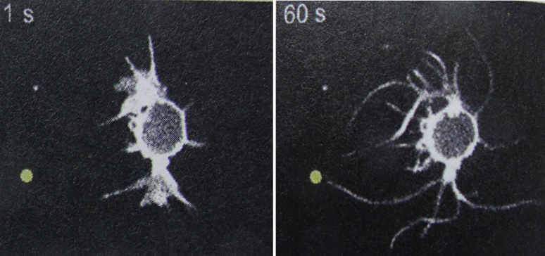
Figure 10: These are enlarged images of a neuron located near a spot illuminated by a laser (light yellow spot).
Comparing both photos allows evaluating the changes in the neuron that occurred within 1 minute [20].
Thus, given that the Poynting vector determines not only the direction of light propagation, but also the direction of movement of positive charges [21], it is very likely that negatively charged fragments of the cytoplasmic membrane of a neuron are drawn to a positively charged spot illuminated by a laser (Figure 10).
One way or another, it was decided to further test the supposed participation of charged surfaces in the formation of filaments; however, it was expected that the results of this test could be extrapolated to the cytoplasmic membranes of neurons.
Waters with the required charges were obtained as in [1].
Since the color and shape of copper salt crystals are particularly sensitive to the electrical potential of the water in which these crystals are formed (Figure 3), these salts were used here as indicators to visualize the electrization of aqueous media, just as in [1].
All salts were purchased from the “Ukrreakhim” (Ukraine).
Initially, it makes sense to analyze some previously obtained results. Thus, it was previously established that in aqueous solutions of salts, which are prepared in negatively charged water and are in contact with glass surfaces, filaments are formed, initially oriented perpendicular to such surfaces (Figures 11, 12) [4].

Figure 11: These are filaments formed in a glass Petri dish after drying a CuCl2 solution prepared in negatively charged water [4].

Figure 12: These are spiral filaments formed at the wall of a glass Petri dish filled with an aqueous solution of CuSO4 prepared in negatively charged water [4].
It is worth noting here that glass sorbs aqueous hydroxyl ions OH– [6], thereby receiving a negative charge, which electrostatically repels the negatively charged components of aqueous-salt solutions, thereby probably orienting the filaments formed from these components (compare Figures 9, 11, 12).
It is also likely that the negative, i.e., identical, charges of these filaments determine their mutual electrostatic repulsion and thereby ensure their separation; at the same time, it is no less likely that the electrical forces created by the glass surface polarize the structural components of such filaments, thereby ensuring their integration due to dipole-dipole interactions [5].
One way or another, all these considerations allowed us to assume that it is the negatively charged surface of the glass that exhaustively provides both the shape and direction of these same filaments (Figures 11, 12).
Given the predominantly negative charge on the outer surfaces of living cells [18], it was concluded that the forces that create filaments on the surface of cells (Figure 13) are of the same electrical nature and direction as the forces that create filaments on glass surfaces (Figures 11, 12); since this conclusion did not fundamentally contradict the previously proposed assumption about the participation of negatively charged media in the formation of neuronal dendrites [3], it seemed completely justified.
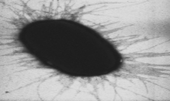
Figure 13: It is a separate cell of E. coli.
However, this conclusion seemed justified only until it was discovered that similar filaments are formed around rhombic crystals immersed in negatively charged water (Figure 14); in this experiment it was assumed that these crystals retained a positive charge on the water in which they formed (Figure 3).

Figure 14: When rhombic crystals are removed from a solution of copper sulfate and introduced into negatively charged water, numerous filaments quickly cover the surfaces of the crystals [2].
When analyzing this result, one should take into account the polymorphism of crystals formed in solutions prepared with differently charged water; in particular, it should be taken into account that rhombic crystals are formed in solutions prepared in positively charged water (Figures 1, 3).
Actually, it was this result (Figure 14) that led to the conclusion that both negatively and positively charged surfaces are involved in the formation of filaments in aqueous media. Over time, this conclusion received experimental confirmation.
Thus, it has been established that the filaments formed in aqueous-salt solutions prepared in negatively charged water and located between the negatively charged surfaces of glass and the positively charged surfaces of corrosion-resistant metals are similar to all previously obtained
filaments (compare Figures 11, 12, 14 and 15); at the same time, the identity, at least visual, of the endings of these same filaments was discovered (Figure 15).
When analyzing the last result (Figure 15), Kyon's rule should be taken into account: upon contact of the two phases, the phase with greater dielectric permeability receives a positive charge and the phase with lower dielectric permeability – negative [6, 22]. Thus, taking into account that the dielectric constant of liquid water varies, depending on temperature, in the range from 55.1 to 88.3 [6], and the dielectric constant of metals is always significantly higher (in theoretical calculations, the dielectric constant of metals is usually taken equal to ꝏ [23]), liquid water upon contact with any metal acquires a negative charge, and this same metal acquires a positive charge.

Figure 15: This is a "copper" coin surrounded by a dried aqueous solution of K2CO3 prepared in negatively charged water.
(Since the dielectric permeability of water changes depending on its temperature [6], the term “dielectric constant” is not applicable to water. In addition, the term "dielectric constant" does not correlate well with the sensitivity of water's dielectric permeability to high-frequency electromagnetic fields [19]; that it is this sensitivity that is important for the topic under discussion will soon become clear.)
Thus, all the results obtained (Figures 11, 12, 14, 15) showed that visually identical filaments are formed in aqueous solutions in contact with both negatively and positively charged surfaces.
Finally, it was discovered that apparently dendritic structures form in aqueous environments around corroding metals (Figure 16).

Figure 16: This is a corroded "copper" coin surrounded by a dried aqueous solution of CuCl2 prepared in negatively charged water.
Since these dendrites are formed from metal corrosion products (compare Figures 4 and 16), their formation is appropriate to compare with the formation of nerve and bacterial filaments, undoubtedly formed from components of cytoplasmic membranes (Figures 8, 10, 13). This, accordingly, suggests that cytoplasmic membranes not only supply “building material” for cellular filaments, but also generate electrical forces that create them.
It is especially worth noting that no auxiliary factors were involved in the formation of these same dendrites (Figure 16); this, accordingly, suggests that electrostatic repulsion is quite sufficient for the formation of any dendrites, including nerve ones (Figure 8).
Perhaps, the effect of skin electrons [23] on salt crystals dendrites should also be mentioned here. So, it is quite obvious that needle-like salt crystals grow from the area where skin electrons enter the studied salt solution (Figure 17).
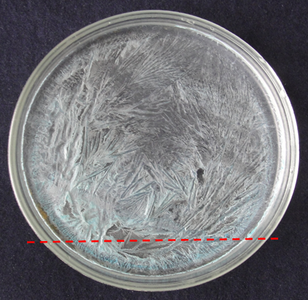
Figure 17: These are needle-shaped crystals formed after the drying of an aqueous solution of CuSO4 located under a wire with a pulsed electric current with a frequency of 2 Hz (dashed red line); it is worth noting that these needles are directed away from the wire.
Apparently, it is also worth noting here that electrons in terrestrial conditions move exclusively downward, like any anions [24]. Therefore, skin electrons fall into the studied salt solution below that part of it, which is located under the wire with an electric current (dashed red line in Figure 17).
Thus, the results obtained (Figures 1 12, 14 – 16) clearly demonstrate the ability of charged surfaces to form filaments in aqueous media that are
collinear to the electric field lines undoubtedly created by these surfaces (Figure 9). At the same time, the obvious similarity of filaments formed on both non-living and living surfaces (Figures 10 – 16) suggests the identity of the electrical forces that create any filaments; this, in turn, suggests that all filaments are visualizations of electric field lines, including extracellular ones.
To ensure the adequacy of these suggestions, it is worth extrapolating them to such a phenomenon as the “corona effect” (Figure 18), characterized by its discoverers as mysterious [25].
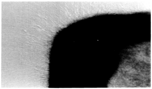
Figure 18: This is a fragment of the cytoplasmic membrane of a neuron with filaments formed under the influence of variable EMFs and creating a “corona effect” [25].
So, it is likely that the clearly visible filaments forming this very “corona”, which apparently consists of negatively charged fragments of the cytoplasmic membrane of the neuron (Figure 18), appear due to their electrostatic repulsion from the negatively charged outer surface of the cytoplasmic membrane; it is also likely that this repulsion is enhanced due to the multiple decrease in dielectric constant of the aqueous environment of the neuron exposed to variable EMFs [19].
Therefore, this very “corona” (Figure 18) can be considered only as a variety of the filaments presented above (Figures 10 – 15), quickly forming in favorable conditions.
At the same time, when analyzing the effect of the laser on the aqueous environment of the neuron (Figure 10), it should also be taken into account that under the influence of visible light, the dielectric constant of water decreases by ~10 times [19].
It should probably be added here that the formation of this very “corona” (Figure 18) can also be considered as a result of the skin effect (Figure 17), which undoubtedly occurs in variable EMFs [23] and manifests itself in this case in the pushing of negatively charged fragments of the cytoplasmic membrane of neuron to the periphery.
One way or another, the proposed analysis of the “corona effect” showed the productivity of the suggestions outlined at the beginning of this discussion, and thus confirmed their adequacy.
It seems appropriate to add here that the proposed explanation of the "corona effect" can also be used to explain the regenerative effect of PEMFs on nervous tissues [8 – 17]. To verify this, two facts should be taken into account:
In view of this, any PEMF that generates individual direct current pulses can reduce the dielectric constant of biological tissues, including nerve tissues (of course, taking into account the undoubted porosity of biological tissues).
Thus, it is likely that it is the electric fields generated by charged surfaces that build both nonliving and living filaments. It is also likely that all these filaments visualize the structures of such electric fields, in fact, their lines of force (Figure 19).

Figure 19: These are visualizations of electric field lines created by spherical (left) and extended (right) bodies used with education aim (compare with Figures 10 – 17).
The structures shown above, both salt and living, appear to be similar to both of these visualizations; this similarity suggests that these same structures are also visualizations of the corresponding electric fields.
This, accordingly, suggests that the natural ability of neurons to self-repair is based on the electrostatic repulsion of neuronal fragments by cytoplasmic membranes. This, in turn, allows anticipating that amplifying this repulsion will contribute to the natural ability of neurons to recreate. Thus, it is very likely that both PEMFs and the laser are means of
amplifying the electrical forces that determine the natural regenerative abilities of neurons.
Despite the unusual nature of the proposed approach, which obviously requires a radical revision of established ideas about the mechanisms of formation of extracellular filaments [30 – 32], it seems to be very productive and therefore requires development.
In particular, it is expected that this approach will make it possible to identify the forces that form networks in water-salt solutions under the influence of PEMFs (Figure 20) with the forces that form neural networks.
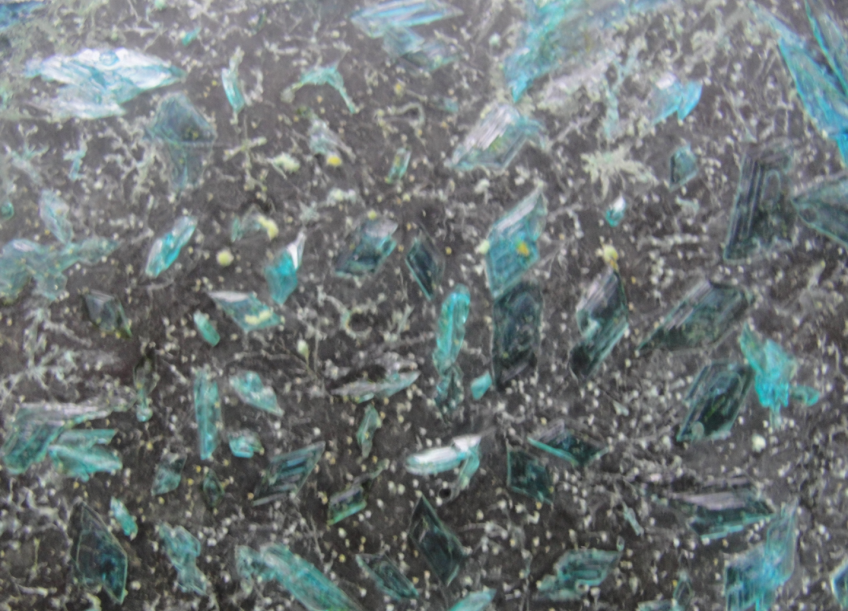
Figure 20: This is a dried aqueous solution of CuSO4, which, until completely dry, was exposed to an EMF pulsating at a frequency of 2 Hz.
The white “beads” connected by white threads are probably quite visible here.
It is clear that these networks were formed in a purely non-living environment, that is, in the absence of any "living force".
Also, developing this same approach, it is worth adding that it can no less successfully be used to explain the restorative effect of PEMFs on bones, the nature of which is considered unclear [33]. Otherwise, the ideas presented both earlier and here can be seen as attempts to respond to Szent-György, who urged biologists not to ignore the existence of "two matrices: water and electromagnetic fields" [29].
Clearly Auctoresonline and particularly Psychology and Mental Health Care Journal is dedicated to improving health care services for individuals and populations. The editorial boards' ability to efficiently recognize and share the global importance of health literacy with a variety of stakeholders. Auctoresonline publishing platform can be used to facilitate of optimal client-based services and should be added to health care professionals' repertoire of evidence-based health care resources.

Journal of Clinical Cardiology and Cardiovascular Intervention The submission and review process was adequate. However I think that the publication total value should have been enlightened in early fases. Thank you for all.

Journal of Women Health Care and Issues By the present mail, I want to say thank to you and tour colleagues for facilitating my published article. Specially thank you for the peer review process, support from the editorial office. I appreciate positively the quality of your journal.

Journal of Clinical Research and Reports I would be very delighted to submit my testimonial regarding the reviewer board and the editorial office. The reviewer board were accurate and helpful regarding any modifications for my manuscript. And the editorial office were very helpful and supportive in contacting and monitoring with any update and offering help. It was my pleasure to contribute with your promising Journal and I am looking forward for more collaboration.

We would like to thank the Journal of Thoracic Disease and Cardiothoracic Surgery because of the services they provided us for our articles. The peer-review process was done in a very excellent time manner, and the opinions of the reviewers helped us to improve our manuscript further. The editorial office had an outstanding correspondence with us and guided us in many ways. During a hard time of the pandemic that is affecting every one of us tremendously, the editorial office helped us make everything easier for publishing scientific work. Hope for a more scientific relationship with your Journal.

The peer-review process which consisted high quality queries on the paper. I did answer six reviewers’ questions and comments before the paper was accepted. The support from the editorial office is excellent.

Journal of Neuroscience and Neurological Surgery. I had the experience of publishing a research article recently. The whole process was simple from submission to publication. The reviewers made specific and valuable recommendations and corrections that improved the quality of my publication. I strongly recommend this Journal.

Dr. Katarzyna Byczkowska My testimonial covering: "The peer review process is quick and effective. The support from the editorial office is very professional and friendly. Quality of the Clinical Cardiology and Cardiovascular Interventions is scientific and publishes ground-breaking research on cardiology that is useful for other professionals in the field.

Thank you most sincerely, with regard to the support you have given in relation to the reviewing process and the processing of my article entitled "Large Cell Neuroendocrine Carcinoma of The Prostate Gland: A Review and Update" for publication in your esteemed Journal, Journal of Cancer Research and Cellular Therapeutics". The editorial team has been very supportive.

Testimony of Journal of Clinical Otorhinolaryngology: work with your Reviews has been a educational and constructive experience. The editorial office were very helpful and supportive. It was a pleasure to contribute to your Journal.

Dr. Bernard Terkimbi Utoo, I am happy to publish my scientific work in Journal of Women Health Care and Issues (JWHCI). The manuscript submission was seamless and peer review process was top notch. I was amazed that 4 reviewers worked on the manuscript which made it a highly technical, standard and excellent quality paper. I appreciate the format and consideration for the APC as well as the speed of publication. It is my pleasure to continue with this scientific relationship with the esteem JWHCI.

This is an acknowledgment for peer reviewers, editorial board of Journal of Clinical Research and Reports. They show a lot of consideration for us as publishers for our research article “Evaluation of the different factors associated with side effects of COVID-19 vaccination on medical students, Mutah university, Al-Karak, Jordan”, in a very professional and easy way. This journal is one of outstanding medical journal.

Dear Hao Jiang, to Journal of Nutrition and Food Processing We greatly appreciate the efficient, professional and rapid processing of our paper by your team. If there is anything else we should do, please do not hesitate to let us know. On behalf of my co-authors, we would like to express our great appreciation to editor and reviewers.

As an author who has recently published in the journal "Brain and Neurological Disorders". I am delighted to provide a testimonial on the peer review process, editorial office support, and the overall quality of the journal. The peer review process at Brain and Neurological Disorders is rigorous and meticulous, ensuring that only high-quality, evidence-based research is published. The reviewers are experts in their fields, and their comments and suggestions were constructive and helped improve the quality of my manuscript. The review process was timely and efficient, with clear communication from the editorial office at each stage. The support from the editorial office was exceptional throughout the entire process. The editorial staff was responsive, professional, and always willing to help. They provided valuable guidance on formatting, structure, and ethical considerations, making the submission process seamless. Moreover, they kept me informed about the status of my manuscript and provided timely updates, which made the process less stressful. The journal Brain and Neurological Disorders is of the highest quality, with a strong focus on publishing cutting-edge research in the field of neurology. The articles published in this journal are well-researched, rigorously peer-reviewed, and written by experts in the field. The journal maintains high standards, ensuring that readers are provided with the most up-to-date and reliable information on brain and neurological disorders. In conclusion, I had a wonderful experience publishing in Brain and Neurological Disorders. The peer review process was thorough, the editorial office provided exceptional support, and the journal's quality is second to none. I would highly recommend this journal to any researcher working in the field of neurology and brain disorders.
Dear Agrippa Hilda, Journal of Neuroscience and Neurological Surgery, Editorial Coordinator, I trust this message finds you well. I want to extend my appreciation for considering my article for publication in your esteemed journal. I am pleased to provide a testimonial regarding the peer review process and the support received from your editorial office. The peer review process for my paper was carried out in a highly professional and thorough manner. The feedback and comments provided by the authors were constructive and very useful in improving the quality of the manuscript. This rigorous assessment process undoubtedly contributes to the high standards maintained by your journal.

International Journal of Clinical Case Reports and Reviews. I strongly recommend to consider submitting your work to this high-quality journal. The support and availability of the Editorial staff is outstanding and the review process was both efficient and rigorous.

Thank you very much for publishing my Research Article titled “Comparing Treatment Outcome Of Allergic Rhinitis Patients After Using Fluticasone Nasal Spray And Nasal Douching" in the Journal of Clinical Otorhinolaryngology. As Medical Professionals we are immensely benefited from study of various informative Articles and Papers published in this high quality Journal. I look forward to enriching my knowledge by regular study of the Journal and contribute my future work in the field of ENT through the Journal for use by the medical fraternity. The support from the Editorial office was excellent and very prompt. I also welcome the comments received from the readers of my Research Article.

Dear Erica Kelsey, Editorial Coordinator of Cancer Research and Cellular Therapeutics Our team is very satisfied with the processing of our paper by your journal. That was fast, efficient, rigorous, but without unnecessary complications. We appreciated the very short time between the submission of the paper and its publication on line on your site.

I am very glad to say that the peer review process is very successful and fast and support from the Editorial Office. Therefore, I would like to continue our scientific relationship for a long time. And I especially thank you for your kindly attention towards my article. Have a good day!

"We recently published an article entitled “Influence of beta-Cyclodextrins upon the Degradation of Carbofuran Derivatives under Alkaline Conditions" in the Journal of “Pesticides and Biofertilizers” to show that the cyclodextrins protect the carbamates increasing their half-life time in the presence of basic conditions This will be very helpful to understand carbofuran behaviour in the analytical, agro-environmental and food areas. We greatly appreciated the interaction with the editor and the editorial team; we were particularly well accompanied during the course of the revision process, since all various steps towards publication were short and without delay".

I would like to express my gratitude towards you process of article review and submission. I found this to be very fair and expedient. Your follow up has been excellent. I have many publications in national and international journal and your process has been one of the best so far. Keep up the great work.

We are grateful for this opportunity to provide a glowing recommendation to the Journal of Psychiatry and Psychotherapy. We found that the editorial team were very supportive, helpful, kept us abreast of timelines and over all very professional in nature. The peer review process was rigorous, efficient and constructive that really enhanced our article submission. The experience with this journal remains one of our best ever and we look forward to providing future submissions in the near future.

I am very pleased to serve as EBM of the journal, I hope many years of my experience in stem cells can help the journal from one way or another. As we know, stem cells hold great potential for regenerative medicine, which are mostly used to promote the repair response of diseased, dysfunctional or injured tissue using stem cells or their derivatives. I think Stem Cell Research and Therapeutics International is a great platform to publish and share the understanding towards the biology and translational or clinical application of stem cells.

I would like to give my testimony in the support I have got by the peer review process and to support the editorial office where they were of asset to support young author like me to be encouraged to publish their work in your respected journal and globalize and share knowledge across the globe. I really give my great gratitude to your journal and the peer review including the editorial office.

I am delighted to publish our manuscript entitled "A Perspective on Cocaine Induced Stroke - Its Mechanisms and Management" in the Journal of Neuroscience and Neurological Surgery. The peer review process, support from the editorial office, and quality of the journal are excellent. The manuscripts published are of high quality and of excellent scientific value. I recommend this journal very much to colleagues.

Dr.Tania Muñoz, My experience as researcher and author of a review article in The Journal Clinical Cardiology and Interventions has been very enriching and stimulating. The editorial team is excellent, performs its work with absolute responsibility and delivery. They are proactive, dynamic and receptive to all proposals. Supporting at all times the vast universe of authors who choose them as an option for publication. The team of review specialists, members of the editorial board, are brilliant professionals, with remarkable performance in medical research and scientific methodology. Together they form a frontline team that consolidates the JCCI as a magnificent option for the publication and review of high-level medical articles and broad collective interest. I am honored to be able to share my review article and open to receive all your comments.

“The peer review process of JPMHC is quick and effective. Authors are benefited by good and professional reviewers with huge experience in the field of psychology and mental health. The support from the editorial office is very professional. People to contact to are friendly and happy to help and assist any query authors might have. Quality of the Journal is scientific and publishes ground-breaking research on mental health that is useful for other professionals in the field”.

Dear editorial department: On behalf of our team, I hereby certify the reliability and superiority of the International Journal of Clinical Case Reports and Reviews in the peer review process, editorial support, and journal quality. Firstly, the peer review process of the International Journal of Clinical Case Reports and Reviews is rigorous, fair, transparent, fast, and of high quality. The editorial department invites experts from relevant fields as anonymous reviewers to review all submitted manuscripts. These experts have rich academic backgrounds and experience, and can accurately evaluate the academic quality, originality, and suitability of manuscripts. The editorial department is committed to ensuring the rigor of the peer review process, while also making every effort to ensure a fast review cycle to meet the needs of authors and the academic community. Secondly, the editorial team of the International Journal of Clinical Case Reports and Reviews is composed of a group of senior scholars and professionals with rich experience and professional knowledge in related fields. The editorial department is committed to assisting authors in improving their manuscripts, ensuring their academic accuracy, clarity, and completeness. Editors actively collaborate with authors, providing useful suggestions and feedback to promote the improvement and development of the manuscript. We believe that the support of the editorial department is one of the key factors in ensuring the quality of the journal. Finally, the International Journal of Clinical Case Reports and Reviews is renowned for its high- quality articles and strict academic standards. The editorial department is committed to publishing innovative and academically valuable research results to promote the development and progress of related fields. The International Journal of Clinical Case Reports and Reviews is reasonably priced and ensures excellent service and quality ratio, allowing authors to obtain high-level academic publishing opportunities in an affordable manner. I hereby solemnly declare that the International Journal of Clinical Case Reports and Reviews has a high level of credibility and superiority in terms of peer review process, editorial support, reasonable fees, and journal quality. Sincerely, Rui Tao.

Clinical Cardiology and Cardiovascular Interventions I testity the covering of the peer review process, support from the editorial office, and quality of the journal.

Clinical Cardiology and Cardiovascular Interventions, we deeply appreciate the interest shown in our work and its publication. It has been a true pleasure to collaborate with you. The peer review process, as well as the support provided by the editorial office, have been exceptional, and the quality of the journal is very high, which was a determining factor in our decision to publish with you.

The peer reviewers process is quick and effective, the supports from editorial office is excellent, the quality of journal is high. I would like to collabroate with Internatioanl journal of Clinical Case Reports and Reviews journal clinically in the future time.

Clinical Cardiology and Cardiovascular Interventions, I would like to express my sincerest gratitude for the trust placed in our team for the publication in your journal. It has been a true pleasure to collaborate with you on this project. I am pleased to inform you that both the peer review process and the attention from the editorial coordination have been excellent. Your team has worked with dedication and professionalism to ensure that your publication meets the highest standards of quality. We are confident that this collaboration will result in mutual success, and we are eager to see the fruits of this shared effort.

Dear Dr. Jessica Magne, Editorial Coordinator 0f Clinical Cardiology and Cardiovascular Interventions, I hope this message finds you well. I want to express my utmost gratitude for your excellent work and for the dedication and speed in the publication process of my article titled "Navigating Innovation: Qualitative Insights on Using Technology for Health Education in Acute Coronary Syndrome Patients." I am very satisfied with the peer review process, the support from the editorial office, and the quality of the journal. I hope we can maintain our scientific relationship in the long term.

Dear Monica Gissare, - Editorial Coordinator of Nutrition and Food Processing. ¨My testimony with you is truly professional, with a positive response regarding the follow-up of the article and its review, you took into account my qualities and the importance of the topic¨.

Dear Dr. Jessica Magne, Editorial Coordinator 0f Clinical Cardiology and Cardiovascular Interventions, The review process for the article “The Handling of Anti-aggregants and Anticoagulants in the Oncologic Heart Patient Submitted to Surgery” was extremely rigorous and detailed. From the initial submission to the final acceptance, the editorial team at the “Journal of Clinical Cardiology and Cardiovascular Interventions” demonstrated a high level of professionalism and dedication. The reviewers provided constructive and detailed feedback, which was essential for improving the quality of our work. Communication was always clear and efficient, ensuring that all our questions were promptly addressed. The quality of the “Journal of Clinical Cardiology and Cardiovascular Interventions” is undeniable. It is a peer-reviewed, open-access publication dedicated exclusively to disseminating high-quality research in the field of clinical cardiology and cardiovascular interventions. The journal's impact factor is currently under evaluation, and it is indexed in reputable databases, which further reinforces its credibility and relevance in the scientific field. I highly recommend this journal to researchers looking for a reputable platform to publish their studies.

Dear Editorial Coordinator of the Journal of Nutrition and Food Processing! "I would like to thank the Journal of Nutrition and Food Processing for including and publishing my article. The peer review process was very quick, movement and precise. The Editorial Board has done an extremely conscientious job with much help, valuable comments and advices. I find the journal very valuable from a professional point of view, thank you very much for allowing me to be part of it and I would like to participate in the future!”

Dealing with The Journal of Neurology and Neurological Surgery was very smooth and comprehensive. The office staff took time to address my needs and the response from editors and the office was prompt and fair. I certainly hope to publish with this journal again.Their professionalism is apparent and more than satisfactory. Susan Weiner

My Testimonial Covering as fellowing: Lin-Show Chin. The peer reviewers process is quick and effective, the supports from editorial office is excellent, the quality of journal is high. I would like to collabroate with Internatioanl journal of Clinical Case Reports and Reviews.

My experience publishing in Psychology and Mental Health Care was exceptional. The peer review process was rigorous and constructive, with reviewers providing valuable insights that helped enhance the quality of our work. The editorial team was highly supportive and responsive, making the submission process smooth and efficient. The journal's commitment to high standards and academic rigor makes it a respected platform for quality research. I am grateful for the opportunity to publish in such a reputable journal.

My experience publishing in International Journal of Clinical Case Reports and Reviews was exceptional. I Come forth to Provide a Testimonial Covering the Peer Review Process and the editorial office for the Professional and Impartial Evaluation of the Manuscript.
