AUCTORES
Globalize your Research

Case Report
*Corresponding Author: Vincent Borderie, Department of Ophthalmology, Clinical Research Group 32, Centre Hospitalier National d’Ophtalmologie des 15-20, Sorbonne Université, 28 rue de Charenton, 75571 Paris cedex 12
Citation: Négrier P, Putterman M, Georgeon C, Beaudouin F, Borderie V. (2020) Corneal pyogenic granuloma secondary to toxic epidermal necrolysis syndrome. Journal of Surgical Case Reports and Images, 3(4): Doi: 10.31579/2690-1897/027
Copyright: © 2020. : Vincent Borderie. This is an open-access article distributed under the terms of the Creative Commons Attribution License, which permits unrestricted use, distribution, and reproduction in any medium, provided the original author and source are credited.
Received: 17 July 2020 | Accepted: 25 August 2020 | Published: 28 September 2020
Keywords: Pyogenic granuloma; trauma; Necrolysis Syndrome
A 30–year man was referred to our institution for progressive bilateral keratoconjunctivitis following toxic epidermal necrolysis. Slit-lamp examination showed an elevated, red, vascularized lesion covering the entire cornea. The lesion was removed by superficial lamellar keratectomy. The histopathological findings confirmed the diagnosis of corneal pyogenic granuloma. These uncommon lesions usually develop in adults after minor trauma or surgery. To our knowledge, this is the first reported case of corneal pyogenic granuloma related to toxic epidermal necrolysis
Pyogenic granuloma (PG) is an exuberant proliferation of granulation tissue that typically develops after minor trauma or surgery. This tissue is similar to that seen in association with wound healing1. Corneal pyogenic granuloma (CPG) can rarely complicate corneal surgeries and there is one report following penetrating keratoplasty2. CPG is rare, only few cases have been reported. The avascular nature of the cornea may explain the rarity of pyogenic granuloma at this site3. PG have been reported in many sites4 including the eyelid skin, conjunctiva, limbus, lacrimal puncta, acquired anophthalmic orbits and veins of ocular adnexa15. A constant clinical finding of these reported corneal lesions is either an epithelial defect in the presence of corneal neovascularization and ocular surface disease or mechanical irritation6.
We report here a case of CPG that occurred 12 years after toxic epidermal necrolysis induced by oral carbamazepine.
A 30-year-old man from Morocco was referred to our institution in 2012 with bilateral progressive fibrotic keratoconjunctivitis developed following toxic epidermal necrolysis in 2005, caused by carbamazepine.
On presentation, visual acuity was hand motion perception in the right eye and finger counting in the left eye. Slit-lamp examination of both eyes showed superior and inferior symblephara, fluorescein-positive corneal epithelial defects, stromal scarring, loss of visible palisades of Vogt, both superficial and deep vascularization, and stage-3 limbal stem cell deficiency (Fig. 1). Anterior chamber examination and fundus were unremarkable. Spectral domain optical coherence tomography (SD-OCT) showed abnormal corneal and limbal epithelium (Fig. 1).
Despite topical cyclosporine and oral mycophenolate mofetyl, the patient underwent progressive worsening of the ocular surface condition with increased vascularization and stromal thinning in both eyes.
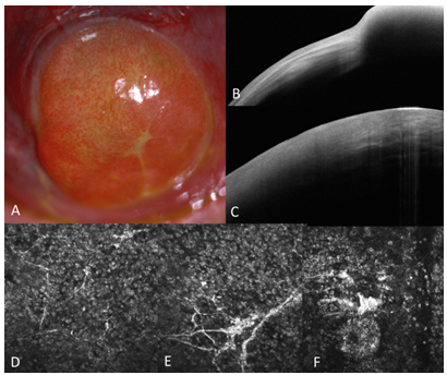
Five years later, slit-lamp examination revealed a solitary red elevated vascularized lesion covering the entire cornea of the right eye (Fig. 2). SD-OCT showed a hyperreflective elevated lesion, with irregular edges, well demarcated from adjacent limbal tissue (Fig. 2). A corneal tumor was suspected. It was surgically removed by superficial lamellar keratectomy combined with amniotic membrane transplantation.
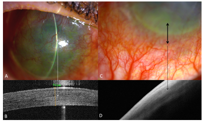
Slit-lamp images show superficial corneal vascularization and opacification of the whole cornea associated with absence of visible palisades of Vogt related to advanced limbal stem cell deficiency. OCT images show hyperreflective and irregular epithelium and loss of normal limbal niche structures featuring flat hyperreflective limbal epithelium
Macroscopically, the excisional red mass measured 5 x 4mm.
Microscopic examination revealed a non-specific pyogenic granuloma featuring neovessels within an edematous chorion and lymphoplasmacytic inflammatory elements with no fibrous reaction (negative staining for epithelial membrane antigen). The inflammatory infiltrate included B-lymphocytes and plasmocytes in addition to rare histiocytes. The lesion was strongly vascularized with positive staining for CD34. Alpha smooth muscle actin staining was negative showing the absence of myofibroblast proliferation.
Pyogenic granuloma (PG) is a common benign vascular lesion. PG in humans was first described in 18977 by Poncet and Dor as “human botryomycosis”. It was thought at this time the lesion was due to a fungal infection, whereas others later thought it was due to pyogenic bacterial infection, usually Staphylococcus aureus8. In 1925, Michelson9 suggested that the term PG should include “all sharply circumscribed granulation tissue growths occurring on cutaneous or mucous membrane surfaces and having the appearance of a tumor”. Although misnamed, the term PG has persisted in the medical literature. In fact, the term is a misnomer; the lesion is not typically pyogenic, unless there is secondary infection. A PG contains neither the inflammatory exudate of polymorphonuclear leukocytes with the resulting tissue proteolysis which is characteristic of a pyogenic reaction, nor the typical epithelioid giant cell reaction characteristic of granulomatous inflammation10. A PG is actually a vasoproliferative inflammatory response composed of granulation tissue. This tissue is the healing response that occurs following inflammation and it is characterized by the presence of fibroblast proliferation, small capillary-like channels that may leak fluid and thereby contribute to the edematous nature of the healing tissue, and scattered inflammatory cells. The cellular infiltrate usually consists of varying amounts of neutrophils, lymphocytes, plasma cells and other mononuclear elements11.
The common sites of its occurrence are the skin of the face, the extremities and mucosal surfaces of the oral cavity. They can also occur at the limbus or on the bulbar conjunctiva, simulating a pterygium12. A CPG is a relatively rare entity.
The occurrence of CPG was first reported by Minckler13 who reported a case which was misdiagnosed as conjunctival squamous cell carcinoma and after enucleation confirmed the diagnostic of CPG.
The typical presentation of the CPG is a rapidly growing lump which, on slit lamp examination, appears as well circumscribed, smooth surfaced or rough irregular surfaced, red, sessile and highly vascularized mass5414. The shape of the lesion varied from a mushroom-like contour with a narrow base to a broad based attachment with the base as broad as the body8.
Our patient had a similar clinical presentation with a rapid course and a presentation within one month of the onset of the growth15. The solitary red elevated fleshy vascularized lesion covered the entire cornea of the right eye.
CPG commonly grows at the sites of pre-existing corneal trauma, infectious corneal ulcer, or surgery. Most authors consider this lesion to be a secondary reaction to these underlying processes1617. Cameron and Mahmood have described an epithelial defect preceding the development of a CPG in over half their patients8. They suggest the lesion resulted from delayed wound healing of the epithelial defect in the presence of corneal vascularization and an ocular surface disease or chronic irritant. Our patient developed his lesion in less than 3 months.
The histopathological findings of our case were consistent with the common histopathological findings of CPG showing an excessive proliferation of granulation tissue with mononuclear cell infiltration5414. An exuberant granulation tissue lesion results from the delayed healing of the epithelial defect in the presence of corneal vascularization and an ocular surface disease or chronic irritant8.
OCT images showed hyperreflective and irregular epithelium and loss of normal limbal niche structures featuring flat hyperreflective limbal epithelium and IVCM showed abnormal epithelial cells with bright nuclei.
CPG is very unusual and can occasionally lead to difficult differential diagnosis of corneal masses. The differential diagnoses include anterior segment choristoma, vascular hamartoma, viral papilloma4 or squamous cell carcinoma (SCC). CPG can be mistaken for a SCC resulting in inappropriate enucleation1213. The history and clinical findings help for distinguish the lesions. The age of onset, history of prior trauma, infection or inflammation, rapid growth and the clinical appearance will often point to the correct diagnosis. CPG are typically preceded by a persistent epithelial defect. SCC is a slower growing lesion and has contact with the limbus with typical corneal extensions. It is generally not as vascular in appearance and the color is not as red as that observed with CPG. Histopathology of the tumor will confirm the diagnosis.
The first treatment of CPG consists in treating the ocular surface disease then remove chronic epithelial defects by medical or surgical methods18. Topical steroids may cause some shrinkage of corneal blood vessels as well as decrease the exudative response of the inflammatory reaction. If there is no response treatment of CPG is mainly surgical, which is excision. After surgical excision of the mass, the cornea may heal with scaring, especially if there is an underlying inflammatory process like our case. In such a condition, patients may benefit from optical keratoplasty. An appropriate clinical management of a patient with a CPG begins with the accurate recognition of the lesion to avoid unnecessarily aggressive treatment. Padadopoulos related a case of a spontaneous resolution3.
Despite its rarity, pyogenic granuloma should be considered in any patient with a fleshy, vascularized, elevated, rapidly growing corneal mass, especially in the setting of corneal infection preceded by a history of accidental and surgical trauma or persistent corneal defect. An excisional biopsy should be performed to make a definite diagnosis.
To our knowledge, this is the first case reported with an association between CPG and toxic epidermal necrolysis.
Clearly Auctoresonline and particularly Psychology and Mental Health Care Journal is dedicated to improving health care services for individuals and populations. The editorial boards' ability to efficiently recognize and share the global importance of health literacy with a variety of stakeholders. Auctoresonline publishing platform can be used to facilitate of optimal client-based services and should be added to health care professionals' repertoire of evidence-based health care resources.

Journal of Clinical Cardiology and Cardiovascular Intervention The submission and review process was adequate. However I think that the publication total value should have been enlightened in early fases. Thank you for all.

Journal of Women Health Care and Issues By the present mail, I want to say thank to you and tour colleagues for facilitating my published article. Specially thank you for the peer review process, support from the editorial office. I appreciate positively the quality of your journal.

Journal of Clinical Research and Reports I would be very delighted to submit my testimonial regarding the reviewer board and the editorial office. The reviewer board were accurate and helpful regarding any modifications for my manuscript. And the editorial office were very helpful and supportive in contacting and monitoring with any update and offering help. It was my pleasure to contribute with your promising Journal and I am looking forward for more collaboration.

We would like to thank the Journal of Thoracic Disease and Cardiothoracic Surgery because of the services they provided us for our articles. The peer-review process was done in a very excellent time manner, and the opinions of the reviewers helped us to improve our manuscript further. The editorial office had an outstanding correspondence with us and guided us in many ways. During a hard time of the pandemic that is affecting every one of us tremendously, the editorial office helped us make everything easier for publishing scientific work. Hope for a more scientific relationship with your Journal.

The peer-review process which consisted high quality queries on the paper. I did answer six reviewers’ questions and comments before the paper was accepted. The support from the editorial office is excellent.

Journal of Neuroscience and Neurological Surgery. I had the experience of publishing a research article recently. The whole process was simple from submission to publication. The reviewers made specific and valuable recommendations and corrections that improved the quality of my publication. I strongly recommend this Journal.

Dr. Katarzyna Byczkowska My testimonial covering: "The peer review process is quick and effective. The support from the editorial office is very professional and friendly. Quality of the Clinical Cardiology and Cardiovascular Interventions is scientific and publishes ground-breaking research on cardiology that is useful for other professionals in the field.

Thank you most sincerely, with regard to the support you have given in relation to the reviewing process and the processing of my article entitled "Large Cell Neuroendocrine Carcinoma of The Prostate Gland: A Review and Update" for publication in your esteemed Journal, Journal of Cancer Research and Cellular Therapeutics". The editorial team has been very supportive.

Testimony of Journal of Clinical Otorhinolaryngology: work with your Reviews has been a educational and constructive experience. The editorial office were very helpful and supportive. It was a pleasure to contribute to your Journal.

Dr. Bernard Terkimbi Utoo, I am happy to publish my scientific work in Journal of Women Health Care and Issues (JWHCI). The manuscript submission was seamless and peer review process was top notch. I was amazed that 4 reviewers worked on the manuscript which made it a highly technical, standard and excellent quality paper. I appreciate the format and consideration for the APC as well as the speed of publication. It is my pleasure to continue with this scientific relationship with the esteem JWHCI.

This is an acknowledgment for peer reviewers, editorial board of Journal of Clinical Research and Reports. They show a lot of consideration for us as publishers for our research article “Evaluation of the different factors associated with side effects of COVID-19 vaccination on medical students, Mutah university, Al-Karak, Jordan”, in a very professional and easy way. This journal is one of outstanding medical journal.

Dear Hao Jiang, to Journal of Nutrition and Food Processing We greatly appreciate the efficient, professional and rapid processing of our paper by your team. If there is anything else we should do, please do not hesitate to let us know. On behalf of my co-authors, we would like to express our great appreciation to editor and reviewers.

As an author who has recently published in the journal "Brain and Neurological Disorders". I am delighted to provide a testimonial on the peer review process, editorial office support, and the overall quality of the journal. The peer review process at Brain and Neurological Disorders is rigorous and meticulous, ensuring that only high-quality, evidence-based research is published. The reviewers are experts in their fields, and their comments and suggestions were constructive and helped improve the quality of my manuscript. The review process was timely and efficient, with clear communication from the editorial office at each stage. The support from the editorial office was exceptional throughout the entire process. The editorial staff was responsive, professional, and always willing to help. They provided valuable guidance on formatting, structure, and ethical considerations, making the submission process seamless. Moreover, they kept me informed about the status of my manuscript and provided timely updates, which made the process less stressful. The journal Brain and Neurological Disorders is of the highest quality, with a strong focus on publishing cutting-edge research in the field of neurology. The articles published in this journal are well-researched, rigorously peer-reviewed, and written by experts in the field. The journal maintains high standards, ensuring that readers are provided with the most up-to-date and reliable information on brain and neurological disorders. In conclusion, I had a wonderful experience publishing in Brain and Neurological Disorders. The peer review process was thorough, the editorial office provided exceptional support, and the journal's quality is second to none. I would highly recommend this journal to any researcher working in the field of neurology and brain disorders.
Dear Agrippa Hilda, Journal of Neuroscience and Neurological Surgery, Editorial Coordinator, I trust this message finds you well. I want to extend my appreciation for considering my article for publication in your esteemed journal. I am pleased to provide a testimonial regarding the peer review process and the support received from your editorial office. The peer review process for my paper was carried out in a highly professional and thorough manner. The feedback and comments provided by the authors were constructive and very useful in improving the quality of the manuscript. This rigorous assessment process undoubtedly contributes to the high standards maintained by your journal.

International Journal of Clinical Case Reports and Reviews. I strongly recommend to consider submitting your work to this high-quality journal. The support and availability of the Editorial staff is outstanding and the review process was both efficient and rigorous.

Thank you very much for publishing my Research Article titled “Comparing Treatment Outcome Of Allergic Rhinitis Patients After Using Fluticasone Nasal Spray And Nasal Douching" in the Journal of Clinical Otorhinolaryngology. As Medical Professionals we are immensely benefited from study of various informative Articles and Papers published in this high quality Journal. I look forward to enriching my knowledge by regular study of the Journal and contribute my future work in the field of ENT through the Journal for use by the medical fraternity. The support from the Editorial office was excellent and very prompt. I also welcome the comments received from the readers of my Research Article.

Dear Erica Kelsey, Editorial Coordinator of Cancer Research and Cellular Therapeutics Our team is very satisfied with the processing of our paper by your journal. That was fast, efficient, rigorous, but without unnecessary complications. We appreciated the very short time between the submission of the paper and its publication on line on your site.

I am very glad to say that the peer review process is very successful and fast and support from the Editorial Office. Therefore, I would like to continue our scientific relationship for a long time. And I especially thank you for your kindly attention towards my article. Have a good day!

"We recently published an article entitled “Influence of beta-Cyclodextrins upon the Degradation of Carbofuran Derivatives under Alkaline Conditions" in the Journal of “Pesticides and Biofertilizers” to show that the cyclodextrins protect the carbamates increasing their half-life time in the presence of basic conditions This will be very helpful to understand carbofuran behaviour in the analytical, agro-environmental and food areas. We greatly appreciated the interaction with the editor and the editorial team; we were particularly well accompanied during the course of the revision process, since all various steps towards publication were short and without delay".

I would like to express my gratitude towards you process of article review and submission. I found this to be very fair and expedient. Your follow up has been excellent. I have many publications in national and international journal and your process has been one of the best so far. Keep up the great work.

We are grateful for this opportunity to provide a glowing recommendation to the Journal of Psychiatry and Psychotherapy. We found that the editorial team were very supportive, helpful, kept us abreast of timelines and over all very professional in nature. The peer review process was rigorous, efficient and constructive that really enhanced our article submission. The experience with this journal remains one of our best ever and we look forward to providing future submissions in the near future.

I am very pleased to serve as EBM of the journal, I hope many years of my experience in stem cells can help the journal from one way or another. As we know, stem cells hold great potential for regenerative medicine, which are mostly used to promote the repair response of diseased, dysfunctional or injured tissue using stem cells or their derivatives. I think Stem Cell Research and Therapeutics International is a great platform to publish and share the understanding towards the biology and translational or clinical application of stem cells.

I would like to give my testimony in the support I have got by the peer review process and to support the editorial office where they were of asset to support young author like me to be encouraged to publish their work in your respected journal and globalize and share knowledge across the globe. I really give my great gratitude to your journal and the peer review including the editorial office.

I am delighted to publish our manuscript entitled "A Perspective on Cocaine Induced Stroke - Its Mechanisms and Management" in the Journal of Neuroscience and Neurological Surgery. The peer review process, support from the editorial office, and quality of the journal are excellent. The manuscripts published are of high quality and of excellent scientific value. I recommend this journal very much to colleagues.

Dr.Tania Muñoz, My experience as researcher and author of a review article in The Journal Clinical Cardiology and Interventions has been very enriching and stimulating. The editorial team is excellent, performs its work with absolute responsibility and delivery. They are proactive, dynamic and receptive to all proposals. Supporting at all times the vast universe of authors who choose them as an option for publication. The team of review specialists, members of the editorial board, are brilliant professionals, with remarkable performance in medical research and scientific methodology. Together they form a frontline team that consolidates the JCCI as a magnificent option for the publication and review of high-level medical articles and broad collective interest. I am honored to be able to share my review article and open to receive all your comments.

“The peer review process of JPMHC is quick and effective. Authors are benefited by good and professional reviewers with huge experience in the field of psychology and mental health. The support from the editorial office is very professional. People to contact to are friendly and happy to help and assist any query authors might have. Quality of the Journal is scientific and publishes ground-breaking research on mental health that is useful for other professionals in the field”.

Dear editorial department: On behalf of our team, I hereby certify the reliability and superiority of the International Journal of Clinical Case Reports and Reviews in the peer review process, editorial support, and journal quality. Firstly, the peer review process of the International Journal of Clinical Case Reports and Reviews is rigorous, fair, transparent, fast, and of high quality. The editorial department invites experts from relevant fields as anonymous reviewers to review all submitted manuscripts. These experts have rich academic backgrounds and experience, and can accurately evaluate the academic quality, originality, and suitability of manuscripts. The editorial department is committed to ensuring the rigor of the peer review process, while also making every effort to ensure a fast review cycle to meet the needs of authors and the academic community. Secondly, the editorial team of the International Journal of Clinical Case Reports and Reviews is composed of a group of senior scholars and professionals with rich experience and professional knowledge in related fields. The editorial department is committed to assisting authors in improving their manuscripts, ensuring their academic accuracy, clarity, and completeness. Editors actively collaborate with authors, providing useful suggestions and feedback to promote the improvement and development of the manuscript. We believe that the support of the editorial department is one of the key factors in ensuring the quality of the journal. Finally, the International Journal of Clinical Case Reports and Reviews is renowned for its high- quality articles and strict academic standards. The editorial department is committed to publishing innovative and academically valuable research results to promote the development and progress of related fields. The International Journal of Clinical Case Reports and Reviews is reasonably priced and ensures excellent service and quality ratio, allowing authors to obtain high-level academic publishing opportunities in an affordable manner. I hereby solemnly declare that the International Journal of Clinical Case Reports and Reviews has a high level of credibility and superiority in terms of peer review process, editorial support, reasonable fees, and journal quality. Sincerely, Rui Tao.

Clinical Cardiology and Cardiovascular Interventions I testity the covering of the peer review process, support from the editorial office, and quality of the journal.

Clinical Cardiology and Cardiovascular Interventions, we deeply appreciate the interest shown in our work and its publication. It has been a true pleasure to collaborate with you. The peer review process, as well as the support provided by the editorial office, have been exceptional, and the quality of the journal is very high, which was a determining factor in our decision to publish with you.

The peer reviewers process is quick and effective, the supports from editorial office is excellent, the quality of journal is high. I would like to collabroate with Internatioanl journal of Clinical Case Reports and Reviews journal clinically in the future time.

Clinical Cardiology and Cardiovascular Interventions, I would like to express my sincerest gratitude for the trust placed in our team for the publication in your journal. It has been a true pleasure to collaborate with you on this project. I am pleased to inform you that both the peer review process and the attention from the editorial coordination have been excellent. Your team has worked with dedication and professionalism to ensure that your publication meets the highest standards of quality. We are confident that this collaboration will result in mutual success, and we are eager to see the fruits of this shared effort.

Dear Dr. Jessica Magne, Editorial Coordinator 0f Clinical Cardiology and Cardiovascular Interventions, I hope this message finds you well. I want to express my utmost gratitude for your excellent work and for the dedication and speed in the publication process of my article titled "Navigating Innovation: Qualitative Insights on Using Technology for Health Education in Acute Coronary Syndrome Patients." I am very satisfied with the peer review process, the support from the editorial office, and the quality of the journal. I hope we can maintain our scientific relationship in the long term.

Dear Monica Gissare, - Editorial Coordinator of Nutrition and Food Processing. ¨My testimony with you is truly professional, with a positive response regarding the follow-up of the article and its review, you took into account my qualities and the importance of the topic¨.

Dear Dr. Jessica Magne, Editorial Coordinator 0f Clinical Cardiology and Cardiovascular Interventions, The review process for the article “The Handling of Anti-aggregants and Anticoagulants in the Oncologic Heart Patient Submitted to Surgery” was extremely rigorous and detailed. From the initial submission to the final acceptance, the editorial team at the “Journal of Clinical Cardiology and Cardiovascular Interventions” demonstrated a high level of professionalism and dedication. The reviewers provided constructive and detailed feedback, which was essential for improving the quality of our work. Communication was always clear and efficient, ensuring that all our questions were promptly addressed. The quality of the “Journal of Clinical Cardiology and Cardiovascular Interventions” is undeniable. It is a peer-reviewed, open-access publication dedicated exclusively to disseminating high-quality research in the field of clinical cardiology and cardiovascular interventions. The journal's impact factor is currently under evaluation, and it is indexed in reputable databases, which further reinforces its credibility and relevance in the scientific field. I highly recommend this journal to researchers looking for a reputable platform to publish their studies.

Dear Editorial Coordinator of the Journal of Nutrition and Food Processing! "I would like to thank the Journal of Nutrition and Food Processing for including and publishing my article. The peer review process was very quick, movement and precise. The Editorial Board has done an extremely conscientious job with much help, valuable comments and advices. I find the journal very valuable from a professional point of view, thank you very much for allowing me to be part of it and I would like to participate in the future!”

Dealing with The Journal of Neurology and Neurological Surgery was very smooth and comprehensive. The office staff took time to address my needs and the response from editors and the office was prompt and fair. I certainly hope to publish with this journal again.Their professionalism is apparent and more than satisfactory. Susan Weiner

My Testimonial Covering as fellowing: Lin-Show Chin. The peer reviewers process is quick and effective, the supports from editorial office is excellent, the quality of journal is high. I would like to collabroate with Internatioanl journal of Clinical Case Reports and Reviews.

My experience publishing in Psychology and Mental Health Care was exceptional. The peer review process was rigorous and constructive, with reviewers providing valuable insights that helped enhance the quality of our work. The editorial team was highly supportive and responsive, making the submission process smooth and efficient. The journal's commitment to high standards and academic rigor makes it a respected platform for quality research. I am grateful for the opportunity to publish in such a reputable journal.

My experience publishing in International Journal of Clinical Case Reports and Reviews was exceptional. I Come forth to Provide a Testimonial Covering the Peer Review Process and the editorial office for the Professional and Impartial Evaluation of the Manuscript.
