AUCTORES
Globalize your Research

Review Article
*Corresponding Author: Anthony Kodzo-Grey Venyo, North Manchester General Hospital Department of Urology Manchester United Kingdom.
Citation: Anthony K-G Venyo. (2020) Amoebiasis of the Penis: A Review and Update. Journal of Clinical Surgery and Research. 1(1) DOI: 10.31579/2768-2757/001
Copyright: © 2020 Anthony Kodzo-Grey Venyo. This is an open-access article distributed under the terms of the Creative Commons Attribution License, which permits unrestricted use, distribution, and reproduction in any medium, provided the original author and source are credited.
Received: 24 September 2020 | Accepted: 23 December 2020 | Published: 27 December 2020
Keywords: amoebiasis of penis; amebiasis of penis; penile amoebiasis; penile amebiasis; entamoeba histolytica; balanoposthitis; trophozoites; microscopic examination; biopsy of penile lesion; metronidazole; anti-amoebic medicament; emetine; chancroid; syphilis
Ulceration of the penis/foreskin which would tend to be painful, Swelling of the penis, Oedema of the penis, Discharge from ulcer or inflamed area of penis that could be purulent or may contain blood, Balanoposthitis, Exudation from a penile ulcer which could be mild, profuse, purulent or bloody, Sloughing off of part of the foreskin and other tissues with resulting hypospadias, indurated swelling of the penis which may initially involve one part of the penis but could quickly spread, A history of homosexual coital activity may be obtained, The spouse of a man who has Amoebiasis of the penis could also have Amoebiasis of vulva, cervix or endometrium, The prepuce may not be retractable, There may be ulceration or swelling of the glans penis that may be irregular. Clinical examination findings in cases of Amoebiasis of the penis could reveal some of the ensuing: The general and systematic examinations may be normal. Examination of the penis may show: Tight non-retractile foreskin, Ulceration of foreskin, Swelling of the foreskin, Swelling of the glans penis, Ulceration and swelling of glans penis., Swelling and inflammation of the shaft of the penis, Ulceration on the shaft of the penis, Development of an iatrogenic hypospadias which was not there before, The inguinal lymph nodes may not be palpable but sometimes they may be enlarged on one side or on both sides, The penile swelling may involve part of the penis but at times on rare occasions the entire penis may be swollen, the penile swelling could on rare occasions extend to the supra-pubic area, On rare occasions the swelling of the penis could extend to include the scrotum but this is extremely rare. Amoebiasis of the penis does mimic various common conditions that affect the penis including: squamous cell carcinoma of the penis, chancroid, primary syphilitic ulcer of the penis, granuloma inguinale, balanoposthitis, and many other lesions affecting the penis. A high-index of suspicion is required to diagnose Amoebiasis of the penis. Clinicians need to be aware that male homosexuals who practice penetrative penis-anal coital activity have a higher risk of developing amoebiasis of the penis especially in Amoebiasis endemic countries. If an individual is suspected to have balanoposthitis or non-specific infection of the penis and is treated with antibiotics but the lesion does not respond to treatment, amoebiasis of the penis should be suspected. Secretions and discharges from the penile ulcer as well as biopsies of the penile lesion should be submitted for pathology examination which would demonstrate trophozoites, entamoebae as well as inflammatory cells. Even if carcinoma of the penis is initially suspected biopsy of the penile lesion would show features of Amoebiasis in the absence of any features of malignancy but in the very rare situation of a combination of Amoebiasis of the penis and carcinoma of the penis microscopic pathology examination of a biopsy specimen of the penile lesion would show features of Amoebiasis and carcinoma of the penis. .Amoebiasis of the penis does quickly and effectively respond to anti-amoebic medicaments.
It has been iterated that Amoebiasis of the penis is a rare clinical entity due to the fact that the penis is an unusual site for the manifestation of amoebiasis. [1] It has additionally been stated that homosexuals tend to have a higher risk for the development of amoebiasis of the penis. [1] Amoebic ulcers tend to mimic cutaneous lesions that had arisen from squamous cell carcinoma of the penis, chancroid, primary syphilis, granuloma inguinale, and many other causes of lesions involving the penis. [1] It had been recommended that Amoebiasis of the penis should be suspected when a patient with a penile lesion which has been clinically provisionally diagnosed as having balanoposthitis and treated with antibiotics has not responded to antibiotic treatment and such a situation biopsy of the penile lesion to isolate trophozoites to confirm the diagnosis of Amoebiasis of the penis should be necessitated. [1]
It has been documented that most of the individuals that are afflicted by Amoebiasis tend to be asymptomatic; nevertheless, they tend to pass cysts and their condition has been referred to as asymptomatic intra-luminal Amoebiasis. [2] This document was stated to be true with regard to Entamoeba Moshkovskii and majority of Entamoeba Dispar and up to 80% of cases of Entamoeba histolytica. [2] It has also been iterated that whilst Entamoeba dispar had generally been understood to be non-pathogenic, it had been reported in 2015 that Entamoeba Dispar could sometimes cause symptoms. [2] [3]
Amoebiasis of the bowel which tends to be associated with diarrhoea without dysentery with absence of mucus or stool. [2]. With regard to Amoebic dysentery or colitis there tends to be an associated mucus with the diarrhoea or visible or non-visible blood [2] some of the manifestations that tend to be associated with the common Amoebic dysentery include:
Amoebiasis of the penis is a very rare condition that is sporadically reported and because of its rarity within Amoebiasis endemic areas clinicians within the Amoebiasis-endemic areas would tend not to be familiar with the manifestations of the infection. Additionally because of global travel and the practice of homosexuality Amoebiasis of the penis would be encountered in non-Amoebiasis endemic areas where most clinicians would not have encountered a case before and would also tend to be unfamiliar with the manifestations and treatment of the disease. Usually people who have Amoebiasis of the penis do not have symptoms of gastro-intestinal Amoebiasis. Amoebiasis of the penis would tend to present with non-specific symptoms including:
Pain in the penis including: the foreskin, glans, or shaft of penis; ulceration of foreskin, glans penis, or shaft of the penis; swelling / induration of penis including foreskin, glans penis, or shaft of penis; discharge of purulent nature or blood from foreskin, glans penis, or shaft of penis; recently developed hypospadias of unknown cause. The swelling and induration of the penis tends to be non-specific causes psychological trauma to most patients because their local practitioners have given them antibiotics presuming the symptoms have been related to infection but the symptoms have remained the same or are getting worse. The next thing is the patients worry about a strong possibility that they may have a malignancy of the penis. The ensuing article contains a review and update of the literature related to case reports, case series, and studies undertaken related to Amoebiasis of the penis as well as an overview documentations related to Amoebiasis in general.
Aim
To review and update the literature on Amoebiasis of the penis.
Internet data bases were used to search for literature on Amoebiasis of the penis including. Google, Google Scholar; Yahoo, and PUB MED. The search words that were used included Amoebiasis of the penis, Amebiasis of penis, penile amoebiasis, penile amebiasis, urogenital amoebiasis, urogenital amebiasis, amoebiasis, amebiasis. Fifty references were identified which were used to write the paper that has been divided into (A) Overview which has discussed various aspects of amoebiasis in general and amoebiasis of the penis in general to provide a bird’s eye view of the subject and (B) Miscellaneous narrations, summations, and discussions from case reports, case series, and studies related to Amoebiasis of the penis.
Result / Review and Update of Literature
Definition and general comments
Terminology
Epidemiology
Sites
Pathophysiology
Clinical presentations
Clinical examination findings
The general and systematic examinations of the patients who have Amoebiasis of the penis may be normal. However examination of the penis, genitalia and inguinal region may show:
The aforementioned features are non-specific and would not be diagnostic of Amoebiasis alone because many conditions would tend to present similarly.
Stool
Haematology investigations
Biochemistry Blood Tests
Radiology Investigations
Computed tomography (CT) scan
Magnetic Resonance Imaging (MRI) scan
Diagnosis
Diagnosis of Amoebiasis of the penis tends to be confirmed by the following:
Staining for Amoebiasis [2]
The positive stains for Amoebiasis include:
Negative stains
The negative stain for Amoebiasis does include:
Update on laboratory diagnosis of amoebiasis: [6]
Amoebiasis caused by Entamoeba histolytica, is a public health problem in many developing countries which does cause up to 100,000 fatal cases globally annually. The detection of pathogenic Entamoeba histolytica and its differentiation from non-pathogenic Entamoeba spp. does play a vital role with regard to the clinical management of patients. Laboratory diagnosis of intestinal amoebiasis within the developing countries of the world does still rely upon labour intensive and insensitive methods that involve staining of samples of the stool and microscopy examination. Newer and more sensitive methods for the diagnosis of amoebiasis do include various antigen detection ELISAs and rapid tests; nevertheless, their diagnostic sensitivity and sensitivity does seem to vary between studies that had been undertaken, and some of the tests have not been able to distinguish from among the Entamoeba species. Molecular detection techniques are highly sensitive and specific and isothermal amplification approaches could be developed into field applicable tests; nevertheless, the cost would tend to be a barrier for their utilization as a routine laboratory method of testing for Amoebiasis in most developing poor countries where Amoebiasis is endemic. [6]
Entamoeba culture methods: [5]
ISO-ENZYME/MODEME ANALYSIS [5]
The ensuing summations have been made with regard to Iso-Enzyme Modeme Analysis: [5]
Serological tests [5]
Antibody detection [5]
Relevant summations related to antibody detection in relation to the diagnosis amoebiasis that had been made include: [5]
Detection of antigen [5]
Summations relating to the detection of antigen in amoebiasis have included the following: [5]
Molecular Methods
Summations related to utilization of molecular methods diagnosis and management of amoebiasis have been summarised as follows: [5]
Conventional polymerase chain reaction (PCR)
Summations related to the utilization of conventional PCR in the detection / diagnosis of amoebiasis include: [5]
REAL-TIME POLYMERASE CHAIN REACTION [5]
Parija et al. [5] summarized salient points related to utilization of Real-Time Polymerase Chain Reaction as follows:
Treatment
Treatment of Amoebiasis of the penis tends to be by utilization of anti-amoebic medicaments which tends to result in quick resolution of the infection and some of the medicaments include:
In situations when destruction of tissue has resulted in the formation of hypospadias then utilization of the most appropriate surgical procedure to repair the hypospadias would need to be adopted including pedicle island flap repair.
Differential Diagnosis of Amoebiasis of penis.
Some of the differential diagnosis of Amoebiasis of the bowel include: [2]
Some of the differential diagnosis of Amoebiasis of the penis include:
Outcome
When Amoebiasis of the penis is diagnosed accurately treatment with utilization of anti-amoebic medicaments for one to two weeks does result in resolution of the infection completely but if there was any structural damage like hypospadias that would need to be repaired surgically.
Thomas and Antony [35] in 1976, reported a case of amoebiasis of the penis. They stated that cases of amoebiasis of the penis are very rare and that cases of amoebiasis of the penis had tended to be mistaken clinically a malignant lesion or cancer as well as an ulcerative venereal disease affecting the penis. Furthermore, Thomas and Antony [35] iterated that prior to the publication of their case report only 8 cases of Amoebiasis of the penis had been reported within the 51 years preceding their publication which had included publications by: Shih, Wu, and Lieu in 1939 [36]; Hermann and Berman in 1942 [37]; Camecho and Beirana in 1959 [8]; Mylius and Ten Seldam in 1962 [39]; Talwaker in 1962 [40]; Quevedo and Elias Dib in 1963 [41]; Purpon Jiminez and Engelking in 1967 [42].
Shih et al. [36] reported a 54-year-old Chinese man who had presented with an ulcerative lesion of his penis of 5 months duration. The clinicians provisionally considered the lesion to be either a malignant lesion or a pyogenic lesion. Shih et al. [36] reported also that examinations of the exudate of the ulcer as well as the superficial layers of the ulcer did reveal amoebae which had the characteristic features of Entamoeba Histolytica. The patient did not have any history of dysentery. Examinations undertaken repeatedly of the patient’s faeces did not show any evidence of Entamoeba. Additionally the patient had denied having had extraneous intercourse. He was treated with utilization of Emetine which resulted in a speedy cure. In view of the fact that his home was too far for his wife to come for examination the possibility of the amoebiasis being an emanation from amoebic dysentery which the wife might have had could not be ruled out and that meant the source of amoebiasis of the man’s penis could not be established. Shih et al. [36] cases of cutaneous amoebiasis usually tend to arise either from an abscess (for example liver abscess) discharging on to the abdominal wall or from extension from the bowel content.
Sosa Camacho and Beirana [38] in 1959 reported a case of dermatosis in which an ulcerated lesion of the penis with abundant secretion was found in young male who had practiced active pederasty. The lesion had clinically been provisionally diagnosed as a carcinoma of the penis. The pathology report and the clinical aspect of the lesion did not correspond to carcinoma and therefore, a culture of the secretion was undertaken and the report came back as showing many Entamoeba histolytica. He was treated by means of emetine and oxyquinoline with successful result. The lesson to learn from this case report is that amoebiasis of the penis could mimic carcinoma of the penis and hence clinicians globally should remember this disease and have a high-index of suspicion for the disease in order to confirm its diagnosis.
Lahiri [43] in 1964, reported a case of Amoebiasis of the penis in the Ghana medical Journal. The lesson to learn from this report is that amoebiasis involving the bowel is common in Ghana and West Africa and though amoebiasis of the penis is not that common clinicians in the Ecowas states should be aware that Amoebiasis of the penis can occur and hence a high index of suspicion for the disease should be on the minds of local clinicians in order to quickly establish the diagnosis of the disease.
Purpon et al. [42] in 1967, reported a 31-year-old white man, who was admitted on March 04 1965. It was reported that 22 days preceding his admission to hospital, he had been intoxicated he had had homosexual interactions whilst he was inebriated. The next day pursuant to his homosexual interactions he had developed a burning sensation in his penis. Many days subsequently he had noticed an extremely painful ulceration on his coronal sulcus. The ulcer did spread rapidly and had encompassed his whole penis with a purulent exudate in non-excessive amounts. Examination of his penis showed an ulcerous strip that measured 2.5 cm wide that surrounded the his glans penis and part of his foreskin (prepuce). The examination also revealed that only a small part of the mucosa by the external urethral meatus had not been damaged. The ulceration had been sunken at the bottom, and it had been covered with a bloody and scanty exudate as well as it had been hollowed out at its edges. His abdominal examination was normal and there was no significant lymph node enlargement. Thorough assessment and laboratory investigation confirmed a diagnosis of amoebiasis of the penis. Purpon et al. [42] did state that amoebiasis of the penis is rare and at the time of publication of their paper, only 7 cases of amoebiasis of the penis had been reported in the global literature [36] [37] [38] [39] [40] [41] [44]; nevertheless, they had been aware of many cases of amoebiasis of the penis that had not been reported to them by means of personal communications (see details from the article). Purpon et al. [42] stated that they had reported their case of amoebiasis of the penis in order to draw attention to a disease which is rare and which in their opinion might be confused with other neoplastic and inflammatory lesions of the penis. Purpon et al. [42] stated that Mylius and Ten Seldam [39] had cited a report of a case of amoebiasis of the penis that had been encountered in a native of New Guinea whose spouse had suffered from amoebic vulvovaginitis .
In 1973, the experience of a pathologist called Cooke who had worked in Papua New Guinea were summated in the Journal of the Medical Association of Thailand summated as follows: [45]
Jayaweera [46] in 1975 reported a case of amoebic ulceration of the cervix in a female as well as a case of amoebiasis of the penis in a male. They stated that amoebiasis of the cervix and amoebiasis of the penis can closely mimic or simulate carcinoma of the cervix and carcinoma of the penis. A lesson to be learnt from this case report is that amoebiasis of the cervix and amoebiasis of the penis do exist and they are treatable diseases and for this reasons clinicians in amoebiasis endemic areas as well as globally show be aware that amoebiasis of the cervix and amoebiasis of the penis though they are generally uncommon because of global travel can be encountered anywhere in the world because of this a high-index of suspicion for amoebiasis of the cervix and the penis should be borne in mind.
Hejase et al. [1] reported a case of amoebiasis of the penis in 1975 in which they summated pertinent aspects related to amoebiasis of the penis as follows:
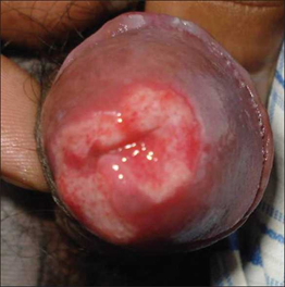
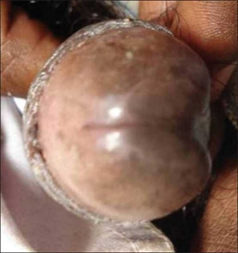
Mohanty et al. [47] reported a 47-year-old man who did present with a very painful ulcer of 15 days duration that had developed over the glans of his penis and which had encompassed his external urethral meatus. The penile ulcer did have a well-demarcated border and a raised erythematous rim (see figure 1). The whole penis was oedematous as well as associated with foul-smelling haemo-purulent exudates. He did have bilateral inguinal lymph node enlargements. The penile lesion did commence as multiple superficial ulcers that had coalesced to constitute a spreading ulcer. He did have a history of homosexuality. His local practitioner had already treated him with utilization of ciprofloxacin and azithromycin without any response of the penile lesion to treatment. The results of his stool examination, urine examination, blood routine haematology and biochemistry tests were normal. The results of his Venereal Disease Research Laboratory (VDRL) as well as Human Immunodeficiency Virus (HIV) tests were also non-reactive. A scrape cytology examination of the penile lesion was undertaken and its examination did reveal presence of inflammatory cells, and no atypia, or dysplastic cells. A wet mount of the haemo-purulent discharge did show presence of trophozoites of Entamoeba histolytica. The stool examination was normal and did not reveal any parasite. He was prescribed a two week course of metronidazole and after one week his ulcer had regressed and complete regression ensued in 2 weeks (see figure 2).
Amoebiasis of the penis is a very rare infection of the penis which is reported sporadically in Amoebiasis endemic and Amoebiasis non-endemic areas of the world. Amoebiasis of the penis does mimic various common conditions that affect the penis including: squamous cell carcinoma of the penis, chancroid, primary syphilitic ulcer of the penis, granuloma inguinale, balanoposthitis, and many other lesions affecting the penis. A high-index of suspicion is required to diagnose Amoebiasis of the penis. Clinicians need to be aware that male homosexuals who practice penetrative penis-anal coital activity have a higher risk of developing amoebiasis of the penis especially in Amoebiasis endemic countries. If an individual is suspected to have balanoposthitis or non-specific infection of the penis is treated with antibiotics but the lesion does not respond to treatment, amoebiasis of the penis should be suspected. Secretions and discharges from the penile ulcer as well as biopsies of the penile lesion should be submitted for pathology examination which would demonstrate trophozoites, entamoebae as well as inflammatory cells. Amoebiasis of the penis does quickly and effectively respond to anti-amoebic medicaments.
Conflict of interest – None
Indian J Sex Transm Dis AIDS for granting permission for reproduction of figures and contents of their journal article under Copyright © Indian Journal of Sexually Transmitted Diseases and AIDS This is an open-access article distributed under the terms of the Creative Commons Attribution License, which permits unrestricted use, distribution, and reproduction in any medium, provided original work is properly cited.
Clearly Auctoresonline and particularly Psychology and Mental Health Care Journal is dedicated to improving health care services for individuals and populations. The editorial boards' ability to efficiently recognize and share the global importance of health literacy with a variety of stakeholders. Auctoresonline publishing platform can be used to facilitate of optimal client-based services and should be added to health care professionals' repertoire of evidence-based health care resources.

Journal of Clinical Cardiology and Cardiovascular Intervention The submission and review process was adequate. However I think that the publication total value should have been enlightened in early fases. Thank you for all.

Journal of Women Health Care and Issues By the present mail, I want to say thank to you and tour colleagues for facilitating my published article. Specially thank you for the peer review process, support from the editorial office. I appreciate positively the quality of your journal.

Journal of Clinical Research and Reports I would be very delighted to submit my testimonial regarding the reviewer board and the editorial office. The reviewer board were accurate and helpful regarding any modifications for my manuscript. And the editorial office were very helpful and supportive in contacting and monitoring with any update and offering help. It was my pleasure to contribute with your promising Journal and I am looking forward for more collaboration.

We would like to thank the Journal of Thoracic Disease and Cardiothoracic Surgery because of the services they provided us for our articles. The peer-review process was done in a very excellent time manner, and the opinions of the reviewers helped us to improve our manuscript further. The editorial office had an outstanding correspondence with us and guided us in many ways. During a hard time of the pandemic that is affecting every one of us tremendously, the editorial office helped us make everything easier for publishing scientific work. Hope for a more scientific relationship with your Journal.

The peer-review process which consisted high quality queries on the paper. I did answer six reviewers’ questions and comments before the paper was accepted. The support from the editorial office is excellent.

Journal of Neuroscience and Neurological Surgery. I had the experience of publishing a research article recently. The whole process was simple from submission to publication. The reviewers made specific and valuable recommendations and corrections that improved the quality of my publication. I strongly recommend this Journal.

Dr. Katarzyna Byczkowska My testimonial covering: "The peer review process is quick and effective. The support from the editorial office is very professional and friendly. Quality of the Clinical Cardiology and Cardiovascular Interventions is scientific and publishes ground-breaking research on cardiology that is useful for other professionals in the field.

Thank you most sincerely, with regard to the support you have given in relation to the reviewing process and the processing of my article entitled "Large Cell Neuroendocrine Carcinoma of The Prostate Gland: A Review and Update" for publication in your esteemed Journal, Journal of Cancer Research and Cellular Therapeutics". The editorial team has been very supportive.

Testimony of Journal of Clinical Otorhinolaryngology: work with your Reviews has been a educational and constructive experience. The editorial office were very helpful and supportive. It was a pleasure to contribute to your Journal.

Dr. Bernard Terkimbi Utoo, I am happy to publish my scientific work in Journal of Women Health Care and Issues (JWHCI). The manuscript submission was seamless and peer review process was top notch. I was amazed that 4 reviewers worked on the manuscript which made it a highly technical, standard and excellent quality paper. I appreciate the format and consideration for the APC as well as the speed of publication. It is my pleasure to continue with this scientific relationship with the esteem JWHCI.

This is an acknowledgment for peer reviewers, editorial board of Journal of Clinical Research and Reports. They show a lot of consideration for us as publishers for our research article “Evaluation of the different factors associated with side effects of COVID-19 vaccination on medical students, Mutah university, Al-Karak, Jordan”, in a very professional and easy way. This journal is one of outstanding medical journal.

Dear Hao Jiang, to Journal of Nutrition and Food Processing We greatly appreciate the efficient, professional and rapid processing of our paper by your team. If there is anything else we should do, please do not hesitate to let us know. On behalf of my co-authors, we would like to express our great appreciation to editor and reviewers.

As an author who has recently published in the journal "Brain and Neurological Disorders". I am delighted to provide a testimonial on the peer review process, editorial office support, and the overall quality of the journal. The peer review process at Brain and Neurological Disorders is rigorous and meticulous, ensuring that only high-quality, evidence-based research is published. The reviewers are experts in their fields, and their comments and suggestions were constructive and helped improve the quality of my manuscript. The review process was timely and efficient, with clear communication from the editorial office at each stage. The support from the editorial office was exceptional throughout the entire process. The editorial staff was responsive, professional, and always willing to help. They provided valuable guidance on formatting, structure, and ethical considerations, making the submission process seamless. Moreover, they kept me informed about the status of my manuscript and provided timely updates, which made the process less stressful. The journal Brain and Neurological Disorders is of the highest quality, with a strong focus on publishing cutting-edge research in the field of neurology. The articles published in this journal are well-researched, rigorously peer-reviewed, and written by experts in the field. The journal maintains high standards, ensuring that readers are provided with the most up-to-date and reliable information on brain and neurological disorders. In conclusion, I had a wonderful experience publishing in Brain and Neurological Disorders. The peer review process was thorough, the editorial office provided exceptional support, and the journal's quality is second to none. I would highly recommend this journal to any researcher working in the field of neurology and brain disorders.
Dear Agrippa Hilda, Journal of Neuroscience and Neurological Surgery, Editorial Coordinator, I trust this message finds you well. I want to extend my appreciation for considering my article for publication in your esteemed journal. I am pleased to provide a testimonial regarding the peer review process and the support received from your editorial office. The peer review process for my paper was carried out in a highly professional and thorough manner. The feedback and comments provided by the authors were constructive and very useful in improving the quality of the manuscript. This rigorous assessment process undoubtedly contributes to the high standards maintained by your journal.

International Journal of Clinical Case Reports and Reviews. I strongly recommend to consider submitting your work to this high-quality journal. The support and availability of the Editorial staff is outstanding and the review process was both efficient and rigorous.

Thank you very much for publishing my Research Article titled “Comparing Treatment Outcome Of Allergic Rhinitis Patients After Using Fluticasone Nasal Spray And Nasal Douching" in the Journal of Clinical Otorhinolaryngology. As Medical Professionals we are immensely benefited from study of various informative Articles and Papers published in this high quality Journal. I look forward to enriching my knowledge by regular study of the Journal and contribute my future work in the field of ENT through the Journal for use by the medical fraternity. The support from the Editorial office was excellent and very prompt. I also welcome the comments received from the readers of my Research Article.

Dear Erica Kelsey, Editorial Coordinator of Cancer Research and Cellular Therapeutics Our team is very satisfied with the processing of our paper by your journal. That was fast, efficient, rigorous, but without unnecessary complications. We appreciated the very short time between the submission of the paper and its publication on line on your site.

I am very glad to say that the peer review process is very successful and fast and support from the Editorial Office. Therefore, I would like to continue our scientific relationship for a long time. And I especially thank you for your kindly attention towards my article. Have a good day!

"We recently published an article entitled “Influence of beta-Cyclodextrins upon the Degradation of Carbofuran Derivatives under Alkaline Conditions" in the Journal of “Pesticides and Biofertilizers” to show that the cyclodextrins protect the carbamates increasing their half-life time in the presence of basic conditions This will be very helpful to understand carbofuran behaviour in the analytical, agro-environmental and food areas. We greatly appreciated the interaction with the editor and the editorial team; we were particularly well accompanied during the course of the revision process, since all various steps towards publication were short and without delay".

I would like to express my gratitude towards you process of article review and submission. I found this to be very fair and expedient. Your follow up has been excellent. I have many publications in national and international journal and your process has been one of the best so far. Keep up the great work.

We are grateful for this opportunity to provide a glowing recommendation to the Journal of Psychiatry and Psychotherapy. We found that the editorial team were very supportive, helpful, kept us abreast of timelines and over all very professional in nature. The peer review process was rigorous, efficient and constructive that really enhanced our article submission. The experience with this journal remains one of our best ever and we look forward to providing future submissions in the near future.

I am very pleased to serve as EBM of the journal, I hope many years of my experience in stem cells can help the journal from one way or another. As we know, stem cells hold great potential for regenerative medicine, which are mostly used to promote the repair response of diseased, dysfunctional or injured tissue using stem cells or their derivatives. I think Stem Cell Research and Therapeutics International is a great platform to publish and share the understanding towards the biology and translational or clinical application of stem cells.

I would like to give my testimony in the support I have got by the peer review process and to support the editorial office where they were of asset to support young author like me to be encouraged to publish their work in your respected journal and globalize and share knowledge across the globe. I really give my great gratitude to your journal and the peer review including the editorial office.

I am delighted to publish our manuscript entitled "A Perspective on Cocaine Induced Stroke - Its Mechanisms and Management" in the Journal of Neuroscience and Neurological Surgery. The peer review process, support from the editorial office, and quality of the journal are excellent. The manuscripts published are of high quality and of excellent scientific value. I recommend this journal very much to colleagues.

Dr.Tania Muñoz, My experience as researcher and author of a review article in The Journal Clinical Cardiology and Interventions has been very enriching and stimulating. The editorial team is excellent, performs its work with absolute responsibility and delivery. They are proactive, dynamic and receptive to all proposals. Supporting at all times the vast universe of authors who choose them as an option for publication. The team of review specialists, members of the editorial board, are brilliant professionals, with remarkable performance in medical research and scientific methodology. Together they form a frontline team that consolidates the JCCI as a magnificent option for the publication and review of high-level medical articles and broad collective interest. I am honored to be able to share my review article and open to receive all your comments.

“The peer review process of JPMHC is quick and effective. Authors are benefited by good and professional reviewers with huge experience in the field of psychology and mental health. The support from the editorial office is very professional. People to contact to are friendly and happy to help and assist any query authors might have. Quality of the Journal is scientific and publishes ground-breaking research on mental health that is useful for other professionals in the field”.

Dear editorial department: On behalf of our team, I hereby certify the reliability and superiority of the International Journal of Clinical Case Reports and Reviews in the peer review process, editorial support, and journal quality. Firstly, the peer review process of the International Journal of Clinical Case Reports and Reviews is rigorous, fair, transparent, fast, and of high quality. The editorial department invites experts from relevant fields as anonymous reviewers to review all submitted manuscripts. These experts have rich academic backgrounds and experience, and can accurately evaluate the academic quality, originality, and suitability of manuscripts. The editorial department is committed to ensuring the rigor of the peer review process, while also making every effort to ensure a fast review cycle to meet the needs of authors and the academic community. Secondly, the editorial team of the International Journal of Clinical Case Reports and Reviews is composed of a group of senior scholars and professionals with rich experience and professional knowledge in related fields. The editorial department is committed to assisting authors in improving their manuscripts, ensuring their academic accuracy, clarity, and completeness. Editors actively collaborate with authors, providing useful suggestions and feedback to promote the improvement and development of the manuscript. We believe that the support of the editorial department is one of the key factors in ensuring the quality of the journal. Finally, the International Journal of Clinical Case Reports and Reviews is renowned for its high- quality articles and strict academic standards. The editorial department is committed to publishing innovative and academically valuable research results to promote the development and progress of related fields. The International Journal of Clinical Case Reports and Reviews is reasonably priced and ensures excellent service and quality ratio, allowing authors to obtain high-level academic publishing opportunities in an affordable manner. I hereby solemnly declare that the International Journal of Clinical Case Reports and Reviews has a high level of credibility and superiority in terms of peer review process, editorial support, reasonable fees, and journal quality. Sincerely, Rui Tao.

Clinical Cardiology and Cardiovascular Interventions I testity the covering of the peer review process, support from the editorial office, and quality of the journal.

Clinical Cardiology and Cardiovascular Interventions, we deeply appreciate the interest shown in our work and its publication. It has been a true pleasure to collaborate with you. The peer review process, as well as the support provided by the editorial office, have been exceptional, and the quality of the journal is very high, which was a determining factor in our decision to publish with you.

The peer reviewers process is quick and effective, the supports from editorial office is excellent, the quality of journal is high. I would like to collabroate with Internatioanl journal of Clinical Case Reports and Reviews journal clinically in the future time.

Clinical Cardiology and Cardiovascular Interventions, I would like to express my sincerest gratitude for the trust placed in our team for the publication in your journal. It has been a true pleasure to collaborate with you on this project. I am pleased to inform you that both the peer review process and the attention from the editorial coordination have been excellent. Your team has worked with dedication and professionalism to ensure that your publication meets the highest standards of quality. We are confident that this collaboration will result in mutual success, and we are eager to see the fruits of this shared effort.

Dear Dr. Jessica Magne, Editorial Coordinator 0f Clinical Cardiology and Cardiovascular Interventions, I hope this message finds you well. I want to express my utmost gratitude for your excellent work and for the dedication and speed in the publication process of my article titled "Navigating Innovation: Qualitative Insights on Using Technology for Health Education in Acute Coronary Syndrome Patients." I am very satisfied with the peer review process, the support from the editorial office, and the quality of the journal. I hope we can maintain our scientific relationship in the long term.

Dear Monica Gissare, - Editorial Coordinator of Nutrition and Food Processing. ¨My testimony with you is truly professional, with a positive response regarding the follow-up of the article and its review, you took into account my qualities and the importance of the topic¨.

Dear Dr. Jessica Magne, Editorial Coordinator 0f Clinical Cardiology and Cardiovascular Interventions, The review process for the article “The Handling of Anti-aggregants and Anticoagulants in the Oncologic Heart Patient Submitted to Surgery” was extremely rigorous and detailed. From the initial submission to the final acceptance, the editorial team at the “Journal of Clinical Cardiology and Cardiovascular Interventions” demonstrated a high level of professionalism and dedication. The reviewers provided constructive and detailed feedback, which was essential for improving the quality of our work. Communication was always clear and efficient, ensuring that all our questions were promptly addressed. The quality of the “Journal of Clinical Cardiology and Cardiovascular Interventions” is undeniable. It is a peer-reviewed, open-access publication dedicated exclusively to disseminating high-quality research in the field of clinical cardiology and cardiovascular interventions. The journal's impact factor is currently under evaluation, and it is indexed in reputable databases, which further reinforces its credibility and relevance in the scientific field. I highly recommend this journal to researchers looking for a reputable platform to publish their studies.

Dear Editorial Coordinator of the Journal of Nutrition and Food Processing! "I would like to thank the Journal of Nutrition and Food Processing for including and publishing my article. The peer review process was very quick, movement and precise. The Editorial Board has done an extremely conscientious job with much help, valuable comments and advices. I find the journal very valuable from a professional point of view, thank you very much for allowing me to be part of it and I would like to participate in the future!”

Dealing with The Journal of Neurology and Neurological Surgery was very smooth and comprehensive. The office staff took time to address my needs and the response from editors and the office was prompt and fair. I certainly hope to publish with this journal again.Their professionalism is apparent and more than satisfactory. Susan Weiner

My Testimonial Covering as fellowing: Lin-Show Chin. The peer reviewers process is quick and effective, the supports from editorial office is excellent, the quality of journal is high. I would like to collabroate with Internatioanl journal of Clinical Case Reports and Reviews.

My experience publishing in Psychology and Mental Health Care was exceptional. The peer review process was rigorous and constructive, with reviewers providing valuable insights that helped enhance the quality of our work. The editorial team was highly supportive and responsive, making the submission process smooth and efficient. The journal's commitment to high standards and academic rigor makes it a respected platform for quality research. I am grateful for the opportunity to publish in such a reputable journal.

My experience publishing in International Journal of Clinical Case Reports and Reviews was exceptional. I Come forth to Provide a Testimonial Covering the Peer Review Process and the editorial office for the Professional and Impartial Evaluation of the Manuscript.
