AUCTORES
Globalize your Research

Research Article
*Corresponding Author: Homady M. H, Department of Biomedical Sciences, College of Science, Cihan University-Erbil, Kurdistan - Iraq.
Citation: Homady M. H.; Juma, A. S. M.; Ubeid, M. H.; Salih, T. S. Al-Jubori, M, M. (2021) Age and Gender in Relation to Colorectal Cancer in Najef Province: A Histopathological Study. Journal of Clinical and Laboratory Research. 2(1) DOI: 10.31579/2768-0487/006
Copyright: ©2021 Homady M. H. This is an open-access article distributed under the terms of the Creative Commons Attribution License, which permits unrestricted use, distribution, and reproduction in any medium, provided the original author and source are credited.
Received: 29 January 2021 | Accepted: 19 February 2021 | Published: 22 February 2021
Keywords: colorectal; age; gender; carcinoma; malignancy
Colorectal cancer (CRC), which is also referred to colorectal adenocarcinoma, occurs when the growth of cells goes out of control in the colon or rectum. A number of histological colorectal carcinoma are listed, such as mucinous, signet ring cell, and moderately differentiated adenocarcinoma. The present study included fifty tissue blocks (16 females and 34 males) of patient groups with CRC and thirty five tissue blocks of colon tissue (ulcerative colitis) which were used as control group. The mean age of patients group was 51.44±16.67 years. The majority of patients with colonic carcinoma were above the age of 40, accounting for 80%, while 20% of cases were below the age of 40 years. A recto-sigmoid location is the most common site for colonic tumors accounting for 60%. Grade of tumor was well differentiated in 56%, and the following features were observed: The tissue appears with multi-layering, back to back arrangement (little intervening stroma), loss of polarity, loss of goblet cells, and invasion of stroma and presence of nuclear criteria of malignancy: hyperchromatism, high N/C ratio visible nucleoli and abnormal mitosis. The present results also showed that in grade I lesion, most of tumor retains glandular pattern, moderately differentiated in 28%, and tumor is nearly equally composed of glandular and solid patterns. However the poorly differentiated was 16% with same cellular criteria of malignancy but almost all the tumor was composed of solid areas. The present findings divided the stage of tumor patients into: 22% stage I; 66% stage II, and 12% stage III.
Colorectal cancer (CRC) is the 2nd most common cancer in females and the 3rd most common cancer in males (Ferlay et al., 2013). This disease is the most common malignancy in men with 75 years of age and over. It has been concluded that over one million people develop CRC annually, where the disease specific mortality rate being in the developed world (Cunningham et al., 2010).
Lifestyle, genetic and environmental factors were found to be of some of the factors that make CRC a multifactorial disease. Even though CRC could be hereditary and non-hereditary, however, the non-hereditary type is the most common and mainly caused by somatic mutations in response to environmental factors. Colorectal tumours appear with a wide variety of abnormal tissue growths (malignant tumours) ranging from benign tumours to infiltrating cancer, and are primarily tumours that developed from epithelial cells (namely, adenomas or adenocarcinomas). Genetic change in the epithelial cells of colon is considered the essential process in the etiology of colorectal carcinoma. However, liver and lung distant metastases in CRC are common (Zarychanski et al., 2007). The peak incidence of CRC is at the age of 60 to 70 years and fewer than 20% of cases occur before age of 50, males being slightly more affected than females. Most CRC occurs sporadically, where 25% of the patients were found to have a family history of the disease, suggesting that shared genes and environment may contribute to the disease (Jasperson et al., 2010). Rates of CRC increase with environmental factors that may represent risk factors (Migliore et al., 2011). Diets high in total fat and meat, both red and white meats, appear to be associated with developing adenomatous polyps and an increased incidence of CRC risk (Meyer & White, 1993; Michels et al., 2000). The use of some drugs and supplements, non-steroidal anti-inflammatory diseases (NSAIDs), estrogens, folic acid, and calcium might prevent the development of CRC (Terry et al., 2001; Church & Simmang, 2003).
The CRC under the various types of adenocarcinoma is: mucinous coiled adenocarcinoma (>50 mucinous), small-cell (oat cell) carcinoma, squamous cell carcinoma, signet-ring carcinoma, adenosquamous carcinoma, medullary carcinoma, and undifferentiated carcinoma (Hamilton et al., 2000). A malignant epithelial tumor is the most common adenocarcinoma, originating from glandular epithelium of the colorectal mucosa. CRC are adenocarcinoma originating from epithelial cells of the colorectal mucosa and represent over 90% (Hamilton et al., 2010). Glandular formation is what characterizes conventional adenocarcinoma, which is the basis for histological tumor grading. More than 95% of the tumor is gland forming in well differentiated adenocarcinoma. 50-95% of moderately differentiated adenocarcinoma shows gland formation. Poorly differentiated adenocarcinoma is mostly solid with less than 50% gland formation (Compton, Fielding, et al., 2000; Compton, Committee, et al., 2000). Large glandular structure with pools of extra cellular mucin is typical of mucinous adenocarcinoma. Many mucinous adenocarcinomas occur in patients with hereditary non polyposis CRC (HNPC or lynch syndrome) (Leopoldo et al. 2008). A prominent intra cytoplasmic mucin vacuole that pushes the nucleus to the periphery is characteristic of signet ring cell (Kang et al., 2005; Makino et al., 2005; Chen et al., 2010).
The incidence of CRC rates are the highest in Africa and Americans (Murphy et al., 2011) in addition to the overall mortality (Wong, 2010) when compared to white male and female patients. Etiologic factors could be the cause of these differences, such as smoking or diabetes mellitus (Alexander et al., 2007). The overall life time risk of CRC for men and women is similar numerically, even though most studies have shown an increased risk for men regarding advanced colorectal neoplasia as well as CRC (Roy & Bianchi, 2009; Zisman et al., 2006). The American Cancer Society (2015) concluded that a person can develop CRC at any age, the risk being increased greatly with age. In fact, more than 90% of colorectal cases are diagnosed in patients over the age of 50.
Therefore, the aim of the present study was to clarify the relation of age and gender with the different types CRC in Najef province.
The present study was conducted in the laboratories of Molecular Biology, Faculty of Science, Kufa University and Al-Sadar Teaching Hospital in Al-Najef province. Fifty tissue blocks embedded in paraffin wax of colorectal cancer (CRC) (16 female and 34 males) were obtained as patients group. Thirty five other tissue blocks also embedded in paraffin wax from colon (ulcerative colitis) were collected randomly during the collection of malignant samples age and sex being matched, used as a control group. Five µm-thick sections were obtained from paraffin embedded tissues. These sections were processed and stained by using Haematoxylin and Eosin, the method described by Al-Jubori, (2015). Digital analysis was performed using image J software, and data were analyzed using two software programs, (SPSS) version 16 and Microsoft Office Excel (2010). For purpose of presentation, numeric variables were expressed in the form of mean + SD (standard deviation). Mean values were compared using independent samples t-test. Chi-square test was used to study association between any two categorical variables.
Age Mean and Age Range in Patients and Control Groups:
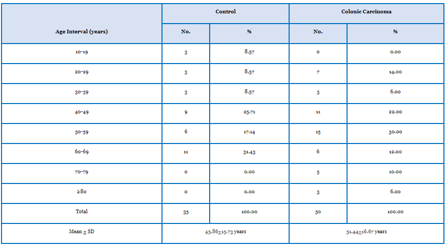
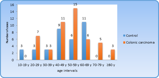
In the present study, the mean age of patients was 51.44±16.67 years and the median was 52 years, whereas the age range in patients group was from 21 years through to 85 years. The mean age of control group was 45.86±15.73 and median age was 49 years, whereas the age range in control group was from 10 to 65 years.
The majority of patients with colonic carcinoma were more than 40 years of age, accounting for 80%, while 20% of cases were below the age of 40, fifteen cases out of 50 (30%) were between the age of 50-59 years, (table 1 and figure 1).
Gender of Patients and Control Groups:
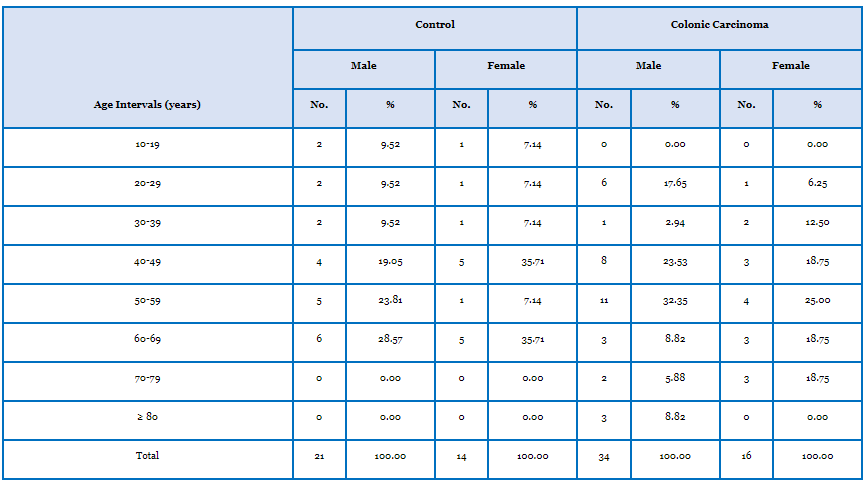
The patients’ group included 34 (68%) males and 16 (32%) females, while control group included 21 (60%) males and 14 (40%) females. Despite these minor differences in gender ratio between patients and control groups, there was no statistical difference.
The ratios of males to females in the present study were 1.5:1 and 2:1 in both control and patients groups, respectively. The mean age of male patients was 50.68±17.48 years, whereas the mean age of female patients was 53.06±15.22 years and there was no statistical difference in the mean age between male and female patients.
The mean age of male control subjects was 45.33±16.97 years, while the mean age of females control subjects was 46.64±14.23 years, which did not reveal any significant difference in the mean age between males and females control subjects. Performing a classification of patients by gender and age intervals revealed that most of the male patients were in the age interval of 50-59 years (32.35%). On the other hand, the female patients revealed the higher frequency in the ages between 50-59 years (25%), as shown in table (2).
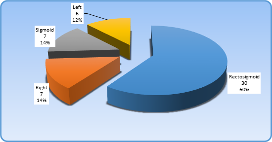
These results revealed the distribution of patients according to site of tumor was as:
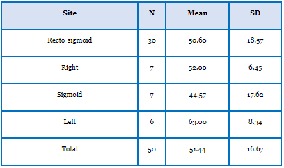
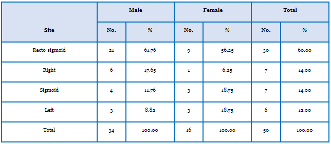
Thirty out of 50 patients had a recto-sigmoid tumor, accounting for 60%.; Seven out of 50 patients had a sigmoid tumor, accounting for 14%.; seven patients had a right sided colonic tumor, accounting for 14%, and six patients had a colonic tumor on the left side, accounting for 12%.
It was obvious that the recto-sigmoid location is the most common site for colonic tumors in patients enrolled in the present study, as shown in figure (2).
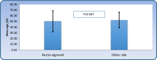
The mean age of patients according to site was as follows: Patients with recto-sigmoid location had a mean age of 50.6±18.57 years, patients with sigmoid location were 44.57±17.62 years of age, patients with right sided colonic tumor had a mean age of 52±6.45 years and patients with left sided colonic tumor had a mean age of 63.0±8.34 years (table 3). The results did not show any significant variation, as shown in figure (3).
Classification of patients according to the site and gender:
The site of colonic tumors in male patients was: 61.76% recto-sigmoid location; 17.65% right sided location; 17.65% sigmoid location and 8.82% left sided location. Additionally the site of colonic tumors in female patients was: 56% recto-sigmoid location; 18.75 % sigmoid location; 18.75% left sided location and 6.25 % right sided location (table 4).
Grade and Stage of Colonic Carcinoma:
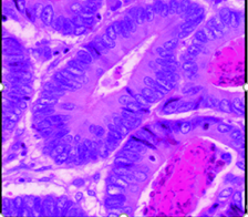
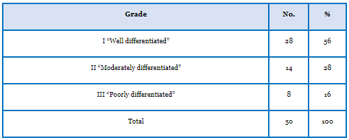
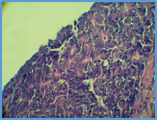
The distribution of patients according to grade of tumor was: 1. Twenty eight patients (56%) showed grade I histological pattern with well differentiated morphology. Grade I is shown in figure (5), when it is compared to normal colonic mucosa figure (4) the following features are observed: the tissue appears with multi-layering, back to back arrangement (little intervening stroma), loss of polarity, loss of goblet cells, invasion of stroma and presence of nuclear criteria of malignancy: hyperchromatism, high N/C ratio visible nucleoli and abnormal mitosis. It should be mentioned that in grade I lesion most of the tumor retains glandular pattern.
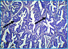
2. Fourteen patients (28%) had grade II pattern with moderately differentiated morphology. Grade II is shown in figure (6), in which the same criteria of grade I are present with tumor nearly equally composed of glandular and solid pattern.
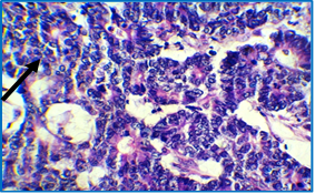
3. Eight patients (16%) had grade III histological pattern with poorly differentiated morphology. Grade III is shown in figure (7). The figure showed same cellular criteria of malignancy but almost all the tumor is composed of solid areas.
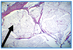
It was clear that the majority of patients exhibited well differentiated grade I tumors (table 5). According to these findings, it should be mentioned that two of the cases of colonic carcinoma were non-conventional adenocarcinoma: one of them exhibited neuroendocrine differentiation, figure (8) and the other one was mucinous figure (9).
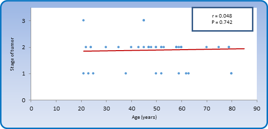
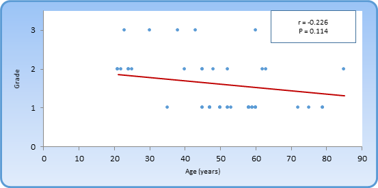
The mean age of patients with grade I histological pattern was 56.11±10.40 years, and the mean age of patients with grade II histological pattern was 41.21±19.73 years, whereas the mean age of patients with grade III histological pattern was 53±22.52 years. The Spearman rank test revealed a non-significant correlation between age and grade (figure10).

The distribution of patients according to grade and gender was:
1. In male patients, there were 17 cases (50%) with grade I well differentiated histological pattern; 12 cases (35.29%) with grade II moderately differentiated histological pattern and 5 patients (14.71%) with grade III poorly differentiated histological pattern.
2. In female patients there were 11 patients (68.75%) with well differentiated histological pattern; 2 patients (12.5%) with moderately differentiated histological pattern and 3 patients (18.75%) with poorly differentiated histological pattern.
Despite these differences in distribution of patients according to grade and gender, there was no significant association between grade and gender.
The distribution of patients according to grade and site of tumor was:
1. Patients with recto-sigmoid location showed the following grades: 53.33% grade I; 30% grade II and 16.17% grade III.
2. Patients with right sided location showed the following grades: 71.43% grade I; 14.29% grade II and 14.29% grade III.
3. Patients with sigmoid location showed the following grades: 28.57% grade I; 57.14% grade II and 14.29% grade III.
4. Patients with Left sided lesions showed the following grades: 83.33% grade I; 0% grade II and 16.67% grade III.
According to these findings there was no significant association between grade and site of tumor.
The distribution of patients according to stage of colonic carcinoma was:
1. There were 11 patients (22%) in stage I disease.
2. There were 33 patients (66%) in stage II disease.
3. There were 6 patients (12%) in stage III disease.
Most of the patients were in stage II (66%). 50.27 ± 21.3 years was the mean age of patients in stage I disease, 51.7 ± 14.22 years being the mean age of patients in stage II disease, while mean age of patients with stage III disease was 52.17 ± 22.79 years.
Figure (11) showed a non-significant correlation between stage of tumor and age of patients (r = 0.048, P > 0.05).
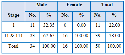
The distribution of patients according to gender and stage of disease was:
1. Male patients showed the following stages: 32.35% stage I disease, 52.94% stage II disease and 14.71 % stage III disease.
2. Female patients showed the following stages: 0% stage I, 93.75% stage II and 6.25% stage III disease.
Table (6) shows a significant association between stage of disease and gender of patients (P < 0.05), in such a way that female patients are more liable to advanced stage disease than male patients.
Both grade and stage are ordinal variables; therefore Kendall's tau-b is the best statistical tool to study correlation between them. Kendall's tau-b (figure 12) yielded a non-significant negative correlation between stage of disease and grade of tumor (r = -0.240, P > 0.05).
The present study revealed that the mean age of patients with colorectal carcinoma was 51.44±16.67 years and the median was 52 years while the age range was from 21 years through to 85 years. On the other hand, the majority of patients with colorectal carcinoma were more than 40 years of age, accounting for 80%, while 20% of cases were less than 40 years of age. A question comes into mind is that why does colorectal carcinoma affect old age subjects more frequently than young age people?
This question can be discussed based upon the fact that carcinogenesis is a multistep process that involves a sequence of mutational events on the level of oncogenes and tumor suppressor genes. The etiology of cancer in general is attributed to two main general causes, the first one being hereditary germ line mutations in genes controlling cell cycle and growth and the second one is environmental factors.
Environmental factors, specifically the interactions between microorganisms in the colonic mucosa and food digestion products may render the colonic mucosa to become malignant due to mutations in genes. The mean age of patients with colorectal carcinoma was 53.98±14.71 years and the median age was 55 years (Iraqi Cancer Registry, 2009). These findings are similar to the results of the present study and the minor differences are clearly due to the difference in sample size which was 701 in Iraqi Cancer Registry, while it was only 50 cases in the present study. It was published in the Iraqi Cancer Registry (2009) that about 82% of colorectal carcinoma patients are above 40 years of age and that around 18% of patients are below the age of 40 years. Similar findings were found in the present study.
Moreover, another study conducted in Iraq stated that the mean age of patients with colorectal carcinoma was 54.5 years, a finding that solidified the result of the present study (Nidal et al., 2014). In another study also done in Iraq, the mean age of patients with colorectal carcinoma was 52.4±16.3 years with an age range of 21-81 years (Ameen et al., 2014); these findings are comparable to those of the present results.
In a recent study done on 968 cases with colorectal carcinoma in Turkey, the mean age was 58.9±12.6 years which is higher than that of the present study, while the age range was 18-85 years which is comparable to the age range of the present study (Aykan et al., 2015). This younger age of Iraqi patients with carcinoma in comparison to Turkish patients may suggest the presence of an environmental carcinogenic agent in Iraq that made the progression of colorectal carcinoma faster and to appear in younger age persons.
The discussion section concerning age, which was stated above, came up with an important question that needs to be answered: why do Iraqi people develop CRC younger than nearby countries? This question can be viewed from two points of view; the first one is racial variation in age incidence and the second one is the presence of environmental factors in Iraqi environment that are not present in other countries, exposure to radiation for instance.
Military radiation exposure was found to be a predisposing factor to colorectal carcinoma (Kaiser et al., 2014). An increase in the incidence of birth defects was also observed, in addition to cancers, which were attributed to the use of depleted uranium in Iraq by the US army (Busby et al., 2010). Radiation exposure during the first and second Gulf war may explain in part the relatively younger age of colorectal carcinoma patients in Iraq in comparison with nearby countries.
Male to Female Ratio of Enrolled Patients
In the present study, the male to female ratio was 2.12:1. One of the hypotheses of why are males more affected than females is because females have a higher estrogen hormone level, much higher than males. Estrogen was found to be associated with the protection against CRC. Estrogens were found to be important in protecting against the initiation and progression of colorectal carcinoma, and this protective effect is most likely facilitated by estrogen receptor β (ERβ) (Hartman & Gustafsson, 2010).
The report of Iraqi Cancer Registry (2010) stated that the male to female ratio of patients with colorectal carcinoma was 1.23:1, whereas the present study gave a higher incidence in male patients than female patients. Majid et al. (2009) reported that the male to female ratio was 1.6:1, which is slightly higher than that of the present study, but again it emphasizes that males are more frequently affected than females. Al-Humadi (2008) reported a male to female ratio of 1.4:1, which is again in accordance with the present study, clarifying a higher frequency of colorectal carcinoma among male patients.
Site of Colorectal Carcinoma within the Colon
The recto-sigmoid region location presented 60% of tumor masses in the current study and 14% of cases were found in the sigmoid region. In combination, rectum and sigmoid area were responsible for 74% of cases. This result should be viewed from two points of view. The first is that screening programs for colonic tumors should make use of colonoscopy procedure as a gold standard test. The second point of view is the question: why do colonic tumors predominantly involve the rectum and sigmoid region? The suggested pathogenesis of colorectal carcinoma is where the answer could be found. It is now well known that most of these malignant tumors arise from premalignant precursors called adenomas (Fredericks et al., 2015). These colon adenomas are categorized into three main types: tubular, villous and tubule-villous adenomas (Qasim et al., 2012). Multistep genetic and epigenetic mutations occur leading to the progression of villous adenomas to adenocarcinoma (Migliore et al., 2011).
Majid et al. (2009) observed that the rectum was responsible for 35% of the locations of colorectal tumors, similar to the results of the current study. The majority of colorectal tumors involved the rectal region and accounted for about 35% (Abdulhussain & Othman, 2013); such results were obtained in the current study.
Grade and Stage of Colorectal Carcinoma
The majority of colorectal carcinoma (56%) had a well differentiated grade I histological pattern in this study. It is known that malignant tumors of the large intestines are well-to-moderately differentiated adenocarcinomas, secreting different amounts of mucin (Rosai & Ackerman, 2011). Others studies showed that the majority of colorectal carcinoma cases (64%) were well differentiated grade I lesions (Mahmodlou et al., 2012). The current study showed similar results.
The present study showed that most of the patients enrolled in the current study (66%) had stage II disease. This can be attributed to the fact that early stage (carcinoma in situ and stage I disease) tumors are often not diagnosed due to lack of proper screening programs like colonoscopy and imaging techniques which can be applied on high risk groups. Most of the patients in the present study had stage II disease is in agreement with many authors: Ali et al. (2014), Abu-Helalah et al. (2013), Abdulhussain & Othman (2013), and Mahmodlou et al. (2012). The majority of the poorly differentiated cases presented at earlier stages of the disease for tumor differentiation, which may be due to non-expectation of the disease.
From the achieved results of this study, the following can be concluded:
There was a significant association between stage of disease and gender of patients and female patients are more liable to advanced stage disease than male patients. Iraqi patients with CRC were at younger ages and had more advanced stages of the disease, presenting mostly in a poorly differentiated type and extra advanced stage than in older patients. These findings make it necessary to conduct a comprehensive awareness program for the control of this type of tumor.
Clearly Auctoresonline and particularly Psychology and Mental Health Care Journal is dedicated to improving health care services for individuals and populations. The editorial boards' ability to efficiently recognize and share the global importance of health literacy with a variety of stakeholders. Auctoresonline publishing platform can be used to facilitate of optimal client-based services and should be added to health care professionals' repertoire of evidence-based health care resources.

Journal of Clinical Cardiology and Cardiovascular Intervention The submission and review process was adequate. However I think that the publication total value should have been enlightened in early fases. Thank you for all.

Journal of Women Health Care and Issues By the present mail, I want to say thank to you and tour colleagues for facilitating my published article. Specially thank you for the peer review process, support from the editorial office. I appreciate positively the quality of your journal.

Journal of Clinical Research and Reports I would be very delighted to submit my testimonial regarding the reviewer board and the editorial office. The reviewer board were accurate and helpful regarding any modifications for my manuscript. And the editorial office were very helpful and supportive in contacting and monitoring with any update and offering help. It was my pleasure to contribute with your promising Journal and I am looking forward for more collaboration.

We would like to thank the Journal of Thoracic Disease and Cardiothoracic Surgery because of the services they provided us for our articles. The peer-review process was done in a very excellent time manner, and the opinions of the reviewers helped us to improve our manuscript further. The editorial office had an outstanding correspondence with us and guided us in many ways. During a hard time of the pandemic that is affecting every one of us tremendously, the editorial office helped us make everything easier for publishing scientific work. Hope for a more scientific relationship with your Journal.

The peer-review process which consisted high quality queries on the paper. I did answer six reviewers’ questions and comments before the paper was accepted. The support from the editorial office is excellent.

Journal of Neuroscience and Neurological Surgery. I had the experience of publishing a research article recently. The whole process was simple from submission to publication. The reviewers made specific and valuable recommendations and corrections that improved the quality of my publication. I strongly recommend this Journal.

Dr. Katarzyna Byczkowska My testimonial covering: "The peer review process is quick and effective. The support from the editorial office is very professional and friendly. Quality of the Clinical Cardiology and Cardiovascular Interventions is scientific and publishes ground-breaking research on cardiology that is useful for other professionals in the field.

Thank you most sincerely, with regard to the support you have given in relation to the reviewing process and the processing of my article entitled "Large Cell Neuroendocrine Carcinoma of The Prostate Gland: A Review and Update" for publication in your esteemed Journal, Journal of Cancer Research and Cellular Therapeutics". The editorial team has been very supportive.

Testimony of Journal of Clinical Otorhinolaryngology: work with your Reviews has been a educational and constructive experience. The editorial office were very helpful and supportive. It was a pleasure to contribute to your Journal.

Dr. Bernard Terkimbi Utoo, I am happy to publish my scientific work in Journal of Women Health Care and Issues (JWHCI). The manuscript submission was seamless and peer review process was top notch. I was amazed that 4 reviewers worked on the manuscript which made it a highly technical, standard and excellent quality paper. I appreciate the format and consideration for the APC as well as the speed of publication. It is my pleasure to continue with this scientific relationship with the esteem JWHCI.

This is an acknowledgment for peer reviewers, editorial board of Journal of Clinical Research and Reports. They show a lot of consideration for us as publishers for our research article “Evaluation of the different factors associated with side effects of COVID-19 vaccination on medical students, Mutah university, Al-Karak, Jordan”, in a very professional and easy way. This journal is one of outstanding medical journal.

Dear Hao Jiang, to Journal of Nutrition and Food Processing We greatly appreciate the efficient, professional and rapid processing of our paper by your team. If there is anything else we should do, please do not hesitate to let us know. On behalf of my co-authors, we would like to express our great appreciation to editor and reviewers.

As an author who has recently published in the journal "Brain and Neurological Disorders". I am delighted to provide a testimonial on the peer review process, editorial office support, and the overall quality of the journal. The peer review process at Brain and Neurological Disorders is rigorous and meticulous, ensuring that only high-quality, evidence-based research is published. The reviewers are experts in their fields, and their comments and suggestions were constructive and helped improve the quality of my manuscript. The review process was timely and efficient, with clear communication from the editorial office at each stage. The support from the editorial office was exceptional throughout the entire process. The editorial staff was responsive, professional, and always willing to help. They provided valuable guidance on formatting, structure, and ethical considerations, making the submission process seamless. Moreover, they kept me informed about the status of my manuscript and provided timely updates, which made the process less stressful. The journal Brain and Neurological Disorders is of the highest quality, with a strong focus on publishing cutting-edge research in the field of neurology. The articles published in this journal are well-researched, rigorously peer-reviewed, and written by experts in the field. The journal maintains high standards, ensuring that readers are provided with the most up-to-date and reliable information on brain and neurological disorders. In conclusion, I had a wonderful experience publishing in Brain and Neurological Disorders. The peer review process was thorough, the editorial office provided exceptional support, and the journal's quality is second to none. I would highly recommend this journal to any researcher working in the field of neurology and brain disorders.
Dear Agrippa Hilda, Journal of Neuroscience and Neurological Surgery, Editorial Coordinator, I trust this message finds you well. I want to extend my appreciation for considering my article for publication in your esteemed journal. I am pleased to provide a testimonial regarding the peer review process and the support received from your editorial office. The peer review process for my paper was carried out in a highly professional and thorough manner. The feedback and comments provided by the authors were constructive and very useful in improving the quality of the manuscript. This rigorous assessment process undoubtedly contributes to the high standards maintained by your journal.

International Journal of Clinical Case Reports and Reviews. I strongly recommend to consider submitting your work to this high-quality journal. The support and availability of the Editorial staff is outstanding and the review process was both efficient and rigorous.

Thank you very much for publishing my Research Article titled “Comparing Treatment Outcome Of Allergic Rhinitis Patients After Using Fluticasone Nasal Spray And Nasal Douching" in the Journal of Clinical Otorhinolaryngology. As Medical Professionals we are immensely benefited from study of various informative Articles and Papers published in this high quality Journal. I look forward to enriching my knowledge by regular study of the Journal and contribute my future work in the field of ENT through the Journal for use by the medical fraternity. The support from the Editorial office was excellent and very prompt. I also welcome the comments received from the readers of my Research Article.

Dear Erica Kelsey, Editorial Coordinator of Cancer Research and Cellular Therapeutics Our team is very satisfied with the processing of our paper by your journal. That was fast, efficient, rigorous, but without unnecessary complications. We appreciated the very short time between the submission of the paper and its publication on line on your site.

I am very glad to say that the peer review process is very successful and fast and support from the Editorial Office. Therefore, I would like to continue our scientific relationship for a long time. And I especially thank you for your kindly attention towards my article. Have a good day!

"We recently published an article entitled “Influence of beta-Cyclodextrins upon the Degradation of Carbofuran Derivatives under Alkaline Conditions" in the Journal of “Pesticides and Biofertilizers” to show that the cyclodextrins protect the carbamates increasing their half-life time in the presence of basic conditions This will be very helpful to understand carbofuran behaviour in the analytical, agro-environmental and food areas. We greatly appreciated the interaction with the editor and the editorial team; we were particularly well accompanied during the course of the revision process, since all various steps towards publication were short and without delay".

I would like to express my gratitude towards you process of article review and submission. I found this to be very fair and expedient. Your follow up has been excellent. I have many publications in national and international journal and your process has been one of the best so far. Keep up the great work.

We are grateful for this opportunity to provide a glowing recommendation to the Journal of Psychiatry and Psychotherapy. We found that the editorial team were very supportive, helpful, kept us abreast of timelines and over all very professional in nature. The peer review process was rigorous, efficient and constructive that really enhanced our article submission. The experience with this journal remains one of our best ever and we look forward to providing future submissions in the near future.

I am very pleased to serve as EBM of the journal, I hope many years of my experience in stem cells can help the journal from one way or another. As we know, stem cells hold great potential for regenerative medicine, which are mostly used to promote the repair response of diseased, dysfunctional or injured tissue using stem cells or their derivatives. I think Stem Cell Research and Therapeutics International is a great platform to publish and share the understanding towards the biology and translational or clinical application of stem cells.

I would like to give my testimony in the support I have got by the peer review process and to support the editorial office where they were of asset to support young author like me to be encouraged to publish their work in your respected journal and globalize and share knowledge across the globe. I really give my great gratitude to your journal and the peer review including the editorial office.

I am delighted to publish our manuscript entitled "A Perspective on Cocaine Induced Stroke - Its Mechanisms and Management" in the Journal of Neuroscience and Neurological Surgery. The peer review process, support from the editorial office, and quality of the journal are excellent. The manuscripts published are of high quality and of excellent scientific value. I recommend this journal very much to colleagues.

Dr.Tania Muñoz, My experience as researcher and author of a review article in The Journal Clinical Cardiology and Interventions has been very enriching and stimulating. The editorial team is excellent, performs its work with absolute responsibility and delivery. They are proactive, dynamic and receptive to all proposals. Supporting at all times the vast universe of authors who choose them as an option for publication. The team of review specialists, members of the editorial board, are brilliant professionals, with remarkable performance in medical research and scientific methodology. Together they form a frontline team that consolidates the JCCI as a magnificent option for the publication and review of high-level medical articles and broad collective interest. I am honored to be able to share my review article and open to receive all your comments.

“The peer review process of JPMHC is quick and effective. Authors are benefited by good and professional reviewers with huge experience in the field of psychology and mental health. The support from the editorial office is very professional. People to contact to are friendly and happy to help and assist any query authors might have. Quality of the Journal is scientific and publishes ground-breaking research on mental health that is useful for other professionals in the field”.

Dear editorial department: On behalf of our team, I hereby certify the reliability and superiority of the International Journal of Clinical Case Reports and Reviews in the peer review process, editorial support, and journal quality. Firstly, the peer review process of the International Journal of Clinical Case Reports and Reviews is rigorous, fair, transparent, fast, and of high quality. The editorial department invites experts from relevant fields as anonymous reviewers to review all submitted manuscripts. These experts have rich academic backgrounds and experience, and can accurately evaluate the academic quality, originality, and suitability of manuscripts. The editorial department is committed to ensuring the rigor of the peer review process, while also making every effort to ensure a fast review cycle to meet the needs of authors and the academic community. Secondly, the editorial team of the International Journal of Clinical Case Reports and Reviews is composed of a group of senior scholars and professionals with rich experience and professional knowledge in related fields. The editorial department is committed to assisting authors in improving their manuscripts, ensuring their academic accuracy, clarity, and completeness. Editors actively collaborate with authors, providing useful suggestions and feedback to promote the improvement and development of the manuscript. We believe that the support of the editorial department is one of the key factors in ensuring the quality of the journal. Finally, the International Journal of Clinical Case Reports and Reviews is renowned for its high- quality articles and strict academic standards. The editorial department is committed to publishing innovative and academically valuable research results to promote the development and progress of related fields. The International Journal of Clinical Case Reports and Reviews is reasonably priced and ensures excellent service and quality ratio, allowing authors to obtain high-level academic publishing opportunities in an affordable manner. I hereby solemnly declare that the International Journal of Clinical Case Reports and Reviews has a high level of credibility and superiority in terms of peer review process, editorial support, reasonable fees, and journal quality. Sincerely, Rui Tao.

Clinical Cardiology and Cardiovascular Interventions I testity the covering of the peer review process, support from the editorial office, and quality of the journal.

Clinical Cardiology and Cardiovascular Interventions, we deeply appreciate the interest shown in our work and its publication. It has been a true pleasure to collaborate with you. The peer review process, as well as the support provided by the editorial office, have been exceptional, and the quality of the journal is very high, which was a determining factor in our decision to publish with you.

The peer reviewers process is quick and effective, the supports from editorial office is excellent, the quality of journal is high. I would like to collabroate with Internatioanl journal of Clinical Case Reports and Reviews journal clinically in the future time.

Clinical Cardiology and Cardiovascular Interventions, I would like to express my sincerest gratitude for the trust placed in our team for the publication in your journal. It has been a true pleasure to collaborate with you on this project. I am pleased to inform you that both the peer review process and the attention from the editorial coordination have been excellent. Your team has worked with dedication and professionalism to ensure that your publication meets the highest standards of quality. We are confident that this collaboration will result in mutual success, and we are eager to see the fruits of this shared effort.

Dear Dr. Jessica Magne, Editorial Coordinator 0f Clinical Cardiology and Cardiovascular Interventions, I hope this message finds you well. I want to express my utmost gratitude for your excellent work and for the dedication and speed in the publication process of my article titled "Navigating Innovation: Qualitative Insights on Using Technology for Health Education in Acute Coronary Syndrome Patients." I am very satisfied with the peer review process, the support from the editorial office, and the quality of the journal. I hope we can maintain our scientific relationship in the long term.

Dear Monica Gissare, - Editorial Coordinator of Nutrition and Food Processing. ¨My testimony with you is truly professional, with a positive response regarding the follow-up of the article and its review, you took into account my qualities and the importance of the topic¨.

Dear Dr. Jessica Magne, Editorial Coordinator 0f Clinical Cardiology and Cardiovascular Interventions, The review process for the article “The Handling of Anti-aggregants and Anticoagulants in the Oncologic Heart Patient Submitted to Surgery” was extremely rigorous and detailed. From the initial submission to the final acceptance, the editorial team at the “Journal of Clinical Cardiology and Cardiovascular Interventions” demonstrated a high level of professionalism and dedication. The reviewers provided constructive and detailed feedback, which was essential for improving the quality of our work. Communication was always clear and efficient, ensuring that all our questions were promptly addressed. The quality of the “Journal of Clinical Cardiology and Cardiovascular Interventions” is undeniable. It is a peer-reviewed, open-access publication dedicated exclusively to disseminating high-quality research in the field of clinical cardiology and cardiovascular interventions. The journal's impact factor is currently under evaluation, and it is indexed in reputable databases, which further reinforces its credibility and relevance in the scientific field. I highly recommend this journal to researchers looking for a reputable platform to publish their studies.

Dear Editorial Coordinator of the Journal of Nutrition and Food Processing! "I would like to thank the Journal of Nutrition and Food Processing for including and publishing my article. The peer review process was very quick, movement and precise. The Editorial Board has done an extremely conscientious job with much help, valuable comments and advices. I find the journal very valuable from a professional point of view, thank you very much for allowing me to be part of it and I would like to participate in the future!”

Dealing with The Journal of Neurology and Neurological Surgery was very smooth and comprehensive. The office staff took time to address my needs and the response from editors and the office was prompt and fair. I certainly hope to publish with this journal again.Their professionalism is apparent and more than satisfactory. Susan Weiner

My Testimonial Covering as fellowing: Lin-Show Chin. The peer reviewers process is quick and effective, the supports from editorial office is excellent, the quality of journal is high. I would like to collabroate with Internatioanl journal of Clinical Case Reports and Reviews.

My experience publishing in Psychology and Mental Health Care was exceptional. The peer review process was rigorous and constructive, with reviewers providing valuable insights that helped enhance the quality of our work. The editorial team was highly supportive and responsive, making the submission process smooth and efficient. The journal's commitment to high standards and academic rigor makes it a respected platform for quality research. I am grateful for the opportunity to publish in such a reputable journal.

My experience publishing in International Journal of Clinical Case Reports and Reviews was exceptional. I Come forth to Provide a Testimonial Covering the Peer Review Process and the editorial office for the Professional and Impartial Evaluation of the Manuscript.
