AUCTORES
Globalize your Research

Research Article
*Corresponding Author: Rahul kumar Vijaykumar Shah, Professor Samileh Noorbakhsh, pediatric infectious diseases Department &Research center, Iran University of Medical Sciences. Tehran, Iran.
Citation: Rahul k.v Shah, Atisha Modi , R Gupta, R. G. Aiyer(2022) Aerodigestive Foreign Bodies: Our Experience Journal of Clinical Otorhinolaryngology 4(3); DOI: 10.31579/2692-9562/049
Copyright: © 2022, Rahul kumar Vijaykumar Shah. This is an open access article distributed under the Creative Commons Attribution License, which permits unrestricted use, distribution, and reproduction in any medium, provided the original work is properly cited.
Received: 15 April 2022 | Accepted: 20 May 2022 | Published: 25 June 2022
Keywords: aerodigestive tract foreign bodies; coins; endoscopy
Foreign body ingestion and aspiration is a common cause of morbidity and mortality, usually occurs in children. A detailed history and physical examination with a high index of suspicion, despite negative imaging are required. Often, a diagnostic endoscopy may be required to definitively investigate the upper aerodigestive tract to rule out foreign body in equivocal findings. Non caustic oesophageal foreign bodies can be observed for a short period of time while Airway foreign bodies require immediate removal in the operating room. A team-based approach with coordination between the trained anaesthetist, surgeon, and operating room staff is required during this foreign body management.
FB- Foreign body, ENT- Ear Nose Throat, PA- posterior-anterior
Killian did first successful bronchial foreign body removal by passing a 9 mm endoscope. After that, in 1905, Chevalier Jackson did bronchial foreign bodies removal and developed instruments for laryngoscopy and bronchoscopy [1], truly known as the father of endoscopic aerodigestive foreign body removal. Aerodigestive foreign body ingestions and aspirations occur more commonly in children under the age of 3 years [2-4], due to their increased mobility, tendency to play and eat at the same time, lack of cognitive recognition of edible versus inedible objects, high propensity for placing objects in the mouth addition, incomplete molars making chewing difficult and an immature or underdeveloped ability to swallow [5].
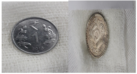
Most foreign bodies are expelled immediately by protective cough and spitting reflexes. Inhaled foreign bodies more commonly include organic materials such as nuts, seeds, vegetable matter, or dried fruits [2,3,6,7] When aspirated, they frequently become lodged in the bronchial tree, with the right main bronchus being more common because of its wider lumen and more vertical path8. Oesophageal foreign bodies lodge commonly in cricopharynx, but most of these pass on to the stomach and may not necessarily require removal [9,10]. Coins and pins are the most commonly ingested items [11]; other common items include batteries, toy parts, bones (fish, chicken), and jewellery [12,13].
A total of 102 patients with definitive history or suspicion of aerodigestive foreign body were admitted or referred to ENT department of Sir Sayajirao General Hospital, Vadodara, Gujarat over a period of two year were reviewed retrospectively [from January 2018 to December 2019]. All these cases are studied in detail for age, sex, type of foreign body, site of lodgement, radiographic evaluation details and treatment given.
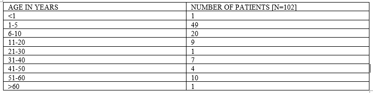
Total number of reviewed patients are 102. The patients’ age distribution is shown in table 1. Children between age of 1 year to 10 years were the most involved (67.64%). The patient’s Sex distribution is shown in table 2. The patient’s male to female ratio was 1.42. In the majority of children, the FB ingestion or aspiration was witnessed or strongly suspected by a bystander after the sudden onset of symptoms.
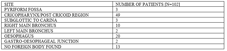
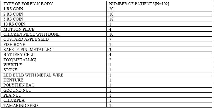
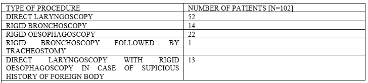
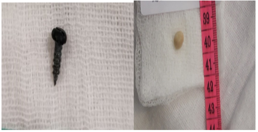
Site of FB lodgment, Type of foreign body found and Type of endoscopy performed is shown in table 3 ,4 and 5 respectively. A total of 102 rigid endoscopies were performed under general anesthesia. Rigid Esophagoscopy was performed in 22 cases (21.56%), Rigid bronchoscopy in 14 cases (13.72%), hypopharyngoscopy and direct laryngoscopy in 52 cases (50.98%). Rigid bronchoscopy followed by tracheostomy was done in 1 patient due to non-retrievable FB via endoscope. Direct laryngoscopy with Rigid Esophagoscopy were done in 13 cases. (12.74%)-cases of no FB found. FB were encountered in 89 patients (87.25%). In 13 patients no FB were found (12.74%). The most common involved sites were the cricopharynx/post cricoid region (48%) followed by the esophagus (19.6%) and the tracheobronchial tree (14.70%). Pyriform fossa FB and Gastro-esophageal junction FB were found in 3 and 2 cases respectively. Coins were the most frequently encountered FB (48%), followed by chicken bone (9.8%), custard apple seed (6.86%), mutton piece (3.9%), battery cell (3.92%), safety pin metal (2.94%), denture (0.98%) and beans, whistle, led bulb with wire, stone etc. The complication rate in our patient series was 2.94% [3 case], two patients had esophageal mucosa erosion after esophagoscopy, one patient had to undergone tracheostomy for FB removal and for airway management. All of these patients were fed with nasogastric tube for a minimal period of 10-14 days and intravenous antibiotic therapy including Cefotaxime and Metronidazole with dexamethasone according to weight were given. All these cases had full recovery.

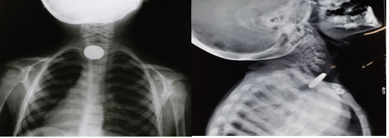

There are three clinical stages of aspiration and ingestion of foreign bodies - The first stage is the impaction of the foreign body causing choking, coughing, or gagging. In second stage symptoms wane as the foreign body settles into a stationary location and the tracheoesophageal reflexes attenuate. This stage lasts for hours to weeks, delaying diagnosis. In third stage complications like obstruction, infection, or perforation [5] occurs. During evaluation of foreign body case, it is advisable to elicit information from the parents like, approximate time of ingestion or inhalation, a history of esophageal dysfunction, severity and duration of swallowing or respiratory dysfunction. In majority of cases, it may be helpful to ask the parents to bring in a similar object from home, particularly for unusual foreign bodies [14]. Foreign body inhalation most commonly causes cough, dyspnea, wheezing, cyanosis, or stridor [3,7] whereas esophageal foreign body ingestion causes drooling, dysphagia, emesis, food refusal, and chest pain. After careful evaluation of history, chest auscultation is must because asymmetry of breath sounds or a prolonged expiratory phase of respiration can give clue to bronchial foreign body, although a normal imaging study does not rule out the presence of a foreign body. Standard frontal and lateral radiographs are the imaging tests of choice for suspected airway foreign bodies [15]. Radiopaque airway foreign bodies are easy to diagnose, whereas organic and other radiolucent airway foreign bodies are more difficult to diagnose. In these cases, other radiographic signs such as unilateral emphysema, hyperinflation, localized atelectasis or infiltrates, and mediastinal or esophageal air trapping may also be indicative of an airway foreign body. In majority of cases, the only evidence of an airway foreign body will be localized air trapping or atelectasis [16]. The classical teaching that sagittal oriented foreign bodies lie in the trachea and coronally positioned foreign bodies are in the esophagus does not hold true in all cases. Esophageal foreign bodies may be found in either the sagittal or coronal configuration. The tracheal foreign bodies more commonly lodge in the sagittal plane because of the longitudinal orientation of the vocal cords and lack of cartilage in the posterior tracheal wall [17]. If the foreign body appears to overlap the tracheal boundaries on a PA view, it is highly unlikely to be in the trachea and a lateral radiograph in this case may confirm that the foreign lies in the esophagus, posterior to the trachea, or demonstrate soft tissue swelling or loss of normal cervical lordosis [18]. Button batteries have a characteristic double contour on lateral view, also known as the “step-off sign,” but may be mistaken for coins on PA views. The characteristic “halo sign” or “double-ring” sign on PA views can help to differentiate a button battery from a coin [14]. Barium swallow is generally not done because it can make subsequent esophageal foreign body removal more difficult.
Low-dose airway CT scans, also known as “virtual bronchoscopy,” are useful when there is a low suspicion for airway foreign body along with a negative chest x-ray and lack of findings on lung auscultation. This Virtual bronchoscopy has high sensitivity so, a negative scan avoids unnecessary bronchoscopy under general anesthesia [19]. For esophageal foreign bodies, standard Posterior-anterior and lateral radiographs are used to identify the presence of and localize multiple foreign bodies [20].
There is a different opinion among surgeons regarding the decision and timing to intervene for an airway foreign body, the choice of anesthesia for bronchoscopy. The three main considerations are (1) method of induction, (2) type of ventilation during bronchoscopy, and (3) maintenance of anesthesia. Rapid sequence techniques is preferred if aspiration of stomach contents is a risk concern [13,23]. Generally, spontaneous ventilation with negative pressure inhalation has been the preferred method because it takes advantage of the natural increase in tracheal and bronchial cross-sectional area during inspiration, and the risk of distal migration of the foreign body with positive pressure ventilation is avoided. However, achieving an adequate depth of anesthesia can be challenging because too deep leads to apnea and consequent hypoxemia and too light risks patient movement and possible bronchial tree injury [18]. Alternatively, controlled jet ventilation ensures a steady level of deep anesthesia and ventilation, which ensures better oxygenation, less coughing or bucking, and less patient movement, but has the risk of displacing the foreign body further down the airway.
The decision to remove an esophageal foreign body depends on factors like type and location of the object, the patient’s age, and time elapsed since the ingestion. An asymptomatic older child with a distal or mid-esophageal object present for less than 24 hours and no history of esophageal disorders may be observed for a period of 8 to 16 hours to see if the object will pass. For young children, foreign bodies present longer than 24 hours, sharp metallic or caustic foreign bodies, or symptomatic patients, urgent endoscopy is warranted; observation for spontaneous passage is not appropriate in these settings. The spontaneous passage rates for esophageal coins in healthy children varies from 9% to 77% [21,22]. For the majority of esophageal foreign bodies, the child should be intubated to minimize the possibility of aspirating the foreign body upon removal and to reduce tracheal compression by the esophagoscope [5].
Postoperatively, if the procedure was uncomplicated, the child can be discharged from the recovery room with regular follow-up to ensure that symptoms have resolved completely. If there is concern regarding foreign body remnant, a repeat endoscopy can be performed [5]. Usually, a postoperative x-ray may be ordered to rule out perforation and mediastinal air.
Most children with aerodigestive foreign body ingestions make a full recovery without permanent sequelae. Complication rates of aerodigestive endoscopy are reported, from 1% to 8% [24]. The risk of complications increases with the duration of time that a foreign body remains in place. The most common complications of rigid bronchoscopy include failure to remove the foreign body, laryngeal edema, pneumothorax, pneumomediastinum, and subcutaneous emphysema. Laryngeal edema may rarely be significant enough to warrant intubation or tracheotomy. Mortality rates in the literature vary from 0.2% to 1.0% [18]. For esophagoscopy, complications include mucosal injury, bleeding and, rarely, perforation, which can cause mediastinitis [10]. In a minority of cases, esophageal endoscopic removal is unsuccessful and requires surgical intervention such as a thoracotomy, esophagotomy, gastrotomy, or jejunotomy [18].
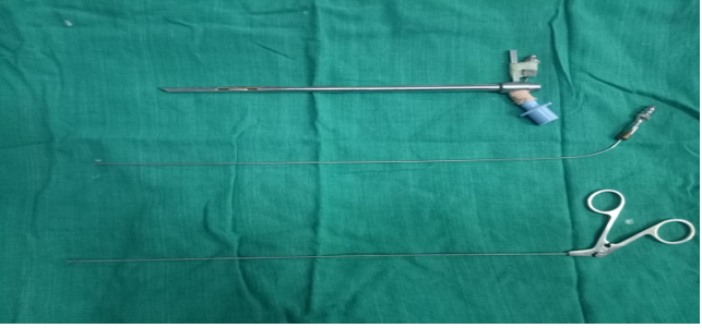
In our retrospective analysis, children below 10 years were most common culprit for foreign body ingestion or inhalation. Standard diagnostic evaluation in form of history, full ENT and chest examination, appropriate x-rays and virtual CT scans were done as and when required. With proper pre-operative preparations endoscopies were performed under general anesthesia. Laryngeal and hypopharyngeal FB were removed under Rigid direct laryngoscope with fiberoptic light carrier. Esophageal FB were removed under rigid esophagoscope with distal illumination. FB in the tracheobronchial tree were removed through the rigid bronchoscope with distal illumination. In our analysis bronchoscope with 4 mm diameter commonly used. All FB were removed with an alligator grasping forceps with double action jaws. Coins were the most common ingested foreign body and custard apple seed were more common inhaled foreign body.
Cricopharynx was most common site of lodgment in ingested foreign body and right main bronchus in inhaled foreign body. Intravenous antibiotic therapy, nebulization and steroids were administrated according to age and weight of patients. Nasogastric feeding tube was put when there where esophageal mucosa erosion or perforation. In immediate postoperative period close monitoring was done, especially after bronchoscopy and chest X-ray was performed after each esophagoscopy or bronchoscopy. Majority of patients were discharged on next morning with follow-up advice.


The ingestion or aspiration of foreign body is a significant cause of morbidity and mortality mainly in the paediatric population. Parental and physician education regarding these dangers are the most important factors in reducing the incidence of this problem. Prompt recognition and a comprehensive history can identify these affected children in a timely fashion. Management is individualized depending on the duration of symptoms, properties of the object or substance ingested and location of the object. An expedient evaluation and workup are important in limiting the number of complications.
There is no conflict of interest among authors.
Clearly Auctoresonline and particularly Psychology and Mental Health Care Journal is dedicated to improving health care services for individuals and populations. The editorial boards' ability to efficiently recognize and share the global importance of health literacy with a variety of stakeholders. Auctoresonline publishing platform can be used to facilitate of optimal client-based services and should be added to health care professionals' repertoire of evidence-based health care resources.

Journal of Clinical Cardiology and Cardiovascular Intervention The submission and review process was adequate. However I think that the publication total value should have been enlightened in early fases. Thank you for all.

Journal of Women Health Care and Issues By the present mail, I want to say thank to you and tour colleagues for facilitating my published article. Specially thank you for the peer review process, support from the editorial office. I appreciate positively the quality of your journal.

Journal of Clinical Research and Reports I would be very delighted to submit my testimonial regarding the reviewer board and the editorial office. The reviewer board were accurate and helpful regarding any modifications for my manuscript. And the editorial office were very helpful and supportive in contacting and monitoring with any update and offering help. It was my pleasure to contribute with your promising Journal and I am looking forward for more collaboration.

We would like to thank the Journal of Thoracic Disease and Cardiothoracic Surgery because of the services they provided us for our articles. The peer-review process was done in a very excellent time manner, and the opinions of the reviewers helped us to improve our manuscript further. The editorial office had an outstanding correspondence with us and guided us in many ways. During a hard time of the pandemic that is affecting every one of us tremendously, the editorial office helped us make everything easier for publishing scientific work. Hope for a more scientific relationship with your Journal.

The peer-review process which consisted high quality queries on the paper. I did answer six reviewers’ questions and comments before the paper was accepted. The support from the editorial office is excellent.

Journal of Neuroscience and Neurological Surgery. I had the experience of publishing a research article recently. The whole process was simple from submission to publication. The reviewers made specific and valuable recommendations and corrections that improved the quality of my publication. I strongly recommend this Journal.

Dr. Katarzyna Byczkowska My testimonial covering: "The peer review process is quick and effective. The support from the editorial office is very professional and friendly. Quality of the Clinical Cardiology and Cardiovascular Interventions is scientific and publishes ground-breaking research on cardiology that is useful for other professionals in the field.

Thank you most sincerely, with regard to the support you have given in relation to the reviewing process and the processing of my article entitled "Large Cell Neuroendocrine Carcinoma of The Prostate Gland: A Review and Update" for publication in your esteemed Journal, Journal of Cancer Research and Cellular Therapeutics". The editorial team has been very supportive.

Testimony of Journal of Clinical Otorhinolaryngology: work with your Reviews has been a educational and constructive experience. The editorial office were very helpful and supportive. It was a pleasure to contribute to your Journal.

Dr. Bernard Terkimbi Utoo, I am happy to publish my scientific work in Journal of Women Health Care and Issues (JWHCI). The manuscript submission was seamless and peer review process was top notch. I was amazed that 4 reviewers worked on the manuscript which made it a highly technical, standard and excellent quality paper. I appreciate the format and consideration for the APC as well as the speed of publication. It is my pleasure to continue with this scientific relationship with the esteem JWHCI.

This is an acknowledgment for peer reviewers, editorial board of Journal of Clinical Research and Reports. They show a lot of consideration for us as publishers for our research article “Evaluation of the different factors associated with side effects of COVID-19 vaccination on medical students, Mutah university, Al-Karak, Jordan”, in a very professional and easy way. This journal is one of outstanding medical journal.

Dear Hao Jiang, to Journal of Nutrition and Food Processing We greatly appreciate the efficient, professional and rapid processing of our paper by your team. If there is anything else we should do, please do not hesitate to let us know. On behalf of my co-authors, we would like to express our great appreciation to editor and reviewers.

As an author who has recently published in the journal "Brain and Neurological Disorders". I am delighted to provide a testimonial on the peer review process, editorial office support, and the overall quality of the journal. The peer review process at Brain and Neurological Disorders is rigorous and meticulous, ensuring that only high-quality, evidence-based research is published. The reviewers are experts in their fields, and their comments and suggestions were constructive and helped improve the quality of my manuscript. The review process was timely and efficient, with clear communication from the editorial office at each stage. The support from the editorial office was exceptional throughout the entire process. The editorial staff was responsive, professional, and always willing to help. They provided valuable guidance on formatting, structure, and ethical considerations, making the submission process seamless. Moreover, they kept me informed about the status of my manuscript and provided timely updates, which made the process less stressful. The journal Brain and Neurological Disorders is of the highest quality, with a strong focus on publishing cutting-edge research in the field of neurology. The articles published in this journal are well-researched, rigorously peer-reviewed, and written by experts in the field. The journal maintains high standards, ensuring that readers are provided with the most up-to-date and reliable information on brain and neurological disorders. In conclusion, I had a wonderful experience publishing in Brain and Neurological Disorders. The peer review process was thorough, the editorial office provided exceptional support, and the journal's quality is second to none. I would highly recommend this journal to any researcher working in the field of neurology and brain disorders.
Dear Agrippa Hilda, Journal of Neuroscience and Neurological Surgery, Editorial Coordinator, I trust this message finds you well. I want to extend my appreciation for considering my article for publication in your esteemed journal. I am pleased to provide a testimonial regarding the peer review process and the support received from your editorial office. The peer review process for my paper was carried out in a highly professional and thorough manner. The feedback and comments provided by the authors were constructive and very useful in improving the quality of the manuscript. This rigorous assessment process undoubtedly contributes to the high standards maintained by your journal.

International Journal of Clinical Case Reports and Reviews. I strongly recommend to consider submitting your work to this high-quality journal. The support and availability of the Editorial staff is outstanding and the review process was both efficient and rigorous.

Thank you very much for publishing my Research Article titled “Comparing Treatment Outcome Of Allergic Rhinitis Patients After Using Fluticasone Nasal Spray And Nasal Douching" in the Journal of Clinical Otorhinolaryngology. As Medical Professionals we are immensely benefited from study of various informative Articles and Papers published in this high quality Journal. I look forward to enriching my knowledge by regular study of the Journal and contribute my future work in the field of ENT through the Journal for use by the medical fraternity. The support from the Editorial office was excellent and very prompt. I also welcome the comments received from the readers of my Research Article.

Dear Erica Kelsey, Editorial Coordinator of Cancer Research and Cellular Therapeutics Our team is very satisfied with the processing of our paper by your journal. That was fast, efficient, rigorous, but without unnecessary complications. We appreciated the very short time between the submission of the paper and its publication on line on your site.

I am very glad to say that the peer review process is very successful and fast and support from the Editorial Office. Therefore, I would like to continue our scientific relationship for a long time. And I especially thank you for your kindly attention towards my article. Have a good day!

"We recently published an article entitled “Influence of beta-Cyclodextrins upon the Degradation of Carbofuran Derivatives under Alkaline Conditions" in the Journal of “Pesticides and Biofertilizers” to show that the cyclodextrins protect the carbamates increasing their half-life time in the presence of basic conditions This will be very helpful to understand carbofuran behaviour in the analytical, agro-environmental and food areas. We greatly appreciated the interaction with the editor and the editorial team; we were particularly well accompanied during the course of the revision process, since all various steps towards publication were short and without delay".

I would like to express my gratitude towards you process of article review and submission. I found this to be very fair and expedient. Your follow up has been excellent. I have many publications in national and international journal and your process has been one of the best so far. Keep up the great work.

We are grateful for this opportunity to provide a glowing recommendation to the Journal of Psychiatry and Psychotherapy. We found that the editorial team were very supportive, helpful, kept us abreast of timelines and over all very professional in nature. The peer review process was rigorous, efficient and constructive that really enhanced our article submission. The experience with this journal remains one of our best ever and we look forward to providing future submissions in the near future.

I am very pleased to serve as EBM of the journal, I hope many years of my experience in stem cells can help the journal from one way or another. As we know, stem cells hold great potential for regenerative medicine, which are mostly used to promote the repair response of diseased, dysfunctional or injured tissue using stem cells or their derivatives. I think Stem Cell Research and Therapeutics International is a great platform to publish and share the understanding towards the biology and translational or clinical application of stem cells.

I would like to give my testimony in the support I have got by the peer review process and to support the editorial office where they were of asset to support young author like me to be encouraged to publish their work in your respected journal and globalize and share knowledge across the globe. I really give my great gratitude to your journal and the peer review including the editorial office.

I am delighted to publish our manuscript entitled "A Perspective on Cocaine Induced Stroke - Its Mechanisms and Management" in the Journal of Neuroscience and Neurological Surgery. The peer review process, support from the editorial office, and quality of the journal are excellent. The manuscripts published are of high quality and of excellent scientific value. I recommend this journal very much to colleagues.

Dr.Tania Muñoz, My experience as researcher and author of a review article in The Journal Clinical Cardiology and Interventions has been very enriching and stimulating. The editorial team is excellent, performs its work with absolute responsibility and delivery. They are proactive, dynamic and receptive to all proposals. Supporting at all times the vast universe of authors who choose them as an option for publication. The team of review specialists, members of the editorial board, are brilliant professionals, with remarkable performance in medical research and scientific methodology. Together they form a frontline team that consolidates the JCCI as a magnificent option for the publication and review of high-level medical articles and broad collective interest. I am honored to be able to share my review article and open to receive all your comments.

“The peer review process of JPMHC is quick and effective. Authors are benefited by good and professional reviewers with huge experience in the field of psychology and mental health. The support from the editorial office is very professional. People to contact to are friendly and happy to help and assist any query authors might have. Quality of the Journal is scientific and publishes ground-breaking research on mental health that is useful for other professionals in the field”.

Dear editorial department: On behalf of our team, I hereby certify the reliability and superiority of the International Journal of Clinical Case Reports and Reviews in the peer review process, editorial support, and journal quality. Firstly, the peer review process of the International Journal of Clinical Case Reports and Reviews is rigorous, fair, transparent, fast, and of high quality. The editorial department invites experts from relevant fields as anonymous reviewers to review all submitted manuscripts. These experts have rich academic backgrounds and experience, and can accurately evaluate the academic quality, originality, and suitability of manuscripts. The editorial department is committed to ensuring the rigor of the peer review process, while also making every effort to ensure a fast review cycle to meet the needs of authors and the academic community. Secondly, the editorial team of the International Journal of Clinical Case Reports and Reviews is composed of a group of senior scholars and professionals with rich experience and professional knowledge in related fields. The editorial department is committed to assisting authors in improving their manuscripts, ensuring their academic accuracy, clarity, and completeness. Editors actively collaborate with authors, providing useful suggestions and feedback to promote the improvement and development of the manuscript. We believe that the support of the editorial department is one of the key factors in ensuring the quality of the journal. Finally, the International Journal of Clinical Case Reports and Reviews is renowned for its high- quality articles and strict academic standards. The editorial department is committed to publishing innovative and academically valuable research results to promote the development and progress of related fields. The International Journal of Clinical Case Reports and Reviews is reasonably priced and ensures excellent service and quality ratio, allowing authors to obtain high-level academic publishing opportunities in an affordable manner. I hereby solemnly declare that the International Journal of Clinical Case Reports and Reviews has a high level of credibility and superiority in terms of peer review process, editorial support, reasonable fees, and journal quality. Sincerely, Rui Tao.

Clinical Cardiology and Cardiovascular Interventions I testity the covering of the peer review process, support from the editorial office, and quality of the journal.

Clinical Cardiology and Cardiovascular Interventions, we deeply appreciate the interest shown in our work and its publication. It has been a true pleasure to collaborate with you. The peer review process, as well as the support provided by the editorial office, have been exceptional, and the quality of the journal is very high, which was a determining factor in our decision to publish with you.

The peer reviewers process is quick and effective, the supports from editorial office is excellent, the quality of journal is high. I would like to collabroate with Internatioanl journal of Clinical Case Reports and Reviews journal clinically in the future time.

Clinical Cardiology and Cardiovascular Interventions, I would like to express my sincerest gratitude for the trust placed in our team for the publication in your journal. It has been a true pleasure to collaborate with you on this project. I am pleased to inform you that both the peer review process and the attention from the editorial coordination have been excellent. Your team has worked with dedication and professionalism to ensure that your publication meets the highest standards of quality. We are confident that this collaboration will result in mutual success, and we are eager to see the fruits of this shared effort.

Dear Dr. Jessica Magne, Editorial Coordinator 0f Clinical Cardiology and Cardiovascular Interventions, I hope this message finds you well. I want to express my utmost gratitude for your excellent work and for the dedication and speed in the publication process of my article titled "Navigating Innovation: Qualitative Insights on Using Technology for Health Education in Acute Coronary Syndrome Patients." I am very satisfied with the peer review process, the support from the editorial office, and the quality of the journal. I hope we can maintain our scientific relationship in the long term.

Dear Monica Gissare, - Editorial Coordinator of Nutrition and Food Processing. ¨My testimony with you is truly professional, with a positive response regarding the follow-up of the article and its review, you took into account my qualities and the importance of the topic¨.

Dear Dr. Jessica Magne, Editorial Coordinator 0f Clinical Cardiology and Cardiovascular Interventions, The review process for the article “The Handling of Anti-aggregants and Anticoagulants in the Oncologic Heart Patient Submitted to Surgery” was extremely rigorous and detailed. From the initial submission to the final acceptance, the editorial team at the “Journal of Clinical Cardiology and Cardiovascular Interventions” demonstrated a high level of professionalism and dedication. The reviewers provided constructive and detailed feedback, which was essential for improving the quality of our work. Communication was always clear and efficient, ensuring that all our questions were promptly addressed. The quality of the “Journal of Clinical Cardiology and Cardiovascular Interventions” is undeniable. It is a peer-reviewed, open-access publication dedicated exclusively to disseminating high-quality research in the field of clinical cardiology and cardiovascular interventions. The journal's impact factor is currently under evaluation, and it is indexed in reputable databases, which further reinforces its credibility and relevance in the scientific field. I highly recommend this journal to researchers looking for a reputable platform to publish their studies.

Dear Editorial Coordinator of the Journal of Nutrition and Food Processing! "I would like to thank the Journal of Nutrition and Food Processing for including and publishing my article. The peer review process was very quick, movement and precise. The Editorial Board has done an extremely conscientious job with much help, valuable comments and advices. I find the journal very valuable from a professional point of view, thank you very much for allowing me to be part of it and I would like to participate in the future!”

Dealing with The Journal of Neurology and Neurological Surgery was very smooth and comprehensive. The office staff took time to address my needs and the response from editors and the office was prompt and fair. I certainly hope to publish with this journal again.Their professionalism is apparent and more than satisfactory. Susan Weiner

My Testimonial Covering as fellowing: Lin-Show Chin. The peer reviewers process is quick and effective, the supports from editorial office is excellent, the quality of journal is high. I would like to collabroate with Internatioanl journal of Clinical Case Reports and Reviews.

My experience publishing in Psychology and Mental Health Care was exceptional. The peer review process was rigorous and constructive, with reviewers providing valuable insights that helped enhance the quality of our work. The editorial team was highly supportive and responsive, making the submission process smooth and efficient. The journal's commitment to high standards and academic rigor makes it a respected platform for quality research. I am grateful for the opportunity to publish in such a reputable journal.

My experience publishing in International Journal of Clinical Case Reports and Reviews was exceptional. I Come forth to Provide a Testimonial Covering the Peer Review Process and the editorial office for the Professional and Impartial Evaluation of the Manuscript.
