AUCTORES
Globalize your Research

Research Article
*Corresponding Author: Shoshi Inooka, Japan Association of Science Specialists.
Citation: Shoshi Inooka (2024), Cell ring-like objects created in agar cultures of synthetic DNA (Streptomyces) crown cells with monolaurin, J Clinical Cardiology and Cardiovascular Interventions, 7(7); DOI: 10.31579/2641-0419/362
Copyright: © 2024, Shoshi Inooka. This is an open access article distributed under the Creative Commons Attribution License, which permits unrestricted use, distribution, and reproduction in any medium, provided the original work is properly cited.
Received: 29 January 2024 | Accepted: 01 July 2024 | Published: 08 July 2024
Keywords: synthetic DNA (Streptomyces) crown cells; agar plate cultures; Sphingosine-DNA; cells; cell ring-like objects; monolaurin
Synthesized DNA crown cells (artificial cells), which can proliferate within egg white in vivo, can be prepared in vitro using sphingosine (Sph)-DNA-adenosine-monolaurin compounds. Synthesized DNA crown cells form assemblies that proliferate in the presence of monolaurin and can be observed in agar cultures. In previous experiments on synthetic DNA (E. coli/Human placenta/Ascidian) crown cells, the proliferation of objects similar in appearance to thalli, double-cells, morning glory and daffodil inflorescences was observed on agar plates.
In the present experiments using synthetic DNA (Streptomyces) crown cells and monolaurin, various objects and examples of cell proliferation were observed on the agar plates. Here, the microscopic appearance of cell ring-like objects is described.
Artificial cells potentially useful for the study of a wide variety of problems in the life science. Until recently, most attempts in generate artificial cells have resulted in only vesicle like cells that package several cellular components such as DNA and enzymes. Such vesicles can do many things that cells can do, including transcribe RNA, translate proteins, and generates ATP, recently, division and replication of DNA have been observed in such vesicles.
Until the first report by the present author in 2012 (1), fully functional (self replicating) cells had not been produced. Moreover, in 2016 (2), the principal methods to prepare generated artificial cells which can be multiply within egg white and cultivated in vitro were reported. In 2016, artificial cells referred to as DNA crown cells were reported by the present author, (3). The cells derive their name from the facts that their exterior consists of DNA.
DNA crown cells were synthesized using four common commercial compounds; Sphigosine (Sph), DNA, adenosine and monolaurin using methods developed based on the author’s experience. of my research. Briefly, Sph, which is the core of sphingolipids, was synthesized using serine and metabolized immediately to form ceramides (4). Non-metabolized Sph (free Sph) exsits within cells in small amount. Free Sph within cells increased as a result of the catabolism of sphingomyelin which consisted of cell membrane and occured with cell injury,
Also, cell injury can increase the amount of of DNA within cells. The chance of binding to DNA of Sph can result in the formation. This free transfer of Sph from cell to cell (5, 6) can cause cell injury (7), because Sph was cytotoxic. Sph-DNA particles were found to be capable of binding to various cell components and cultivable particles were formed.(7, 8).
The method to prepare DNA crown cells is as follows. 1) Sph and DNA were mixture. 2) adenosine was added to the mixtures. 3) monolaurin is then added and results in the formation of synthetic DNA crown cells 4) DNA crown cells were formed from synthetic DNA crown cells within egg white.
Thus, synthetic DNA crown cells can be prepared using Sph-DNA and adenosine-monolaurin (A-M) compounds, and these can develop into DNA crown cells within egg white or in vitro cultures (9, 10).
Such DNA crown cells formed assemblies or cells that proliferated in cultures with monolaurin in test tubes (11-13) and the generated cells could also be cultivated on agar plates (14,15). In previous studies (16) , various types of objects, as well as cell proliferation, were observed when monolaurin-treated synthetic DNA (E. coli/Human placenta/Ascidian) crown cells were cultivated with or without egg white on agar plates and examined microscopically.
The present experiments examined whether such objects were formed when synthetic DNA (Streptomyces) crown cells were cultured with monolaurin on agar plates. Numerous kinds of objects and cell proliferation were observed. Here, the microscopic appearance of objects that are similar in appearance to cell rings are described.
Materials
The materials used were the same as those employed in previous studies (9, 10): Sph (Tokyo Kasei, Japan), DNA (From Streptomyces), adenosine (Sigma-Aldrich; Wako, Japan), monolaurin (Tokyo Kasei), and adenosine-monolaurin (A-M), a compound synthesized from a mixture of adenosine and monolaurin (9, 10). Monolaurin solutions were prepared to a final concentration of 0.1 M in distilled water. Agar plates were prepared using standard agar medium (SMA) (AS ONE, Japan).
Methods
Preparation of synthetic DNA (Streptomyces) crown cells (9, 10).
Step.1 180 µL of Sph (10 mM) and 50 µL of DNA (0.05 µg/µL) were combined, and the mixture was heated and cooled twice.
Step 2. A-M solution (100 µL) was added and the mixture was incubated at 37°C for 15 min.
Step3. 30 µL of monolaurin solution was added and the mixture was incubated at 37°C for another 5 min.
Step. 4 The resulting suspension was used as the synthetic DNA (Streptomyces) crown cells. The culture of monolaurin with DNA (Streptomyces) crown cells on agar plates was performed as follows:
Microscopic observations
Objects on plates were observed directly under a light microscope and with the naked eye.
Figure 1 shows a photograph of an agar plate (No. 1) of synthetic DNA (Streptomyces) crown cells after 7 days of monolaurin culture. Circular brownish objects (arrow), visible with the naked eye, were observed in the center of the Petri dish, which had a diameter of 8.0 cm.
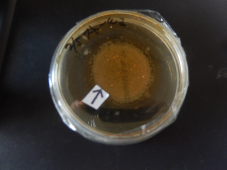
Figure 1. Photograph of an agar plate (No. 1) containing synthetic DNA (Streptomyces) crown cells after 7 days of monolaurin culture. Brownish objects (arrow) can be seen in the center of the Petri dish, which has a diameter of 8.0 cm.
Figure 2 shows a photograph of another agar plate (No. 3) containing synthetic DNA (Streptomyces) crown cells after 7 days of monolaurin culture. Cloudy, dot-like or round objects (arrow), visible with the naked eye, were observed on the Petri dish, which had a diameter of 8.0 cm.
Figure 3 shows the microscopic appearance of synthetic DNA (Streptomyces) crown cells shown in Fig. 1 after 5 days of culture with monolaurin. A ring-like object consisting of numerous cell-like objects was observed (arrow a). An independent cell (approximately 7–8 mm, arrow b) was also observed, and large objects were observed within the ring (arrow c). The approximate size in diameter of the ring-like object (arrow a) was 700–720 mm.
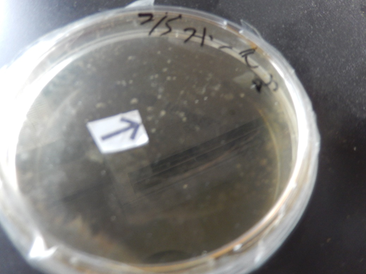
Figure 2. Photograph of an agar plate (No. 3) containing synthetic DNA (Streptomyces) crown cells after 7 days of monolaurin culture. Dot-like or round (arrow) objects can be seen in the Petri dish, which has a diameter of 8.0 cm.
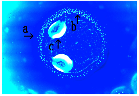
Figure 3. Micrograph of the synthetic DNA (Streptomyces) crown cells shown in Figure. 1 after 5 days of monolaurin culture.
A ring-like object (arrow a) and a cell are shown (arrow b). Objects can be observed within the ring (arrow c). The approximate diameter of the ring-like object (arrow a) is 700–720 mm. The cell (arrow b) measures approximately 7–8 mm.
Figure 4 shows the microscopic appearance of synthetic DNA (Streptomyces) crown cells shown in Fig. 1 after 5 days of culture with monolaurin. In addition, an object consisting of numerous cell-like objects was observed (arrow a). The cell-like objects (arrow c) appeared as though they were flowing and as being aggregated. These cell-like objects (arrow b) were approximately 7–8 mm in diameter
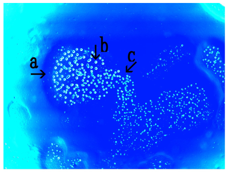
Figure 4. Microscopic appearance of the synthetic DNA (Streptomyces) crown cells shown in Figure. 1 after 5 days of monolaurin culture. An object is shown (arrow a). Cell-like objects (arrow c) are shown. The diameter of the object (arrow b) is approximately 7–8 mm.
Figure 5 shows the microscopic appearance of synthetic DNA (Streptomyces) crown cells shown in Fig. 2 after 7 days of culture with monolaurin. Cell ring-like objects (arrow a), which appeared to be composed of numerous cell-like objects (arrow b) were observed. The objects (arrow a) were approximately 540–600 mm in diameter.
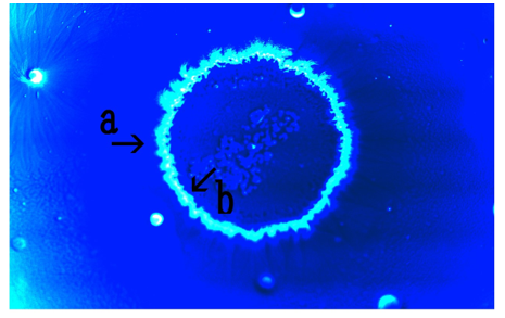
Figure 5. Microscopic appearance of synthetic DNA (Streptomyces) crown cells shown in Fig. 2 after 7 days of monolaurin culture.
Ring-like objects (arrow a) constructed of many cell-like objects (arrow b) are shown. The diameter of the object (arrow a) is approximately 540–600 mm.
Figure 6 shows the microscopic appearance of synthetic DNA (Streptomyces) crown cells shown in Figure. 2 after 7 days of culture with monolaurin. Objects similar in appearance to a divided ring, consisting of numerous cell-like objects were observed (arrows a). The objects, which had an approximate diameter of 500–520 mm (arrow a), appear empty (arrow c).
The single cell-like object (arrow c) measured approximately 18–20 mm.
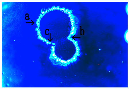
Figure 6. Microscopic appearance of synthetic DNA (Streptomyces) crown cells shown in Figure. 2 after 7 days of monolaurin culture.
An object like divided ring which consists of numerous cell-like objects (arrow b) is shown (arrow a). The diameter of the object is approximately 500–520 mm (arrow a). The size of the object (arrow c) is approximately 18–20 mm.
Figure 7 shows the microscopic appearance of synthetic DNA (Streptomyces) crown cells at 7 days after the addition of monolaurin (Plate was not shown).
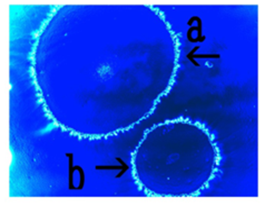
Figure 7: Microscopic appearance of synthetic DNA (Streptomyces) crown cells after 7 days of monolaurin culture.
Two objects are shown (arrows a and b). The diameter of the objects are approximately 440–460 mm (arrow a) and 280–290 mm (arrow b), respectively.
Two ring-like objects consisting of numerous cell-like objects were observed (arrows a and b); the approximate diameter of these objects was 440–460 mm (arrow a) and 280–290 mm (arrow b), respectively.
Figure 8 shows the microscopic appearance of synthetic DNA (Streptomyces) crown cells in egg white cultures for 7 days after the treatment of monolaurin (The method was not described). Objects consisting of numerous bamboo broom-like objects (arrows b and c) were observed (arrow a). Objects similar in appearance to single cells (approximate size 5–8 mm) were also observed (arrow d). These objects measured approximately 6.1–7.4 mm in diameter (arrow a)
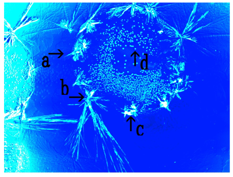
Figure 8. Microscopic appearance of synthetic DNA (Streptomyces) crown cells after 7 days of culture in monolaurin and egg white.
An object consisting of many bamboo broom-like objects (arrows b and c) is shown (arrow a). A single cell-like object measuring approximately 5–8 mm is shown (arrow d).
An object measuring approximately 6.1–7.4 mm in diameter (arrow a) is shown.
In previous experiments using synthetic DNA (E. coli/Human placenta/Ascidian) crown cells, objects similar in appearance to thalli, double cells, morning glory and daffodils were observed on agar plates (16,17, 18). The present experiments examined whether similar objects were observed in agar cultures of synthetic DNA (Streptomyces) crown cells in monolaurin.
Objects, visible with naked eye were observed on all plates (three plates); however, the appearance of the objects differed between plates. The reasons for these differences were not clear, because there is no evidence of the mechanisms underlying object formation. However, it was certain that they formed as a result of associations between the synthetic DNA crown cells and their related components, including monolaurin and egg white.
In this case, the formation of the assemblies in the different cultures of crown cells occurred immediately after the samples were prepared, as described previously. Therefore, ring-like objects (Figure. 1) may form as a result of the association of not cells, but assemblies with monolaurin, which then subsequently increase in size.
Ring-like objects (Figure. 3, arrow a) possibly consisting of numerous cell-like objects were observed (Figure. 4, arrows b and c). There was no evidence of the mechanisms underlying the formation of ring-like objects.
However, round objects that proliferated in the culture were often observed in cultures of synthetic DNA crown cells in monolaurin. Hence, the proliferated cells form rings and may form ring-like objects (Figure. 3, arrow a). Here, these objects are referred to as proliferated cell rings (pr-cell rings).
Large objects were observed within the ring-like objects; however, the identity of these objects is unclear (Figure. 3 arrow c). It is possible that
cell ring-like objects may enclose a kind of assembly composed of Sph/DNA: adenosine-monolaurin. Also, another type of cell ring-like object that consists of numerous cell-like objects (Figure. 5, arrow b) was observed (Figure. 5, arrow a). There were differences between both types of cell ring-like objects.
Cell-like objects (Figure. 5 b) that were constructed of ring-like objects (Figure. 5 a) were often observed in cells that were prepared as cell samples. Though there is no evidence, these findings imply that the objects are not constructed of proliferated cells, but original cells. Here, to distinguish between these two ring-like objects, they are named original cell ring (or-cell ring) and proliferated cells (pr-cell ring). Or-cell rings may be formed by binding of original synthetic DNA (Streptomyces) crown cells to form rings.
The ring may grow and divide (Figure. 6, arrow a) to form a new ring (Fig. 7 arrows a and b), implying that or-cell rings multiply and form new ring-like objects. However, this remains hypothetical.
Thus, two types of ring-like objects are introduced. One type consists of objects that are comprised of proliferated cells, the other type consists of original cells. On the other hand, various assemblies and/or objects of various sizes and shapes were formed when synthetic DNA crown cells were mixed with monolaurin or egg white. Here, although it is not described, another type of ring-like object was observed (Figure. 8). The outside was comprised of objects that were similar in appearance to bamboo brooms. Such objects were often observed in the mixtures of monolaurin-treated synthetic DNA (Streptomyces) crown cells cultured with egg white. Large spaces could be observed with the naked eye (Figs. 1 and 2), and numerous kinds of objects or the numerous features of cell proliferation could be observed within these spaces. Not all aspects of cell-proliferation could be described here.
Most synthetic DNA crown cells formed objects and proliferated. However, the formation of such ring-like objects appears to be rare, as these characteristics were not observed in cultures of other synthetic DNA (E. coli/Human placenta/Ascidian) crown cells. It was not clarified in the present study whether this type of ring formation is characteristic to synthetic DNA (Streptomyces) crown cells.
As described previously, it is important to clarify whether the objects being observed are living. To date, including this study, it has been demonstrated that characteristic objects were formed in four kinds of synthetic DNA (E. coli/Human placenta/Ascidian) crown cells.
Future research will clarify whether synthetic DNA (tumor cells) crown cells form characteristic objects. In addition, experiments will be conducted to clarify whether living cells were contained in the objects observed with the naked eye (Figure. 1 and 2).
These objects have been named as follows: Pro-cell ring Bacteria (Streptomyces) SDCCM (p CR) and Or-cell ring Bacteria (Streptomyces) SDCCM (o-CR). Here, Streptomyces in brackets is the name of the DNA source, SDCC refers to synthetic DNA (crown cells), and M refers to monolaurin. If cells were treated with both monolaurin and egg white, then they are named SDCCME.
Unlimited types of DNA crown cells can be prepared and cultivated (1922).
These cells could be used in a variety of practical fields, including industry, life science, medical science, and may contribute to the welfare of human beings.
The experiments described in this Special Discussion aim to clarify whether such cells can be applied to practical fields, as this is currently unclear. Potential applications in the field of clinical medicine are considered, including specific applications to humans, neurite elongation and intestinal bacteria.
Potential application to humans (23)
There were many chance of binding to DNA with Sph within nucleus or exosome.
In the nucleus, Sph may be derived from shingomyerin in the nucleus membrane and this Sph bind to DNA within nucleus.
In addition, many substances such as protein, DNA, and m-RNA exist in the exosome, and there is a possibility of these molecules bindings to DNA bound to Sph from the membrane. Sph-DNA particles may be released from nucleus and with or without exosome. These conditions could be favorable for the development of DNA crown cells in certain locations, such as where specific lipids (as monolaurin) are present.
Potential applications in neurite elongation
When Sph-DNA particles are cultivated with cells, tube like objects was formed. (7). Objects in synthetic DNA crown cells also became elongated and form long, thin fiber-like tubes (13). Synthetic DNA crown cells have be shown to cause elongation of Bacillus subtilis (24).
These findings indicate that components of DNA crown cells have the abilities to elongate part of organisms, forming objects similar to axons.
Potential application to intestinal bacteria.
The gut flora of animals and human comprises thousand of different kinds of bacteria, many of which have not been identified. Similarity, a wide variety of bacteria exist on Earth, suggesting that many kinds of new bacteria are produced in nature. Previous studies, as well as the present experiments (16-18), suggested that new organisms that are similar to bacteria, which as synthetic DNA crown cells, can arise when culture conditions are favorable. These new organisms can inhabit enviorments such as the gut or other habitats on Earth. New findings in this field could therefore benefit both human and the plant we inhabit.
Clearly Auctoresonline and particularly Psychology and Mental Health Care Journal is dedicated to improving health care services for individuals and populations. The editorial boards' ability to efficiently recognize and share the global importance of health literacy with a variety of stakeholders. Auctoresonline publishing platform can be used to facilitate of optimal client-based services and should be added to health care professionals' repertoire of evidence-based health care resources.

Journal of Clinical Cardiology and Cardiovascular Intervention The submission and review process was adequate. However I think that the publication total value should have been enlightened in early fases. Thank you for all.

Journal of Women Health Care and Issues By the present mail, I want to say thank to you and tour colleagues for facilitating my published article. Specially thank you for the peer review process, support from the editorial office. I appreciate positively the quality of your journal.

Journal of Clinical Research and Reports I would be very delighted to submit my testimonial regarding the reviewer board and the editorial office. The reviewer board were accurate and helpful regarding any modifications for my manuscript. And the editorial office were very helpful and supportive in contacting and monitoring with any update and offering help. It was my pleasure to contribute with your promising Journal and I am looking forward for more collaboration.

We would like to thank the Journal of Thoracic Disease and Cardiothoracic Surgery because of the services they provided us for our articles. The peer-review process was done in a very excellent time manner, and the opinions of the reviewers helped us to improve our manuscript further. The editorial office had an outstanding correspondence with us and guided us in many ways. During a hard time of the pandemic that is affecting every one of us tremendously, the editorial office helped us make everything easier for publishing scientific work. Hope for a more scientific relationship with your Journal.

The peer-review process which consisted high quality queries on the paper. I did answer six reviewers’ questions and comments before the paper was accepted. The support from the editorial office is excellent.

Journal of Neuroscience and Neurological Surgery. I had the experience of publishing a research article recently. The whole process was simple from submission to publication. The reviewers made specific and valuable recommendations and corrections that improved the quality of my publication. I strongly recommend this Journal.

Dr. Katarzyna Byczkowska My testimonial covering: "The peer review process is quick and effective. The support from the editorial office is very professional and friendly. Quality of the Clinical Cardiology and Cardiovascular Interventions is scientific and publishes ground-breaking research on cardiology that is useful for other professionals in the field.

Thank you most sincerely, with regard to the support you have given in relation to the reviewing process and the processing of my article entitled "Large Cell Neuroendocrine Carcinoma of The Prostate Gland: A Review and Update" for publication in your esteemed Journal, Journal of Cancer Research and Cellular Therapeutics". The editorial team has been very supportive.

Testimony of Journal of Clinical Otorhinolaryngology: work with your Reviews has been a educational and constructive experience. The editorial office were very helpful and supportive. It was a pleasure to contribute to your Journal.

Dr. Bernard Terkimbi Utoo, I am happy to publish my scientific work in Journal of Women Health Care and Issues (JWHCI). The manuscript submission was seamless and peer review process was top notch. I was amazed that 4 reviewers worked on the manuscript which made it a highly technical, standard and excellent quality paper. I appreciate the format and consideration for the APC as well as the speed of publication. It is my pleasure to continue with this scientific relationship with the esteem JWHCI.

This is an acknowledgment for peer reviewers, editorial board of Journal of Clinical Research and Reports. They show a lot of consideration for us as publishers for our research article “Evaluation of the different factors associated with side effects of COVID-19 vaccination on medical students, Mutah university, Al-Karak, Jordan”, in a very professional and easy way. This journal is one of outstanding medical journal.

Dear Hao Jiang, to Journal of Nutrition and Food Processing We greatly appreciate the efficient, professional and rapid processing of our paper by your team. If there is anything else we should do, please do not hesitate to let us know. On behalf of my co-authors, we would like to express our great appreciation to editor and reviewers.

As an author who has recently published in the journal "Brain and Neurological Disorders". I am delighted to provide a testimonial on the peer review process, editorial office support, and the overall quality of the journal. The peer review process at Brain and Neurological Disorders is rigorous and meticulous, ensuring that only high-quality, evidence-based research is published. The reviewers are experts in their fields, and their comments and suggestions were constructive and helped improve the quality of my manuscript. The review process was timely and efficient, with clear communication from the editorial office at each stage. The support from the editorial office was exceptional throughout the entire process. The editorial staff was responsive, professional, and always willing to help. They provided valuable guidance on formatting, structure, and ethical considerations, making the submission process seamless. Moreover, they kept me informed about the status of my manuscript and provided timely updates, which made the process less stressful. The journal Brain and Neurological Disorders is of the highest quality, with a strong focus on publishing cutting-edge research in the field of neurology. The articles published in this journal are well-researched, rigorously peer-reviewed, and written by experts in the field. The journal maintains high standards, ensuring that readers are provided with the most up-to-date and reliable information on brain and neurological disorders. In conclusion, I had a wonderful experience publishing in Brain and Neurological Disorders. The peer review process was thorough, the editorial office provided exceptional support, and the journal's quality is second to none. I would highly recommend this journal to any researcher working in the field of neurology and brain disorders.
Dear Agrippa Hilda, Journal of Neuroscience and Neurological Surgery, Editorial Coordinator, I trust this message finds you well. I want to extend my appreciation for considering my article for publication in your esteemed journal. I am pleased to provide a testimonial regarding the peer review process and the support received from your editorial office. The peer review process for my paper was carried out in a highly professional and thorough manner. The feedback and comments provided by the authors were constructive and very useful in improving the quality of the manuscript. This rigorous assessment process undoubtedly contributes to the high standards maintained by your journal.

International Journal of Clinical Case Reports and Reviews. I strongly recommend to consider submitting your work to this high-quality journal. The support and availability of the Editorial staff is outstanding and the review process was both efficient and rigorous.

Thank you very much for publishing my Research Article titled “Comparing Treatment Outcome Of Allergic Rhinitis Patients After Using Fluticasone Nasal Spray And Nasal Douching" in the Journal of Clinical Otorhinolaryngology. As Medical Professionals we are immensely benefited from study of various informative Articles and Papers published in this high quality Journal. I look forward to enriching my knowledge by regular study of the Journal and contribute my future work in the field of ENT through the Journal for use by the medical fraternity. The support from the Editorial office was excellent and very prompt. I also welcome the comments received from the readers of my Research Article.

Dear Erica Kelsey, Editorial Coordinator of Cancer Research and Cellular Therapeutics Our team is very satisfied with the processing of our paper by your journal. That was fast, efficient, rigorous, but without unnecessary complications. We appreciated the very short time between the submission of the paper and its publication on line on your site.

I am very glad to say that the peer review process is very successful and fast and support from the Editorial Office. Therefore, I would like to continue our scientific relationship for a long time. And I especially thank you for your kindly attention towards my article. Have a good day!

"We recently published an article entitled “Influence of beta-Cyclodextrins upon the Degradation of Carbofuran Derivatives under Alkaline Conditions" in the Journal of “Pesticides and Biofertilizers” to show that the cyclodextrins protect the carbamates increasing their half-life time in the presence of basic conditions This will be very helpful to understand carbofuran behaviour in the analytical, agro-environmental and food areas. We greatly appreciated the interaction with the editor and the editorial team; we were particularly well accompanied during the course of the revision process, since all various steps towards publication were short and without delay".

I would like to express my gratitude towards you process of article review and submission. I found this to be very fair and expedient. Your follow up has been excellent. I have many publications in national and international journal and your process has been one of the best so far. Keep up the great work.

We are grateful for this opportunity to provide a glowing recommendation to the Journal of Psychiatry and Psychotherapy. We found that the editorial team were very supportive, helpful, kept us abreast of timelines and over all very professional in nature. The peer review process was rigorous, efficient and constructive that really enhanced our article submission. The experience with this journal remains one of our best ever and we look forward to providing future submissions in the near future.

I am very pleased to serve as EBM of the journal, I hope many years of my experience in stem cells can help the journal from one way or another. As we know, stem cells hold great potential for regenerative medicine, which are mostly used to promote the repair response of diseased, dysfunctional or injured tissue using stem cells or their derivatives. I think Stem Cell Research and Therapeutics International is a great platform to publish and share the understanding towards the biology and translational or clinical application of stem cells.

I would like to give my testimony in the support I have got by the peer review process and to support the editorial office where they were of asset to support young author like me to be encouraged to publish their work in your respected journal and globalize and share knowledge across the globe. I really give my great gratitude to your journal and the peer review including the editorial office.

I am delighted to publish our manuscript entitled "A Perspective on Cocaine Induced Stroke - Its Mechanisms and Management" in the Journal of Neuroscience and Neurological Surgery. The peer review process, support from the editorial office, and quality of the journal are excellent. The manuscripts published are of high quality and of excellent scientific value. I recommend this journal very much to colleagues.

Dr.Tania Muñoz, My experience as researcher and author of a review article in The Journal Clinical Cardiology and Interventions has been very enriching and stimulating. The editorial team is excellent, performs its work with absolute responsibility and delivery. They are proactive, dynamic and receptive to all proposals. Supporting at all times the vast universe of authors who choose them as an option for publication. The team of review specialists, members of the editorial board, are brilliant professionals, with remarkable performance in medical research and scientific methodology. Together they form a frontline team that consolidates the JCCI as a magnificent option for the publication and review of high-level medical articles and broad collective interest. I am honored to be able to share my review article and open to receive all your comments.

“The peer review process of JPMHC is quick and effective. Authors are benefited by good and professional reviewers with huge experience in the field of psychology and mental health. The support from the editorial office is very professional. People to contact to are friendly and happy to help and assist any query authors might have. Quality of the Journal is scientific and publishes ground-breaking research on mental health that is useful for other professionals in the field”.

Dear editorial department: On behalf of our team, I hereby certify the reliability and superiority of the International Journal of Clinical Case Reports and Reviews in the peer review process, editorial support, and journal quality. Firstly, the peer review process of the International Journal of Clinical Case Reports and Reviews is rigorous, fair, transparent, fast, and of high quality. The editorial department invites experts from relevant fields as anonymous reviewers to review all submitted manuscripts. These experts have rich academic backgrounds and experience, and can accurately evaluate the academic quality, originality, and suitability of manuscripts. The editorial department is committed to ensuring the rigor of the peer review process, while also making every effort to ensure a fast review cycle to meet the needs of authors and the academic community. Secondly, the editorial team of the International Journal of Clinical Case Reports and Reviews is composed of a group of senior scholars and professionals with rich experience and professional knowledge in related fields. The editorial department is committed to assisting authors in improving their manuscripts, ensuring their academic accuracy, clarity, and completeness. Editors actively collaborate with authors, providing useful suggestions and feedback to promote the improvement and development of the manuscript. We believe that the support of the editorial department is one of the key factors in ensuring the quality of the journal. Finally, the International Journal of Clinical Case Reports and Reviews is renowned for its high- quality articles and strict academic standards. The editorial department is committed to publishing innovative and academically valuable research results to promote the development and progress of related fields. The International Journal of Clinical Case Reports and Reviews is reasonably priced and ensures excellent service and quality ratio, allowing authors to obtain high-level academic publishing opportunities in an affordable manner. I hereby solemnly declare that the International Journal of Clinical Case Reports and Reviews has a high level of credibility and superiority in terms of peer review process, editorial support, reasonable fees, and journal quality. Sincerely, Rui Tao.

Clinical Cardiology and Cardiovascular Interventions I testity the covering of the peer review process, support from the editorial office, and quality of the journal.

Clinical Cardiology and Cardiovascular Interventions, we deeply appreciate the interest shown in our work and its publication. It has been a true pleasure to collaborate with you. The peer review process, as well as the support provided by the editorial office, have been exceptional, and the quality of the journal is very high, which was a determining factor in our decision to publish with you.

The peer reviewers process is quick and effective, the supports from editorial office is excellent, the quality of journal is high. I would like to collabroate with Internatioanl journal of Clinical Case Reports and Reviews journal clinically in the future time.

Clinical Cardiology and Cardiovascular Interventions, I would like to express my sincerest gratitude for the trust placed in our team for the publication in your journal. It has been a true pleasure to collaborate with you on this project. I am pleased to inform you that both the peer review process and the attention from the editorial coordination have been excellent. Your team has worked with dedication and professionalism to ensure that your publication meets the highest standards of quality. We are confident that this collaboration will result in mutual success, and we are eager to see the fruits of this shared effort.

Dear Dr. Jessica Magne, Editorial Coordinator 0f Clinical Cardiology and Cardiovascular Interventions, I hope this message finds you well. I want to express my utmost gratitude for your excellent work and for the dedication and speed in the publication process of my article titled "Navigating Innovation: Qualitative Insights on Using Technology for Health Education in Acute Coronary Syndrome Patients." I am very satisfied with the peer review process, the support from the editorial office, and the quality of the journal. I hope we can maintain our scientific relationship in the long term.
