
Case Report | DOI: https://doi.org/10.31579/2640-1053/224
1Guelmim Faculty of Medicine and Pharmacy - Ibnou Zohr Agadir University.
2Guelmim Military Hospital Moulay El Hassan General Surgery Department.
3Al AMAL Pathological Anatomy Laboratory, Guelmim.
*Corresponding Author: Imane BOUJGUENNA. Guelmim Faculty of Medicine and Pharmacy - Ibnou Zohr Agadir University.
Citation: Imane BOUJGUENNA, Hichame Krimou, Fatima BOUKIS, (2024), Xanthogranulomatous Cholecystitis with Gallstones: A Case Report, J Cancer Research and Cellular Therapeutics, 8(9); DOI:10.31579/2640-1053/224
Copyright: © 2024, Imane BOUJGUENNA. This is an open access article distributed under the Creative Commons Attribution License, which permits unrestricted use, distribution, and reproduction in any medium, provided the original work is properly cited.
Received: 06 December 2024 | Accepted: 12 December 2024 | Published: 23 December 2024
Keywords: Xanthogranulomatous cholecystitis; gallstones; chronic cholecystitis; histopathology; differential diagnosis
Xanthogranulomatous cholecystitis (XGC) is a rare, destructive inflammatory disease of the gallbladder, considered a variant of chronic cholecystitis. Its prevalence ranges from less than 1% to 9%. XGC is often mistaken for gallbladder carcinoma prior to histological analysis. We report a case of a 69-year-old patient presenting with chronic hepatic colic, diagnosed with lithiasic xanthogranulomatous cholecystitis confirmed by histopathological examination.
Xanthogranulomatous cholecystitis (XGC) is a rare, destructive inflammatory disease of the gallbladder, considered a variant of chronic cholecystitis. Its prevalence varies between less than 1% and 9% (1). XGC is an aggressive form of chronic cholecystitis, often mistaken for gallbladder carcinoma before histological examination.
A 69-year-old male patient, with no significant medical history, presented with chronic hepatic colic persisting for several months. Physical examination revealed a lithiasic gallbladder. A cholecystectomy was performed. Microscopic examination showed a gallbladder measuring 6 cm in length and 2.6 cm in width at the fundus. The wall was thickened but supple. Upon opening, the mucosal surface was extensively abraded with the presence of gallstones. Microscopic analysis revealed a hypertrophic gallbladder wall. The surface epithelial lining was largely ulcerated and replaced by polymorphic granulation tissue composed of lymphocytes, plasma cells, numerous foamy histiocytes, and multinucleated giant cells of the Müller type, with the presence of neutrophils indicating an acute phase. This inflammatory infiltrate was pan-parietal with areas of fibrosis (Figures 1, 2, and 3). The final diagnosis was lithiasic xanthogranulomatous cholecystitis in the acute phase without signs of malignancy. Postoperative recovery was uneventful.
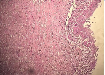
Figure 1: Surface epithelial lining was largely ulcerated and replaced by polymorphic granulation

Figure 2 : Polymorphic granulation

Figure 3 : Histiocytes in the gallbladder wall and fibrosis
Xanthogranulomatous cholecystitis is a rare variant of chronic cholecystitis. XGC is a benign but sometimes aggressive disease resulting from chronic inflammation of the gallbladder wall. Its prevalence ranges from 1% to 9%. It typically occurs in individuals over 50 years of age, with no sex predilection [1-4]. Clinically, it presents with signs of chronic or acute cholecystitis. Ultrasound imaging shows diffuse thickening of the gallbladder wall, intramural hypoechoic nodules, an indistinct liver-gallbladder interface, and the presence of gallstones in most cases. Similar findings are observed on CT and MRI, with involvement of surrounding structures [5-7]. The main differential diagnosis at this stage is neoplastic pathology. Histologically, there is the presence of xanthogranulomas with foamy histiocytes in the gallbladder wall and fibrosis without signs of malignancy. Histological differential diagnoses include gallbladder adenocarcinoma with signet ring cells, marked by anti-cytokeratin antibodies, and malakoplakia, characterized by histiocytes with Michaelis-Gutmann bodies positive on PAS and von Kossa special stains [8-10].
Xanthogranulomatous cholecystitis is a rare benign condition that can be mistaken for neoplastic pathology. Definitive diagnosis is histological, and optimal management relies on anatomo-clinical correlation.
Yes
Consent for publication
Yes
Availability of data and material
Data availability.
Competing interests
The authors declare no conflicts of interest.
Funding
No funding
Authors' contributions
All the authors contributed to the conduct of this work.
Acknowledgments
To anyone who has participated in the care of this patient directly or indirectly
Dear Editorial Team, Clinical Medical Reviews and Reports. My experience with the journal was highly positive. The peer-review process was rigorous, constructive, and completed in a timely manner. The reviewers provided valuable comments that helped improve the quality and clarity of our manuscript. The editorial office was professional, responsive, and supportive throughout all stages of the publication process. Communication was clear and efficient, and any questions were addressed promptly. Overall, I found the journal to maintain high scientific standards and an excellent publication workflow. I would be pleased to consider submitting future work to this journal. Best wishes from, Elena Popa.

It was my pleasure to submit my testimonial concerning the Reviewer Board of our Scientific Journal “Brain and Neurological Disorders”. The Reviewers focused on some modifications and their contribution was helpful. The ladies of our Editorial Office were also supported my efforts. It was my honor to have such a co-operation and I am looking forward for more collaboration.

Dear Grace Pierce, Editorial Coordinator of Journal of Clinical Research and Reports, Thank you for the speedy and efficient peer review process. I appreciate the fact that your peer reviewers do not take months to respond like with some other journals. I would also like to thank the editorial office for responding quickly to my questions. It is an excellent journal. I plan to submit more manuscripts in the future. Best wishes from, Robert W. McGee

Dear Grace Pierce, Editorial Coordinator of Journal of Clinical Research and Reports, Working with you and your team on our recent publication in JCRR has been a truly wonderful and enjoyable experience. The responses were prompt, and the reviewers were patient, constructive, and highly professional. One reviewer in particular gave me the feeling that a professor was carefully reading and commenting on my coursework, which was deeply touching. The entire process was straightforward and hassle‑free, with no tedious online forms to complete. I highly recommend this journal. Best wishes from, DR Aibing Rao, Head of R&D

I Appreciate the Opportunity to Share my Experience with the Journal of Clinical Research and Reports. The peer review process was timely and constructive, and the feedback provided helped improve the quality of our manuscript. The editorial office was professional, responsive, and supportive throughout the process, ensuring smooth communication and efficient handling of the submission. Overall, it was a positive experience collaborating with your team.

Dear Mercy Grace, Editorial Coordinator of Obstetrics Gynecology and Reproductive Sciences, We would like to express our gratitude for your help at all stages of publishing and editing the article. The editors of the magazine answer all the necessary questions and help at every stage. We will definitely continue to cooperate and publish other works in the Obstetrics Gynecology and Reproductive Sciences! Best wishes from, Alla Konstantinovna Politova,
