
Mini-Review | DOI: https://doi.org/10.31579/2640-1053/235
Research and Training Center ‘Physical and Chemical Materials Science’ Under Kyiv Taras Shevchenko University and NAS of Ukraine, Kiev, Ukraine.
*Corresponding Author: Yuri Pivovarenko. Research and Training Centre ‘Physical and Chemical Materials Science’ Under Kyiv Taras Shevchenko University and NAS of Ukraine, Kiev, Ukraine.
Citation: Yuri Pivovarenko, (2025), UV Absorption Spectra Could Replace Nanoparticles in Cancer Diagnostics, J Cancer Research and Cellular Therapeutics, 9(2); DOI:10.31579/2640-1053/235
Copyright: © 2025, Yuri Pivovarenko. This is an open-access article distributed under the terms of the Creative Commons Attribution License, which permits unrestricted use, distribution, and reproduction in any medium, provided the original author and source are credited.
Received: 25 March 2025 | Accepted: 03 April 2025 | Published: 11 April 2025
Keywords: cancer; diagnostics; nanoparticles; UV absorption spectrometry
The ability of nanoparticles to accumulate in cancer-affected areas has made them a key tool in modern cancer diagnostics. At the same time, the toxicity of nanoparticles makes one think about the admissibility of their use in this diagnostic. Taking this into account, it is proposed to replace cancer diagnostics using nanoparticles with cancer diagnostics using UV absorption spectra of human biological fluids. The rationale for this proposal is presented and discussed here
Since various nanoparticles accumulate preferentially in cancer-affected areas, they are often positioned as a cancer diagnostic tool [1-7]. However, the toxicity of nanoparticles [1, 8 – 10], which is the cause of unwanted side effects in patients, makes one wonder about the admissibility of their use in these diagnostics. At the same time, this same toxicity stimulates the search for more acceptable methods of cancer diagnosis. Thus, the proposal to diagnose cancer using UV absorption spectra of human biological fluids can be seen as a result of this search. To make this proposal more understandable for oncologists, some spectral phenomena are discussed in detail here.
First of all, it is necessary to realize the fact that positive electrification of water significantly increases its hydrating capacity, in particular, in relation to biopolymers and fats [11, 12]. This feature of positively charged water acquired a particularly important significance over time, since it was this feature that allowed assuming that it is the positive electrification of the internal environment of the human body that stimulates the continuous proliferation of its cells [12 – 14]. It later became clear that this assumption correlated well with McIntyre's hypothesis that it is precisely increased cellular hydration that promotes both tumor growth and metastasis [15, 16]. Moreover, it became clear that this same assumption allows explaining a number of facts related to cancer [12,17-19]. Taking all this into account, this assumption was considered quite adequate. Accordingly, a conclusion was made about the need to create means that would allow the detection of positively charged water in the human body.
After some thought, it was assumed that such a means could be the UV absorption spectra of human biological fluids. This assumption did not come out of nowhere. First of all, this assumption was based on the difference in the UV absorption spectra of uncharged water and oppositely electrified waters (Figure 1). Secondly, this assumption was based on the difference in the UV absorption spectra of DNA and RNA solutions prepared using oppositely charged waters (Figure 2). Finally, this assumption took into account the difference in the UV absorption spectra of lymphocytes (or rather, their identical suspensions) isolated from the blood of healthy people and from the blood of patients with B-cell chronic lymphocytic leukemia (Figure 3).
Thus, a comparison of all these spectra (Figures 1-3) allowed initially concluding that all peaks in the 200 – 240 nm regions reflect the absorption of UV radiation by water. At the same time, a comparison of all these spectra (Figures 1-3) allowed concluding that the environment of lymphocytes in healthy people is negatively charged, while the environment of lymphocytes in patients with this type of leukemia is positively charged [20].
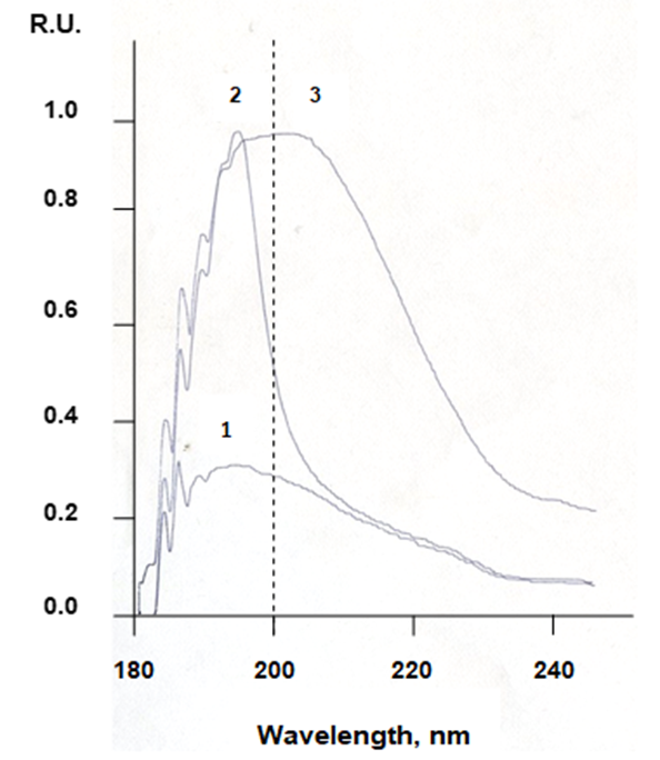
Figure 1. UV absorption spectra of water against air: 1 – uncharged water; 2 – water with a potential of –150 mV; 3 – water with a potential of +250 mV; all spectra were not processed [11].
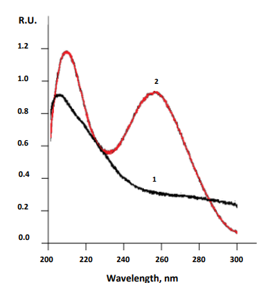
Figure 2. UV absorption spectra of aqueous RNA solutions prepared with oppositely charged waters: 1 – spectrum of RNA prepared with negatively charged water: 2 – spectrum of RNA prepared with positively charged water [20].
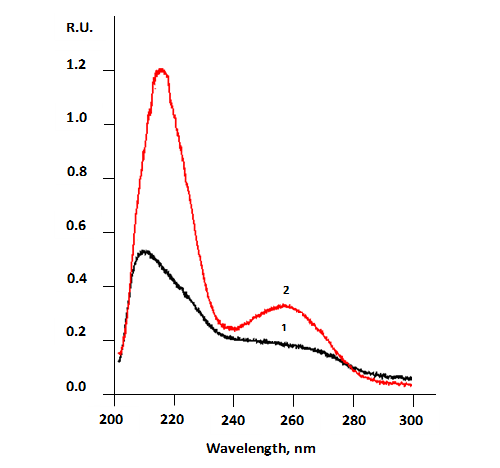
Figure 3. UV absorption spectra of aqueous suspensions of lymphocytes: 1 – suspension of lymphocytes obtained from a healthy person: 2 – suspension of lymphocytes obtained from a patient with B-cell chronic lymphocytic leukemia [20]
Therefore, the proposal to use UV absorption spectra of human biological fluids in the diagnosis of oncological diseases seems entirely justified.
It is probably worth adding here that a comparison of the last two spectra (Figures 2, 3) also allowed concluding that the absorption of UV radiation by lymphocyte suspensions is mainly provided by the absorption of UV radiation by their nucleic acids [20].
Thus, saturation of cancer-affected areas with positively charged water, which has been repeatedly demonstrated [12 – 14, 17 – 20], makes it possible to offer not only a comfortable, but also absolutely non-toxic method for diagnosing oncological diseases for patients. This, in turn, indicates that the adoption of the proposed point of view on the nature of cancer [13 – 17] is equally promising for both its diagnosis and treatment.
Dear Editorial Team, Clinical Medical Reviews and Reports. My experience with the journal was highly positive. The peer-review process was rigorous, constructive, and completed in a timely manner. The reviewers provided valuable comments that helped improve the quality and clarity of our manuscript. The editorial office was professional, responsive, and supportive throughout all stages of the publication process. Communication was clear and efficient, and any questions were addressed promptly. Overall, I found the journal to maintain high scientific standards and an excellent publication workflow. I would be pleased to consider submitting future work to this journal. Best wishes from, Elena Popa.

It was my pleasure to submit my testimonial concerning the Reviewer Board of our Scientific Journal “Brain and Neurological Disorders”. The Reviewers focused on some modifications and their contribution was helpful. The ladies of our Editorial Office were also supported my efforts. It was my honor to have such a co-operation and I am looking forward for more collaboration.

Dear Grace Pierce, Editorial Coordinator of Journal of Clinical Research and Reports, Thank you for the speedy and efficient peer review process. I appreciate the fact that your peer reviewers do not take months to respond like with some other journals. I would also like to thank the editorial office for responding quickly to my questions. It is an excellent journal. I plan to submit more manuscripts in the future. Best wishes from, Robert W. McGee

Dear Grace Pierce, Editorial Coordinator of Journal of Clinical Research and Reports, Working with you and your team on our recent publication in JCRR has been a truly wonderful and enjoyable experience. The responses were prompt, and the reviewers were patient, constructive, and highly professional. One reviewer in particular gave me the feeling that a professor was carefully reading and commenting on my coursework, which was deeply touching. The entire process was straightforward and hassle‑free, with no tedious online forms to complete. I highly recommend this journal. Best wishes from, DR Aibing Rao, Head of R&D

I Appreciate the Opportunity to Share my Experience with the Journal of Clinical Research and Reports. The peer review process was timely and constructive, and the feedback provided helped improve the quality of our manuscript. The editorial office was professional, responsive, and supportive throughout the process, ensuring smooth communication and efficient handling of the submission. Overall, it was a positive experience collaborating with your team.

Dear Mercy Grace, Editorial Coordinator of Obstetrics Gynecology and Reproductive Sciences, We would like to express our gratitude for your help at all stages of publishing and editing the article. The editors of the magazine answer all the necessary questions and help at every stage. We will definitely continue to cooperate and publish other works in the Obstetrics Gynecology and Reproductive Sciences! Best wishes from, Alla Konstantinovna Politova,
