
Research Article | DOI: https://doi.org/10.31579/2690-8808/208
1Junior Resident, Department of Oral & Maxillofacial Surgery, MGPGIDS, Puducherry.
2Professor & Registrar (Academic), Head of IT Wing and Telemedicine, Department of Plastic Surgery & Telemedicine, JIPMER, Pondicherry.
3General Surgery, Senior Resident, Department of Plastic Surgery, JIPMER, Puducherry.
4General Surgery, MCh Plastic Surgery, Assistant Professor, Department of Plastic Surgery, JIPMER, Puducherry.
5General Surgery, Senior Resident, Department of Plastic Surgery, JIPMER, Puducherry.
*Corresponding Author: Ravi Kumar Chittoria, Senior Professor and Associate Dean (Academic), Head of IT Wing and Telemedicine, Department of Plastic, Surgery and Telemedicine, JIPMER. Pondicherry, India.
Citation: Nishant V. Dumont, Ravi K. Chittoria, K. Gupta, Jacob A. Chakiat and Padmalakshmi B. Mohan,et al, (2024), Utilizing Amniotic Membrane Allograft as regenerative therapy for Treating Burns, J, Clinical Case Reports and Studies, 5(6); DOI:10.31579/2690-8808/208
Copyright: ©, 2024, Ravi Kumar Chittoria. This is an open access article distributed under the Creative Commons Attribution License, which permits unrestricted use, distribution, and reproduction in any medium, provided the original work is properly cited.
Received: 26 July 2024 | Accepted: 05 August 2024 | Published: 12 August 2024
Keywords: amniotic membrane allograft; wound healing; regenerative therapy
Burn injuries are prevalent among all age groups and can result from thermal, scald, or electrical sources. Currently, various scaffolds are employed to enhance the healing process and minimize scar formation. Collagen serves as a scaffold, facilitating tissue regeneration and promoting the formation of new blood vessels. Additionally, scaffolds such as amniotic membrane aid in proper epithelialization and scar reduction, boasting unique anti-inflammatory and bacteriostatic properties. In our study, we utilized amniotic membrane allograft as a biological dressing for burn wounds on an adult patient's right thigh.
The process of wound healing is a biological response to various forms of injury, encompassing physical, chemical, mechanical, or thermal damage. This process typically involves several sequential phases: homeostasis, inflammation, proliferation/granulation, and remodeling/maturation [1]. However, deviations from the normal healing trajectory often result in stagnation during the inflammatory phase, particularly evident in cases of burns where standard healing mechanisms may be compromised [2]. In contemporary medicine, the utilization of scaffolds, whether natural or synthetic, has gained traction and recognition. An optimal scaffold should possess specific characteristics: appropriate physical and mechanical properties, a physiological background conducive to cellular adhesion, proliferation, and differentiation, high porosity, a favorable surface area to volume ratio, flexibility to conform to wound shapes, and ideally, biocompatibility and biodegradability [3]. Collagen, whether synthetic or natural, serves as a surrogate for the dermal matrix, facilitating epithelialization during wound healing [4]. Additionally, collagen degradation during this process contributes to neovascularization, thereby promoting angiogenesis. Amniotic membrane, a natural scaffold, exhibits distinctive properties such as anti-inflammatory, bacteriostatic, anti-fibrotic, anti-scarring, and epithelization-promoting qualities [6]. Its low immunogenicity and presence of progenitor cells make it an appealing choice for scaffold applications in wound healing. Silicone functions as a barrier, reducing mechanical friction and transepidermal water loss, factors associated with infection severity. In vitro studies suggest that silicone may modulate inflammatory growth factors implicated in fibrosis and support acute wound healing, including key inflammatory markers like TNF-α, TGF-β, IL-1, and IL-6.
This research was carried out at a Tertiary Care Centre within the Department of Plastic Surgery following approval from the department's ethical committee. Informed consent was duly obtained. The subject of this study was a eighty two years old female who sustained accidental second-degree thermal burns affecting the right thigh. Despite being promptly taken to a nearby hospital, initial resuscitation was inadequate within 30 minutes. He subsequently developed blistering and swelling around the gluteal region the following day and presented to our center with a delay of 12 hours. Upon admission to the tertiary burn care unit, he received initial resuscitation involving intravenous fluids, analgesics, and prophylactic antibiotics. On the fourth day post-burn, a three-layered scaffold dressing was prepared and applied to the deeper burn areas following dermabrasion-assisted tangential excision. This scaffold comprised sterile amniotic membrane, dry collagen sheet, and silicone sheet, with the amniotic membrane layer directly contacting the wound. The amniotic membrane (fig 1) used was obtained from a freshly delivered placenta, thoroughly washed, and stored in an antibiotic solution in a refrigerator. The dressing remained in place for seven days, during which time the amniotic membrane was completely absorbed, and the silicone sheet layer was also removed (fig 2).

Figure 1: Figure showing amniotic membrane which was harvested from human placenta

Figure 2: Figure showing amniotic membrane which was harvested from human placenta being applied over the wound over the gluteal region
Throughout both the intraoperative and postoperative phases, the patient experienced no notable events. Upon opening the dressing on the seventh day post-operation, substantial areas of re-epithelialization and healing were observed. Complete healing of all second-degree superficial burn wounds was achieved without any complications or adverse effects noted throughout the entire procedure. BJWAT wound score improved from 32 at presentation to 25 after utilization of amniotic membrane regenerative therapy (fig 3 & 4).
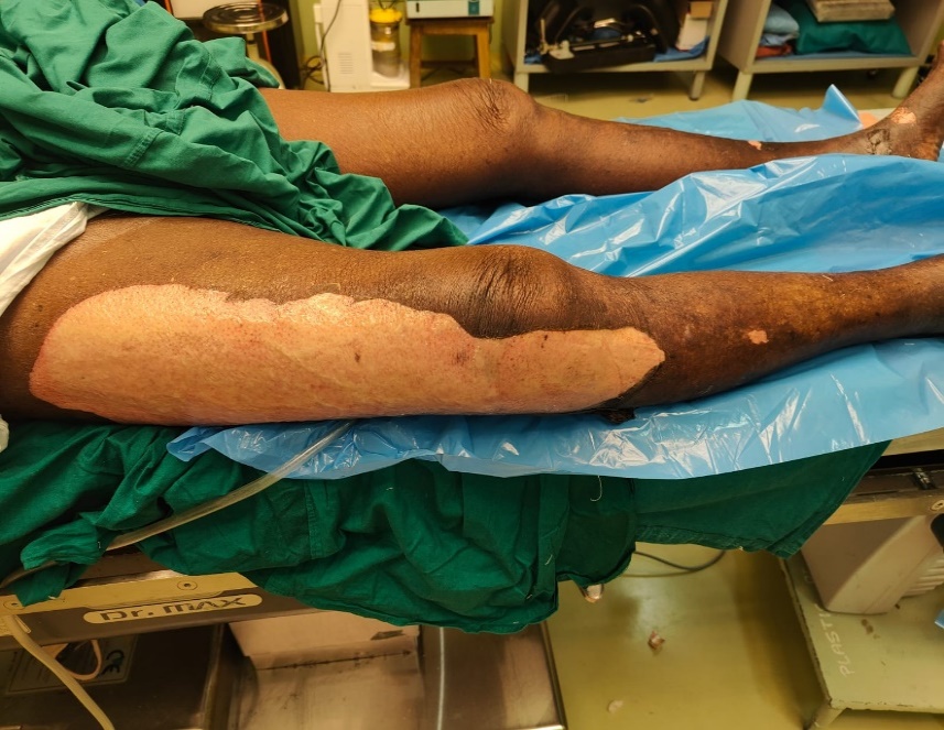
Figure 3: Figure showing condition of the wound at time of presentation (BJWAT wound score - 32)
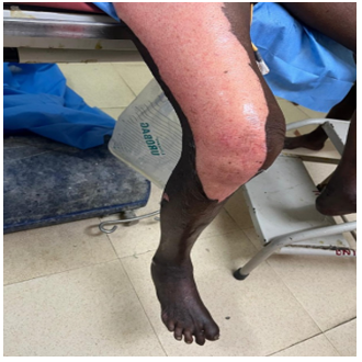
Figure 4: Figure showing condition of the wound after utilization of amniotic membrane regenerative therapy (BJWAT wound score -25).
Partial-thickness burn injuries have the potential to heal spontaneously, whereas full-thickness burns typically necessitate skin grafting to achieve complete wound closure. Historically, split-thickness skin autograft has been the preferred method for closing excised full-thickness burn wounds [5]. However, patients with extensive burn injuries may face challenges due to limited availability of donor sites for autograft harvesting, prompting consideration of alternative options such as skin substitutes. Engineered skin substitutes offer temporary coverage for wounds until donor sites are available for autografting or, if containing autologous cells, can provide permanent closure [7]. While the availability of permanent skin substitutes remains limited, advancements in tissue engineering are anticipated to yield improved models, thus enhancing the options for managing burn wounds. For instance, the commercially available Dermal Regeneration Template comprises a two-layered system. The outer layer, composed of a thin silicone film, serves as a protective barrier against infection and regulates heat and moisture loss. The inner layer, consisting of a collagen glycosaminoglycan (GAG) thermal layer, acts as a biodegradable template facilitating dermal tissue regeneration. The porous nature of the template aids in skin regeneration by serving as a scaffold for tissue growth. Following regeneration of the dermal layer, the outer layer of the template is replaced with a thin epidermal skin graft, allowing for flexible, permanent skin regeneration and promoting faster wound healing with minimal scarring. Seeking to emulate this mechanism, we have developed an indigenous dermal regeneration scaffold using silicone sheet, dry collagen sheets, and amnion3. This cost-effective scaffold can be readily prepared and utilized, particularly in hospital settings within developing countries where the affordability of commercial regeneration templates may be a concern.
The efficacy of employing a cost-effective regenerative scaffold dressing based on amniotic membrane in treating second-degree scald burns has been demonstrated in this investigation. It accelerates the overall healing process of both superficial and deep second-degree wounds to less than a week, thereby reducing hospitalization duration and infection rates. Nevertheless, further extensive multicenter, double-blinded controlled research incorporating statistical analysis is warranted.
Dear Editorial Team, Clinical Medical Reviews and Reports. My experience with the journal was highly positive. The peer-review process was rigorous, constructive, and completed in a timely manner. The reviewers provided valuable comments that helped improve the quality and clarity of our manuscript. The editorial office was professional, responsive, and supportive throughout all stages of the publication process. Communication was clear and efficient, and any questions were addressed promptly. Overall, I found the journal to maintain high scientific standards and an excellent publication workflow. I would be pleased to consider submitting future work to this journal. Best wishes from, Elena Popa.

It was my pleasure to submit my testimonial concerning the Reviewer Board of our Scientific Journal “Brain and Neurological Disorders”. The Reviewers focused on some modifications and their contribution was helpful. The ladies of our Editorial Office were also supported my efforts. It was my honor to have such a co-operation and I am looking forward for more collaboration.

Dear Grace Pierce, Editorial Coordinator of Journal of Clinical Research and Reports, Thank you for the speedy and efficient peer review process. I appreciate the fact that your peer reviewers do not take months to respond like with some other journals. I would also like to thank the editorial office for responding quickly to my questions. It is an excellent journal. I plan to submit more manuscripts in the future. Best wishes from, Robert W. McGee

Dear Grace Pierce, Editorial Coordinator of Journal of Clinical Research and Reports, Working with you and your team on our recent publication in JCRR has been a truly wonderful and enjoyable experience. The responses were prompt, and the reviewers were patient, constructive, and highly professional. One reviewer in particular gave me the feeling that a professor was carefully reading and commenting on my coursework, which was deeply touching. The entire process was straightforward and hassle‑free, with no tedious online forms to complete. I highly recommend this journal. Best wishes from, DR Aibing Rao, Head of R&D

I Appreciate the Opportunity to Share my Experience with the Journal of Clinical Research and Reports. The peer review process was timely and constructive, and the feedback provided helped improve the quality of our manuscript. The editorial office was professional, responsive, and supportive throughout the process, ensuring smooth communication and efficient handling of the submission. Overall, it was a positive experience collaborating with your team.

Dear Mercy Grace, Editorial Coordinator of Obstetrics Gynecology and Reproductive Sciences, We would like to express our gratitude for your help at all stages of publishing and editing the article. The editors of the magazine answer all the necessary questions and help at every stage. We will definitely continue to cooperate and publish other works in the Obstetrics Gynecology and Reproductive Sciences! Best wishes from, Alla Konstantinovna Politova,
