AUCTORES
Globalize your Research

Review Article | DOI: https://doi.org/10.31579/2641-0419/254
1 BSc, PhD, Specialized professional Public Health Research Division National Institute of Health Avenue Street 26 No 51-20 CAN Bogotá, D.C., Colombia.
*Corresponding Author: María Luz Gunturiz A, BSc, PhD, Specialized professional Public Health Research Division National Institute of Health Avenue Street 26 No 51-20 CAN Bogotá, D.C., Colombia.
Citation: María Luz Gunturiz A (2022). Use of Potential Markers in the Management of Myocardial Damage and COVID-19. J. Clinical Cardiology and Cardiovascular Interventions, 5(3); Doi:10.31579/2641-0419/254
Copyright: © 2022 María Luz Gunturiz A, This is an open-access article distributed under the terms of the Creative Commons Attribution License, which permits unrestricted use, distribution, and reproduction in any medium, provided the original author and source are credited.
Received: 18 February 2022 | Accepted: 17 March 2022 | Published: 28 March 2022
Keywords: coronavirus; myocardial injuy; cardiovascular disease; biomarkers; covid-19
Several biomarkers such as cardiac troponin (cTn), D-Dimer, C-reactive protein (CRP) or lactate dehydrogenase (LDH) have been related to the severity and progression of COVID-19. However, the determination of which of these biomarkers can provide greater prognostic value will depend on the clinical background of each patient, comorbidities, among others.
Understanding the pathophysiological processes associated with cardiovascular damage and the metabolic processes associated with the critical course of the infection allows us to infer the prognostic and predictive value of the different biomarkers used both for COVID-19 and at the cardiac level.
It is well described that most of them are not only predictive of the severity of the disease, but are also useful for the clinical management of these pathologies, allowing in turn the stratification of positive COVID-19 patients in risk categories.
The objective of this review is to understand the role of known biomarkers for COVID-19 infection, and those used to predict cardiovascular diseases in these times of pandemic.
Coronaviruses affect the cardiovascular system and the complication and mortality rates of COVID-19 have been shown to be higher in patients with pre-existing cardiovascular risk factors or cardiovascular disease [1-3].
Previous studies have shown that myocardial injury and cardiovascular risk factors are associated with a worse prognosis in patients with COVID-19 in 2 Chinese cohorts, therefore, it has been suggested that the virus can cause heart damage, but the data on this topic is scarce and the clinical and prognostic consequences are still not fully clarified [4,5].
The pathophysiology of myocardial injury caused by SARS-CoV-2 is still under study, although some hypotheses and mechanisms of action have been described. The first is related to direct myocardial injury, where viral RNA was found in heart muscle cells in 35% of SARS-infected subjects in 2005 [6-8]. During these pandemic months, it has been suggested that SARS-CoV-2 can enter myocardial cells by binding to type 2 angiotensin converting enzyme (ACE) receptors on their surface [9-12].
Among the pathogenic mechanisms triggered by COVID-19 are inflammatory cascades, cytokine storms and activation of signaling pathways involved in coagulation, which are common in systemic vasculitis and lead to serious and even fatal complications, such as sepsis, disseminated intravascular coagulation and acute cardiovascular events [13].
Patients affected by COVID-19 produce cytokines that enter the systemic circulation, stimulating macrophages within the plaque to increase local cytokine production and cause an increase in tissue factor expression that makes lesions more thrombogenic [14]. If COVID-19 patients also have severe underlying atherosclerotic diseases, extreme cases of acute myocardial infarction are likely to occur during the course of the disease. COVID-19 patients are likely to suffer from atherosclerosis, leading to insufficient coronary blood supply and causing damage to the myocardium [15,16].
Constant increase in cardiac markers is a predictor of disease worsening and in most cases patients should be transferred to an intensive care unit (ICU) ward for treatment. Although the evidence that patients with hypertension and cardiovascular diseases are more susceptible to SARS-CoV-2 infection is still insufficient, the truth is that patients with hypertension and cardiovascular diseases are more likely to develop severe cases, in addition to the fact that patients with SARS-CoV-2 are more prone to cardiovascular complications [17-19].
Some publications have shown that myocardial damage in patients with COVID-19 is more frequent among patients with more serious disease and who entered ICU. It is worth mentioning that the increase in markers of myocardial damage in these patients regardless of pre-existing diseases could be related to increase myocardial oxygen demand in COVID-19 positive patients with or without known coronary artery disease [20-23]. For another way, SARS-CoV-2 infection, could lead to a significant increase in coronary flow and oxygen demand, causing myocardial ischemia, especially in patients with coronary disease.
The increase in myocardial metabolic activity causes a great expenditure of oxygen in the arterial blood, requiring the generation of flow increases from regulation mechanisms, capable of guaranteeing a balance between supply and demand, involving nervous, humoral, mechanical phenomena and electrical. Tachycardia and severe hypertension contribute to breaking this balance, causing damage to myocardial cells [23].
Definition of myocardial injury
Damage caused to cardiac cells by SARS-CoV-2 infection is relatively common in severe forms of the disease, however, the mechanisms by which these cardiac cell alterations occur are still under study, although whether knows that an imbalance occurs between oxygen supply and demand, systemic inflammatory response, hypoxia, microvascular dysfunction and direct myocardial damage caused by the virus. What is clear is that there is a relationship between myocardial damage and the future evolution of the patient and short-term mortality. For example, in a study that included 416 positive patients for COVID-19, elevated troponin I values above the reference value were found in those patients with an average age of 64 years (19.7%) [24].
Myocardial injury or damage is defined as the detection of a cardiac troponin value (T or I) above the 99th percentile of the upper reference limit, which can respond to acute or chronic damage depending on the values of the enzyme curve. Markers such as creatine kinase MB fraction (CK-MB) could be used in the detection of cardiac cell damage, although their sensitivity and specificity is lower than that of troponins [25,26].
On the other hand, higher in-hospital mortality has been reported in patients with myocardial damage. Guo et al [27] described that in 187 admitted patients, positive for SARS-CoV-2, 27.8% presented elevated troponin T values and mortality 52%, compared to 8.9% of those with enzyme values within the normal range.
In patients with myocardial injury with elevated levels of cardiac enzymes, electrocardiographic alterations of the ST segment and the T wave, regional motility disorders of the left ventricular walls and cardiac function, identified during the echocardiogram, also generally occur [24].
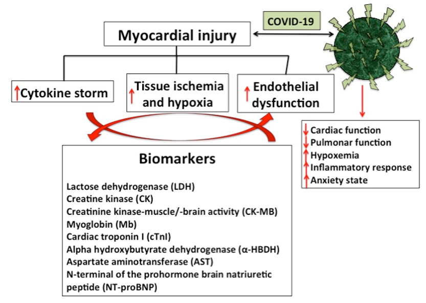
A study have shown that there is damage between 8% and 20% of cardiac cells associated with SARS-CoV-2 infection [28], especially when the disease occurs in its severe forms. Additionally, it has been described that in Chinese patients from the province of Wuhan, with an average age of 64 years positive for COVID-19, about 20% presented troponin I values above the reference value [24]. Although much progress has been made in the understanding of and the relationship between cardiovascular disease and COVID-19, the mechanisms by which alterations occur in the cardiac cell are not entirely clear. Among the factors involved in cardiac injury caused by COVID-19, the imbalance between oxygen supply and demand, systemic inflammatory response, hypoxia, microvascular dysfunction and direct myocardial damage caused by the virus, among others, could be considered [29]. (Figure 1).
However, many authors agree on the relationship between myocardial damage, patient evolution, and short-term mortality. It has been reported that patients with myocardial damage presented a quantitatively higher hospital mortality than those without this condition. Similarly, Guo et al [27], reported that mortality was greater than 50% in admitted patients, positive for SARS-CoV-2, with elevated troponin T values compared to patients who presented enzyme values within the normal range [23, 24].
It should be remembered that myocardial injury or damage is defined as the detection of a cardiac troponin value (T or I) above the 99th percentile of the upper reference limit, which can respond to acute or chronic damage depending on the values of the enzymatic curve. Biomarkers such as CK-MB could also be used in the detection of cardiac cell damage, although with lower sensitivity and specificity [25,26].
On the other hand, patients with myocardial injury, in addition to presenting high levels of cardiac enzymes, have electrocardiographic abnormalities of the ST segment and the T wave, as well as regional motility disorders of the left ventricular walls and cardiac function, identified by echocardiogram. Knowing how much enzyme elevation corresponds to primary damage to cardiac cells, and how much is secondary to critical states of the disease, is the challenge that clinical cardiologists are facing in these times of pandemic [23,24].
It is important to note that due to the difficulty in performing cardiac magnetic resonance imaging or endomyocardial biopsy, the diagnosis for cardiac affectation is based mainly on the elevation of troponin in association with echocardiographic data compatible with acute myocarditis (i.e., segmental wall motion abnormalities, left ventricular ejection fraction (ðLVEFÞ) < 50> 10mm and/or pericardial effusion) and ECG changes (ST elevation or ST/T segment changes) [13, 30,31].
Several authors have reported that high plasma levels of hyperhomocysteinemia (Hcy) significantly increase the incidence of vascular damage and that concentrations above the 90th percentile are associated with an increased risk of degenerative and atherosclerotic processes in the coronary, cerebral, and peripheral circulatory systems [32-34].
Hyperhomocysteinemia is defined as a medical condition characterized by an abnormally high level (>15 μmol/L) of Hcy in the blood [35,36]. The total Hcy concentration in plasma of healthy humans is low, between 5.0 and 15.0 μmol/L when assessed with the use of high performance liquid chromatography (HPLC), or 5.0–12.0 lmol/L when using immunoassay methods [37]. When the level is between 16 and 30 μmol/L it is classified as moderate, 31-100 μmol/L is considered intermediate, and a value >100 μmol/L is classified as severe hyperhomocysteinemia [38,39]. Recent publications have linked hyperhomocysteinemia with cardiovascular diseases, diabetes, CKD, and fatty liver disease [35,36,39]. Although Hcy is an effective biomarker of cardiovascular risk and it is known that cardiovascular complications are critical in hospitalized patients with COVID-19, this biomarker has not been used or studied in the clinical setting or in published prospective studies focused on useful laboratory markers for clinical evaluation of COVID-19 [13].
In line with the Fourth Universal Definition of Myocardial Infarction (2018), cardiac injury was diagnosed if serum levels of cardiac biomarkers (eg,hs-cTnT) were above the 99th percentile upper reference limit, (>14.0ng/L), as recommended by the manufacturer [26,40] and regardless of new abnormalities on electrocardiography and echocardiography. Among the cardiac markers, lactose dehydrogenase (LDH), creatine kinase (CK), creatinine kinase-muscle / brain activity (CK-MB), myoglobin (Mb), cardiac troponin I (cTnI), alpha hydroxybutyrate dehydrogenase (α- HBDH), aspartate aminotransferase (AST) and N-terminal prohormonal brain natriuretic peptide (NT-proBNP) increase their levels in different proportions in patients with COVID-19. Although LDH, CK, α-HBDH, and AST are cardiac enzymes, their increases cannot specifically represent myocardial injury, due in part to damage to the lungs, liver, kidneys, or other organs [30, 41-44].
On the other hand, it has been described that the anti-aging gene Sirtuin 1 has been linked to the risk for cardiovascular disease as well as various chronic diseases such as obesity, NAFLD, diabetes and neurodegenerative diseases. COVID-19 has been strongly linked to Sirtuin 1 inactivation and cardiovascular disease. According to this, within the selection of markers for the diagnosis and treatment of cardiovascular disease may need to involve the plasma measurement of Sirtuin 1 in COVID-19 individuals [45-47].
However, specific myocardial markers including CK-MB, cTnI, Mb, and NT-proBNP are increased to varying degrees in COVID-19 patients, especially in ICU and critically ill patients [48,49].
It is important to note that COVID-19 increases the risk of cardiovascular events, such as acute myocardial infarction, myocarditis and arrhythmias. Cardiac injuries as a result of COVID-19 are associated with higher mortality. It is not yet known whether there will be a long-term elevated cardiovascular risk as a result of COVID-19. In this context, the WHO [48] indicates that in patients with severe COVID-19, laboratory tests usually show a decrease in the number of leukocytes, in particular lymphocytopenia, as well as an increase in the number of neutrophils, inflammatory markers, D-dimer and elevated blood urea and creatinine concentrations. Chest computed tomography usually shows ground-glass opacities, with or without consolidative lesions. They are also more likely to be bilateral, have a peripheral distribution, and involve the lower lobes [48].
In studies with confirmed COVID-19 patients, biomarkers of cardiac injury have been reported to rise above normal during hospitalization and rise sharply immediately before death. It has also been reported that increased troponin, CK-MB, NTproBNP can and be considered indicators of possible cardiac damage during SARS-CoV-2 infection [30,50]. (Table 1)
For NT-proBNP, positivity was considered if serum levels were above the limit for ruling out heart failure in the acute setting, which is <300>
Acute respiratory distress syndrome was diagnosed according to the Berlin criteria as acute-onset hypoxemia (ratio of arterial oxygen partial pressure to fractional inspired oxygen expressed as a Fraction <300>
For the rapid propagation of the COVID-19, it is relevant to have biomarkers available to the progression of this pathology that allow patient risks stratification and identify those patients who will suffer a rapid progression of serious complications and death. The knowledge of new biomarkers for the detection, clinical management and prevention of serious complications should be based on the understanding of the molecular and cellular mechanisms involved in viral pathogenesis [13].
Several studies report that within the main laboratory findings in survivors and non-surviving patients with severe or fatal COVID-19, significant increase in total bilirubin and CK, together with serum ferritin, leukocyte count and IL -6 in non-survivors compared to survivors [20,55,56]. In addition to the strong association between thromboembolism, Covid 19 and myocardial injury, the diagnosis of D-dimer and cardiac markers is crucial in the follow-up of patients with COVID-19. On the other hand, the markers of muscular and cardiac injury were high in patients with both severe and deadly Covid-19 [43,52,57,58]. Additionally, in mortal chaos were found significantly higher cardiac troponin levels probably due to viral myocarditis and cardiac injury due to the progression of the disease to multiple organic insufficiency, also characterized by the significant elevation of liver enzymes (Alanine aminotransferase (ALT) and aminotransferase aspartate (AST)) and with critical changes in the parameters of renal function (blood ureic nitrogen, creatinine) and coagulation markers [13, 59].
Troponin has been used to determine the extent of cardiac injury, but determination of the level NT-proBNP on admission of COVID-19 patients could also help to better stratify the risk of in-hospital mortality or MV [52, 60].
C-reactive protein (CRP) is produced by the liver and its level rises when there is inflammation. LDL cholesterol not only coats the walls of the arteries, but also damages them by causing inflammation that the body tries to cure by sending out a "response team" of proteins called "acute phase reactants" within which CRP is found. Some studies report that testing for CRP levels is a better indicator of cardiovascular disease (CVD) than testing for LDL. However, it is important to mention that a CRP test is not a test for heart disease but rather a test for inflammation in the body. A high level could also be a sign of cancer, infection, inflammatory bowel disease, lupus, rheumatoid arthritis, tuberculosis, or another disease [44,61].
a-Hydroxybutyrate dehydrogenase (HBDH) is an isoenzyme of lactate dehydrogenase (LDH). Heart muscles contain high concentrations of HBDH. The serum concentration of HBDH is elevated in myocardial damage [62,63].
Cardiac markers |
Reference value | Value in patients with pre-existing cardiovascular diseases |
Observations |
Lactose dehydrogenase (LDH) | 120–250 U/L | In Covid patients values of ≥334U/L were reported
| This marker was initially used to aid in the diagnosis and monitoring of acute coronary syndromes. However, as it is a non-specific marker of tissue injury, only of the heart, it is currently hardly measured, and has been replaced by much more specific markers of diseases of cardiovascular origin, such as troponins. This marker rises 12-18 hours after the onset of symptoms and usually normalizes in the first week [44,61]. |
Creatine kinase (CK) |
50–310 U/L | In patients with Covid who did not survive the infection, it was observed that among other markers, CK and CK-MB were greatly increased compared to the values found in patients under the same health conditions but who managed to overcome the disease. | -Poor cardiospecificity and sensitivity -Not very useful for predicting cardiovascular risk -Recommended only if measurement of CK-MB or troponin concentration is not available. However, until the availability of other markers, total CK has been the most widely used biological marker for the diagnosis of myocardial and musculoskeletal disorders. Currently, it still has a relevant role in the follow-up of myocardial infarction in its sub-acute phase [13,16, 30, 48-50] |
Creatinine kinase-muscle/-brain activity (CK-MB) | 0–5 ng/mL | - It is the best alternative if there is no availability to analyze troponins - Detects reinfarction early - Not specific to heart tissue [30, 48-50] | |
Myoglobin (Mb) | 25 a 72 ng/mL (1.28 a 3.67 nmol/L | Myoglobin is considered one of the biomarkers of cardiac damage that, in patients with this pathology, is significantly associated with increased mortality from COVID-19, in more than half of the affected patients. | - Appears very early, less than two hours after the heart attack - It is useful when the patient goes to the doctor as soon as he notices the symptoms - It has a high sensitivity - It is not specific to cardiac tissue, but it is useful to rule out a heart attack [48,49]. |
Cardiac troponin I (cTnI)
| >14 ng/L 0.04 ng/mL | In patients with mycarditis, values of 0.12 ng/mL were reported In patients with Covid-19, the recorded cut-off points associated with a worse prognosis are >25,6 pg/mL(hs-TnI) and ≥21ng/L | - It is the most suitable for detecting acute myocardial infarction. - Detects minimal myocardial damage - Of high clinical value for the choice of treatment - Higher sensitivity and specificity than CK-MB - It is a marker of evolution [30, 48-50] |
C reactive protein (CRP) | hs-CRP level of lower than 1.0 mg/L -- low risk of CVD (heart disease)
hs-CRP level of 1.0 mg/L and 3.0 mg/L -- moderate risk of CVD
hs-CRP level of more than 3.0 mg/L -- high risk of CVD | In patients with Covid-19, the recorded cut-off points associated with a worse prognosis are ≥10mg/dL |
-It is a marker of evolution [44,61] |
D-Dimer | The reference value should be less than 100 ng/ml. Values greater than 500 ng/ml strongly suggest disseminated intravascular coagulation. (Quantitative methods). | The increase in D-dimer and lactate dehydrogenase (LDH) is considered a risk factor for the development of acute respiratory distress syndrome and its progression to death in patients with COVID-19. In China in a cohort of patients with cardiovascular diseases and Covid-19, values of D-dimer of ≥1112ng/mL were observed. | D-dimer is a fragment of fibrin that contains an intermolecular bond between the gamma chains of two fibrin monomers but not fibrinogen. It comes from the action of plasmin on stabilized insoluble fibrin. D-dimer has a negative predictive value of over 90%, meaning that a negative result excludes coagulation activation and consequently fibrinolysis [57] |
Hcy | Normal between 5.0 and 15.0 μmol/L when assessed with the use of high performance liquid chromatography (HPLC), or 5.0–12.0 lmol/L when using immunoassay methods | Above the 90th percentile are associated with an increased risk of degenerative and atherosclerotic processes in the coronary, cerebral, and peripheral circulatory systems. Abnormally high level (>15 μmol/L) of Hcy in the blood. Recent publications have linked hyperhomocysteinemia with cardiovascular diseases, diabetes, CKD, and fatty liver disease. | Although Hcy is an effective biomarker of cardiovascular risk and it is known that cardiovascular complications are critical in hospitalized patients with COVID-19, this biomarker has not been used or studied in the clinical setting or in published prospective studies focused on useful laboratory markers for clinical evaluation of COVID-19 [32-34, 38,39]. |
Aspartate aminotransferase (AST) | Ranges depend on multiple factors and vary slightly between different laboratories. The test report must include the reference ranges used in the laboratory that performed the analysis.
Men: 14 – 20 units perliter (U/L) Women: 10 – 36 U/L
| The amount of AST in the blood is directly proportional to the amount of tissue injured. AST elevation begins 6–10 hours after injury and remains elevated for about 4 days.
In patients with Covid-19 who also presented liver complications, the values observed in many cases doubled the reference values. | The activity of transaminases in the blood has traditionally been used as an indicator of liver injury, and since the heart and muscles produce a fairly high AST, it has also been used in the diagnosis and monitoring of myocardial infarction, although its role has been relegated to the background due to its low specificity and the existence of better markers. Because AST can be elevated for different reasons, in the event of altered values, it is usually necessary to carry out additional tests to correctly interpret the results [41-44] |
N-terminal of the prohormone brain natriuretic peptide (NT-proBNP) | 300 pg/mL | Individuals with severe SARS-CoV-2 infection frequently have elevated levels of BNP and NT-proBNP27. In some patients with severe COVID-19 pneumonia, the prognostic value of NT-proBNP levels was greater than 88.6 pg/ml and was associated with a higher risk of in-hospital death; after adjusting for sex and age. Additionally, patients with elevated COVID-19 and NT-proBNP had high blood pressure and coronary heart disease more frequently. | - High diagnostic value - Helps to stratify risk - It is very useful to establish a treatment - It is a marker of evolution - Functionality of the left ventricle [13,16,50,52] |
Table 1: Cardiac marker and COVID-19
It is well described that inflammation plays an important role in the pathogenesis and prognosis of COVID-19, so many studies are focusing on studying the cytokine storm phenomenon and investigating effective, cost-effective and easily accessible inflammatory biomarkers [64, 65].
Patients with more severe presentations have been shown to trigger an acute systemic inflammatory response with fatal and fulminant hypercytokinemia; which is what has been called a “cytokine storm” [66,67].
Cytokine storm is a generalized inflammatory response characterized by an increase in circulating levels of pro-inflammatory cytokines: interferon gamma (γ), tumor necrosis factor alpha (TNFα), interleukins (IL-1β, IL-6, IL-12) and chemokines. In SARS, it is associated with pulmonary inflammation and extensive lung involvement very similar to that found in MESR-CoV38 infection. Additionally, they are also associated with myocardial damage and cardiac remodeling [68,69].
As already described, inflammation plays an important role in the pathogenesis of COVID-19, but it is also an important driver of pathological cardiac responses to stress, including after myocardial infarction and the transition to heart failure, where immune cells infiltrate the myocardium and release proinflammatory cytokines that regulate maladaptive cardiac remodeling [70,71].
TNFα is a cytokine secreted into cardiac tissue by macrophages, endothelial cells, and cardiomyocytes, and has an effect on reducing the contractile force of the myocardium, in addition to its role in calcium hemostasis, excitation-contraction junction, and oxide metabolism nitric and signaling through second messengers. It can also facilitate cell apoptosis, once chemical damage occurs, and contribute to cardiac dilation [72-78].
TNFα, IL-1β and IL-6 affect the myocardium, due to a decrease in blood pressure and peripheral resistance, while in the long term nitric oxide has a deleterious action on cardiac contractility. For their part, endotoxins and cytokines inhibit cytosolic calcium movement in isolated cardiac myocytes and open potassium-dependent ATP channels, shortening
action potentials and reducing intracellular calcium availability, which decreases calcium reserve and contraction [79-81].
In COVID-19, the inflammatory response caused by the infection has a depressant effect on contractile force, as well as a direct action on the vessels of the heart and arteriosclerotic disease. Increased inflammatory activity in the walls of coronary vessels and inside atherosclerotic plaques contributes to making unstable plaques more susceptible to rupture, increased blood procoagulant activity, contributing to the formation of Occlusive thrombi on a fractured coronary plaque [82-84].
In severe forms of COVID-19 infection, serum levels of inflammation mediators are increased, secondary to a dysfunctional and uncontrolled immune response, causing serious damage to cardiac function. Abnormalities in hematological, biochemical, inflammatory and immunological biomarkers have been observed in patients with COVID-19 and severe or mild systemic disease, which could be used to generate risk stratification models [13, 84].
Infection by COVID-19, in addition to the possible binding of the virus to an ACE2 receptor on myocardial cells, could favor the internalization and subsequent replication of the capsid proteins and the viral genome with a direct effect of the virus on the tissue myocardial. In this context, it is likely that the cytokine storm triggered by the COVID-19 infection is responsible for myocarditis, and in its most severe presentation with circulatory failure and high mortality [85,86].
Within hematologic biomarkers used to to classify COVID-19 patients include white blood cell, lymphocyte, and neutrophil counts, lymphocyte-to-neutrophil ratio (NLR), platelet count, eosinophil count, and hemoglobin. Lymphopenia, excessive activation of the inflammatory cascade, and cardiac involvement have been reported to be crucial features of COVID-19 disease and have high prognostic value, however, the understanding of the mechanisms involved is still under study. Based on results obtained from clinical practice, it has been proposed that coronaviruses can directly infect bone marrow precursors, generating abnormal hematopoiesis and triggering an autoimmune response against blood cells [13, 87-89].
In summary, SARS-CoV-2 damages myocardial cells and induces changes in cardiac laboratory markers to varying degrees through mechanisms that include, among others, direct infection of the myocardial lesion, specific binding to functional receptors on cardiomyocytes and immune-mediated myocardial injury. In this context, in patients with COVID-19, it is relevant to prevent myocardial injury to reduce the possibility of irreversible myocardial remodeling and the appearance of congestive heart failure [31].
During the pandemic, it has been shown that myocardial injury is a common finding in patients hospitalized for COVID-19. In this context, the selection and proper use of cardiovascular injury markers that allow risk stratification and anticipate the need for advanced treatments is relevant. In any case, it is worth mentioning that myocardial injury is also observed in critically ill patients in other circumstances and affected by inflammatory processes that ultimately lead to perfusion defects and myocardial injury. Therefore, the proper selection and incorporation of the different biomarkers can be useful in the diagnosis, early detection and prediction of cardiovascular diseases in times of COVID-19.
The author wishes to acknowledge the financial support provided by the National Institute of Health of Colombia.
Abbreviations
The following abbreviations are used in this manuscript
SARS | Severe Acute Respiratory Syndrome |
COVID-19 | New coronavirus of 2019 (Wuhan virus) |
ICU | Intensive care unit |
LDH | Lactose dehydrogenase |
CK | Creatine kinase |
CK-MB | Creatinine kinase-muscle/-brain activity |
Mb | Myoglobin |
cTnI | Cardiac troponin I |
CRP | C reactive-protein |
α-HBDH | Alpha hydroxybutyrate dehydrogenase |
AST | Aspartate aminotransferase |
NT-proBNP | N-terminal of the prohormone brain natriuretic peptide |
CVD | Cardiovascular disease |
IL | Interleukins |
WHO | World Health Organization |
ACE | Angiotensin converting enzyme |
Clearly Auctoresonline and particularly Psychology and Mental Health Care Journal is dedicated to improving health care services for individuals and populations. The editorial boards' ability to efficiently recognize and share the global importance of health literacy with a variety of stakeholders. Auctoresonline publishing platform can be used to facilitate of optimal client-based services and should be added to health care professionals' repertoire of evidence-based health care resources.

Journal of Clinical Cardiology and Cardiovascular Intervention The submission and review process was adequate. However I think that the publication total value should have been enlightened in early fases. Thank you for all.

Journal of Women Health Care and Issues By the present mail, I want to say thank to you and tour colleagues for facilitating my published article. Specially thank you for the peer review process, support from the editorial office. I appreciate positively the quality of your journal.

Journal of Clinical Research and Reports I would be very delighted to submit my testimonial regarding the reviewer board and the editorial office. The reviewer board were accurate and helpful regarding any modifications for my manuscript. And the editorial office were very helpful and supportive in contacting and monitoring with any update and offering help. It was my pleasure to contribute with your promising Journal and I am looking forward for more collaboration.

We would like to thank the Journal of Thoracic Disease and Cardiothoracic Surgery because of the services they provided us for our articles. The peer-review process was done in a very excellent time manner, and the opinions of the reviewers helped us to improve our manuscript further. The editorial office had an outstanding correspondence with us and guided us in many ways. During a hard time of the pandemic that is affecting every one of us tremendously, the editorial office helped us make everything easier for publishing scientific work. Hope for a more scientific relationship with your Journal.

The peer-review process which consisted high quality queries on the paper. I did answer six reviewers’ questions and comments before the paper was accepted. The support from the editorial office is excellent.

Journal of Neuroscience and Neurological Surgery. I had the experience of publishing a research article recently. The whole process was simple from submission to publication. The reviewers made specific and valuable recommendations and corrections that improved the quality of my publication. I strongly recommend this Journal.

Dr. Katarzyna Byczkowska My testimonial covering: "The peer review process is quick and effective. The support from the editorial office is very professional and friendly. Quality of the Clinical Cardiology and Cardiovascular Interventions is scientific and publishes ground-breaking research on cardiology that is useful for other professionals in the field.

Thank you most sincerely, with regard to the support you have given in relation to the reviewing process and the processing of my article entitled "Large Cell Neuroendocrine Carcinoma of The Prostate Gland: A Review and Update" for publication in your esteemed Journal, Journal of Cancer Research and Cellular Therapeutics". The editorial team has been very supportive.

Testimony of Journal of Clinical Otorhinolaryngology: work with your Reviews has been a educational and constructive experience. The editorial office were very helpful and supportive. It was a pleasure to contribute to your Journal.

Dr. Bernard Terkimbi Utoo, I am happy to publish my scientific work in Journal of Women Health Care and Issues (JWHCI). The manuscript submission was seamless and peer review process was top notch. I was amazed that 4 reviewers worked on the manuscript which made it a highly technical, standard and excellent quality paper. I appreciate the format and consideration for the APC as well as the speed of publication. It is my pleasure to continue with this scientific relationship with the esteem JWHCI.

This is an acknowledgment for peer reviewers, editorial board of Journal of Clinical Research and Reports. They show a lot of consideration for us as publishers for our research article “Evaluation of the different factors associated with side effects of COVID-19 vaccination on medical students, Mutah university, Al-Karak, Jordan”, in a very professional and easy way. This journal is one of outstanding medical journal.

Dear Hao Jiang, to Journal of Nutrition and Food Processing We greatly appreciate the efficient, professional and rapid processing of our paper by your team. If there is anything else we should do, please do not hesitate to let us know. On behalf of my co-authors, we would like to express our great appreciation to editor and reviewers.

As an author who has recently published in the journal "Brain and Neurological Disorders". I am delighted to provide a testimonial on the peer review process, editorial office support, and the overall quality of the journal. The peer review process at Brain and Neurological Disorders is rigorous and meticulous, ensuring that only high-quality, evidence-based research is published. The reviewers are experts in their fields, and their comments and suggestions were constructive and helped improve the quality of my manuscript. The review process was timely and efficient, with clear communication from the editorial office at each stage. The support from the editorial office was exceptional throughout the entire process. The editorial staff was responsive, professional, and always willing to help. They provided valuable guidance on formatting, structure, and ethical considerations, making the submission process seamless. Moreover, they kept me informed about the status of my manuscript and provided timely updates, which made the process less stressful. The journal Brain and Neurological Disorders is of the highest quality, with a strong focus on publishing cutting-edge research in the field of neurology. The articles published in this journal are well-researched, rigorously peer-reviewed, and written by experts in the field. The journal maintains high standards, ensuring that readers are provided with the most up-to-date and reliable information on brain and neurological disorders. In conclusion, I had a wonderful experience publishing in Brain and Neurological Disorders. The peer review process was thorough, the editorial office provided exceptional support, and the journal's quality is second to none. I would highly recommend this journal to any researcher working in the field of neurology and brain disorders.
Dear Agrippa Hilda, Journal of Neuroscience and Neurological Surgery, Editorial Coordinator, I trust this message finds you well. I want to extend my appreciation for considering my article for publication in your esteemed journal. I am pleased to provide a testimonial regarding the peer review process and the support received from your editorial office. The peer review process for my paper was carried out in a highly professional and thorough manner. The feedback and comments provided by the authors were constructive and very useful in improving the quality of the manuscript. This rigorous assessment process undoubtedly contributes to the high standards maintained by your journal.

International Journal of Clinical Case Reports and Reviews. I strongly recommend to consider submitting your work to this high-quality journal. The support and availability of the Editorial staff is outstanding and the review process was both efficient and rigorous.

Thank you very much for publishing my Research Article titled “Comparing Treatment Outcome Of Allergic Rhinitis Patients After Using Fluticasone Nasal Spray And Nasal Douching" in the Journal of Clinical Otorhinolaryngology. As Medical Professionals we are immensely benefited from study of various informative Articles and Papers published in this high quality Journal. I look forward to enriching my knowledge by regular study of the Journal and contribute my future work in the field of ENT through the Journal for use by the medical fraternity. The support from the Editorial office was excellent and very prompt. I also welcome the comments received from the readers of my Research Article.

Dear Erica Kelsey, Editorial Coordinator of Cancer Research and Cellular Therapeutics Our team is very satisfied with the processing of our paper by your journal. That was fast, efficient, rigorous, but without unnecessary complications. We appreciated the very short time between the submission of the paper and its publication on line on your site.

I am very glad to say that the peer review process is very successful and fast and support from the Editorial Office. Therefore, I would like to continue our scientific relationship for a long time. And I especially thank you for your kindly attention towards my article. Have a good day!

"We recently published an article entitled “Influence of beta-Cyclodextrins upon the Degradation of Carbofuran Derivatives under Alkaline Conditions" in the Journal of “Pesticides and Biofertilizers” to show that the cyclodextrins protect the carbamates increasing their half-life time in the presence of basic conditions This will be very helpful to understand carbofuran behaviour in the analytical, agro-environmental and food areas. We greatly appreciated the interaction with the editor and the editorial team; we were particularly well accompanied during the course of the revision process, since all various steps towards publication were short and without delay".

I would like to express my gratitude towards you process of article review and submission. I found this to be very fair and expedient. Your follow up has been excellent. I have many publications in national and international journal and your process has been one of the best so far. Keep up the great work.

We are grateful for this opportunity to provide a glowing recommendation to the Journal of Psychiatry and Psychotherapy. We found that the editorial team were very supportive, helpful, kept us abreast of timelines and over all very professional in nature. The peer review process was rigorous, efficient and constructive that really enhanced our article submission. The experience with this journal remains one of our best ever and we look forward to providing future submissions in the near future.

I am very pleased to serve as EBM of the journal, I hope many years of my experience in stem cells can help the journal from one way or another. As we know, stem cells hold great potential for regenerative medicine, which are mostly used to promote the repair response of diseased, dysfunctional or injured tissue using stem cells or their derivatives. I think Stem Cell Research and Therapeutics International is a great platform to publish and share the understanding towards the biology and translational or clinical application of stem cells.

I would like to give my testimony in the support I have got by the peer review process and to support the editorial office where they were of asset to support young author like me to be encouraged to publish their work in your respected journal and globalize and share knowledge across the globe. I really give my great gratitude to your journal and the peer review including the editorial office.

I am delighted to publish our manuscript entitled "A Perspective on Cocaine Induced Stroke - Its Mechanisms and Management" in the Journal of Neuroscience and Neurological Surgery. The peer review process, support from the editorial office, and quality of the journal are excellent. The manuscripts published are of high quality and of excellent scientific value. I recommend this journal very much to colleagues.

Dr.Tania Muñoz, My experience as researcher and author of a review article in The Journal Clinical Cardiology and Interventions has been very enriching and stimulating. The editorial team is excellent, performs its work with absolute responsibility and delivery. They are proactive, dynamic and receptive to all proposals. Supporting at all times the vast universe of authors who choose them as an option for publication. The team of review specialists, members of the editorial board, are brilliant professionals, with remarkable performance in medical research and scientific methodology. Together they form a frontline team that consolidates the JCCI as a magnificent option for the publication and review of high-level medical articles and broad collective interest. I am honored to be able to share my review article and open to receive all your comments.

“The peer review process of JPMHC is quick and effective. Authors are benefited by good and professional reviewers with huge experience in the field of psychology and mental health. The support from the editorial office is very professional. People to contact to are friendly and happy to help and assist any query authors might have. Quality of the Journal is scientific and publishes ground-breaking research on mental health that is useful for other professionals in the field”.

Dear editorial department: On behalf of our team, I hereby certify the reliability and superiority of the International Journal of Clinical Case Reports and Reviews in the peer review process, editorial support, and journal quality. Firstly, the peer review process of the International Journal of Clinical Case Reports and Reviews is rigorous, fair, transparent, fast, and of high quality. The editorial department invites experts from relevant fields as anonymous reviewers to review all submitted manuscripts. These experts have rich academic backgrounds and experience, and can accurately evaluate the academic quality, originality, and suitability of manuscripts. The editorial department is committed to ensuring the rigor of the peer review process, while also making every effort to ensure a fast review cycle to meet the needs of authors and the academic community. Secondly, the editorial team of the International Journal of Clinical Case Reports and Reviews is composed of a group of senior scholars and professionals with rich experience and professional knowledge in related fields. The editorial department is committed to assisting authors in improving their manuscripts, ensuring their academic accuracy, clarity, and completeness. Editors actively collaborate with authors, providing useful suggestions and feedback to promote the improvement and development of the manuscript. We believe that the support of the editorial department is one of the key factors in ensuring the quality of the journal. Finally, the International Journal of Clinical Case Reports and Reviews is renowned for its high- quality articles and strict academic standards. The editorial department is committed to publishing innovative and academically valuable research results to promote the development and progress of related fields. The International Journal of Clinical Case Reports and Reviews is reasonably priced and ensures excellent service and quality ratio, allowing authors to obtain high-level academic publishing opportunities in an affordable manner. I hereby solemnly declare that the International Journal of Clinical Case Reports and Reviews has a high level of credibility and superiority in terms of peer review process, editorial support, reasonable fees, and journal quality. Sincerely, Rui Tao.

Clinical Cardiology and Cardiovascular Interventions I testity the covering of the peer review process, support from the editorial office, and quality of the journal.

Clinical Cardiology and Cardiovascular Interventions, we deeply appreciate the interest shown in our work and its publication. It has been a true pleasure to collaborate with you. The peer review process, as well as the support provided by the editorial office, have been exceptional, and the quality of the journal is very high, which was a determining factor in our decision to publish with you.

The peer reviewers process is quick and effective, the supports from editorial office is excellent, the quality of journal is high. I would like to collabroate with Internatioanl journal of Clinical Case Reports and Reviews journal clinically in the future time.

Clinical Cardiology and Cardiovascular Interventions, I would like to express my sincerest gratitude for the trust placed in our team for the publication in your journal. It has been a true pleasure to collaborate with you on this project. I am pleased to inform you that both the peer review process and the attention from the editorial coordination have been excellent. Your team has worked with dedication and professionalism to ensure that your publication meets the highest standards of quality. We are confident that this collaboration will result in mutual success, and we are eager to see the fruits of this shared effort.

Dear Dr. Jessica Magne, Editorial Coordinator 0f Clinical Cardiology and Cardiovascular Interventions, I hope this message finds you well. I want to express my utmost gratitude for your excellent work and for the dedication and speed in the publication process of my article titled "Navigating Innovation: Qualitative Insights on Using Technology for Health Education in Acute Coronary Syndrome Patients." I am very satisfied with the peer review process, the support from the editorial office, and the quality of the journal. I hope we can maintain our scientific relationship in the long term.

Dear Monica Gissare, - Editorial Coordinator of Nutrition and Food Processing. ¨My testimony with you is truly professional, with a positive response regarding the follow-up of the article and its review, you took into account my qualities and the importance of the topic¨.

Dear Dr. Jessica Magne, Editorial Coordinator 0f Clinical Cardiology and Cardiovascular Interventions, The review process for the article “The Handling of Anti-aggregants and Anticoagulants in the Oncologic Heart Patient Submitted to Surgery” was extremely rigorous and detailed. From the initial submission to the final acceptance, the editorial team at the “Journal of Clinical Cardiology and Cardiovascular Interventions” demonstrated a high level of professionalism and dedication. The reviewers provided constructive and detailed feedback, which was essential for improving the quality of our work. Communication was always clear and efficient, ensuring that all our questions were promptly addressed. The quality of the “Journal of Clinical Cardiology and Cardiovascular Interventions” is undeniable. It is a peer-reviewed, open-access publication dedicated exclusively to disseminating high-quality research in the field of clinical cardiology and cardiovascular interventions. The journal's impact factor is currently under evaluation, and it is indexed in reputable databases, which further reinforces its credibility and relevance in the scientific field. I highly recommend this journal to researchers looking for a reputable platform to publish their studies.

Dear Editorial Coordinator of the Journal of Nutrition and Food Processing! "I would like to thank the Journal of Nutrition and Food Processing for including and publishing my article. The peer review process was very quick, movement and precise. The Editorial Board has done an extremely conscientious job with much help, valuable comments and advices. I find the journal very valuable from a professional point of view, thank you very much for allowing me to be part of it and I would like to participate in the future!”

Dealing with The Journal of Neurology and Neurological Surgery was very smooth and comprehensive. The office staff took time to address my needs and the response from editors and the office was prompt and fair. I certainly hope to publish with this journal again.Their professionalism is apparent and more than satisfactory. Susan Weiner

My Testimonial Covering as fellowing: Lin-Show Chin. The peer reviewers process is quick and effective, the supports from editorial office is excellent, the quality of journal is high. I would like to collabroate with Internatioanl journal of Clinical Case Reports and Reviews.

My experience publishing in Psychology and Mental Health Care was exceptional. The peer review process was rigorous and constructive, with reviewers providing valuable insights that helped enhance the quality of our work. The editorial team was highly supportive and responsive, making the submission process smooth and efficient. The journal's commitment to high standards and academic rigor makes it a respected platform for quality research. I am grateful for the opportunity to publish in such a reputable journal.

My experience publishing in International Journal of Clinical Case Reports and Reviews was exceptional. I Come forth to Provide a Testimonial Covering the Peer Review Process and the editorial office for the Professional and Impartial Evaluation of the Manuscript.

I would like to offer my testimony in the support. I have received through the peer review process and support the editorial office where they are to support young authors like me, encourage them to publish their work in your esteemed journals, and globalize and share knowledge globally. I really appreciate your journal, peer review, and editorial office.

Dear Agrippa Hilda- Editorial Coordinator of Journal of Neuroscience and Neurological Surgery, "The peer review process was very quick and of high quality, which can also be seen in the articles in the journal. The collaboration with the editorial office was very good."

I would like to express my sincere gratitude for the support and efficiency provided by the editorial office throughout the publication process of my article, “Delayed Vulvar Metastases from Rectal Carcinoma: A Case Report.” I greatly appreciate the assistance and guidance I received from your team, which made the entire process smooth and efficient. The peer review process was thorough and constructive, contributing to the overall quality of the final article. I am very grateful for the high level of professionalism and commitment shown by the editorial staff, and I look forward to maintaining a long-term collaboration with the International Journal of Clinical Case Reports and Reviews.

To Dear Erin Aust, I would like to express my heartfelt appreciation for the opportunity to have my work published in this esteemed journal. The entire publication process was smooth and well-organized, and I am extremely satisfied with the final result. The Editorial Team demonstrated the utmost professionalism, providing prompt and insightful feedback throughout the review process. Their clear communication and constructive suggestions were invaluable in enhancing my manuscript, and their meticulous attention to detail and dedication to quality are truly commendable. Additionally, the support from the Editorial Office was exceptional. From the initial submission to the final publication, I was guided through every step of the process with great care and professionalism. The team's responsiveness and assistance made the entire experience both easy and stress-free. I am also deeply impressed by the quality and reputation of the journal. It is an honor to have my research featured in such a respected publication, and I am confident that it will make a meaningful contribution to the field.

"I am grateful for the opportunity of contributing to [International Journal of Clinical Case Reports and Reviews] and for the rigorous review process that enhances the quality of research published in your esteemed journal. I sincerely appreciate the time and effort of your team who have dedicatedly helped me in improvising changes and modifying my manuscript. The insightful comments and constructive feedback provided have been invaluable in refining and strengthening my work".

I thank the ‘Journal of Clinical Research and Reports’ for accepting this article for publication. This is a rigorously peer reviewed journal which is on all major global scientific data bases. I note the review process was prompt, thorough and professionally critical. It gave us an insight into a number of important scientific/statistical issues. The review prompted us to review the relevant literature again and look at the limitations of the study. The peer reviewers were open, clear in the instructions and the editorial team was very prompt in their communication. This journal certainly publishes quality research articles. I would recommend the journal for any future publications.

Dear Jessica Magne, with gratitude for the joint work. Fast process of receiving and processing the submitted scientific materials in “Clinical Cardiology and Cardiovascular Interventions”. High level of competence of the editors with clear and correct recommendations and ideas for enriching the article.

We found the peer review process quick and positive in its input. The support from the editorial officer has been very agile, always with the intention of improving the article and taking into account our subsequent corrections.

My article, titled 'No Way Out of the Smartphone Epidemic Without Considering the Insights of Brain Research,' has been republished in the International Journal of Clinical Case Reports and Reviews. The review process was seamless and professional, with the editors being both friendly and supportive. I am deeply grateful for their efforts.

To Dear Erin Aust – Editorial Coordinator of Journal of General Medicine and Clinical Practice! I declare that I am absolutely satisfied with your work carried out with great competence in following the manuscript during the various stages from its receipt, during the revision process to the final acceptance for publication. Thank Prof. Elvira Farina

Dear Jessica, and the super professional team of the ‘Clinical Cardiology and Cardiovascular Interventions’ I am sincerely grateful to the coordinated work of the journal team for the no problem with the submission of my manuscript: “Cardiometabolic Disorders in A Pregnant Woman with Severe Preeclampsia on the Background of Morbid Obesity (Case Report).” The review process by 5 experts was fast, and the comments were professional, which made it more specific and academic, and the process of publication and presentation of the article was excellent. I recommend that my colleagues publish articles in this journal, and I am interested in further scientific cooperation. Sincerely and best wishes, Dr. Oleg Golyanovskiy.

Dear Ashley Rosa, Editorial Coordinator of the journal - Psychology and Mental Health Care. " The process of obtaining publication of my article in the Psychology and Mental Health Journal was positive in all areas. The peer review process resulted in a number of valuable comments, the editorial process was collaborative and timely, and the quality of this journal has been quickly noticed, resulting in alternative journals contacting me to publish with them." Warm regards, Susan Anne Smith, PhD. Australian Breastfeeding Association.

Dear Jessica Magne, Editorial Coordinator, Clinical Cardiology and Cardiovascular Interventions, Auctores Publishing LLC. I appreciate the journal (JCCI) editorial office support, the entire team leads were always ready to help, not only on technical front but also on thorough process. Also, I should thank dear reviewers’ attention to detail and creative approach to teach me and bring new insights by their comments. Surely, more discussions and introduction of other hemodynamic devices would provide better prevention and management of shock states. Your efforts and dedication in presenting educational materials in this journal are commendable. Best wishes from, Farahnaz Fallahian.

Dear Maria Emerson, Editorial Coordinator, International Journal of Clinical Case Reports and Reviews, Auctores Publishing LLC. I am delighted to have published our manuscript, "Acute Colonic Pseudo-Obstruction (ACPO): A rare but serious complication following caesarean section." I want to thank the editorial team, especially Maria Emerson, for their prompt review of the manuscript, quick responses to queries, and overall support. Yours sincerely Dr. Victor Olagundoye.

Dear Ashley Rosa, Editorial Coordinator, International Journal of Clinical Case Reports and Reviews. Many thanks for publishing this manuscript after I lost confidence the editors were most helpful, more than other journals Best wishes from, Susan Anne Smith, PhD. Australian Breastfeeding Association.

Dear Agrippa Hilda, Editorial Coordinator, Journal of Neuroscience and Neurological Surgery. The entire process including article submission, review, revision, and publication was extremely easy. The journal editor was prompt and helpful, and the reviewers contributed to the quality of the paper. Thank you so much! Eric Nussbaum, MD

Dr Hala Al Shaikh This is to acknowledge that the peer review process for the article ’ A Novel Gnrh1 Gene Mutation in Four Omani Male Siblings, Presentation and Management ’ sent to the International Journal of Clinical Case Reports and Reviews was quick and smooth. The editorial office was prompt with easy communication.

Dear Erin Aust, Editorial Coordinator, Journal of General Medicine and Clinical Practice. We are pleased to share our experience with the “Journal of General Medicine and Clinical Practice”, following the successful publication of our article. The peer review process was thorough and constructive, helping to improve the clarity and quality of the manuscript. We are especially thankful to Ms. Erin Aust, the Editorial Coordinator, for her prompt communication and continuous support throughout the process. Her professionalism ensured a smooth and efficient publication experience. The journal upholds high editorial standards, and we highly recommend it to fellow researchers seeking a credible platform for their work. Best wishes By, Dr. Rakhi Mishra.

Dear Jessica Magne, Editorial Coordinator, Clinical Cardiology and Cardiovascular Interventions, Auctores Publishing LLC. The peer review process of the journal of Clinical Cardiology and Cardiovascular Interventions was excellent and fast, as was the support of the editorial office and the quality of the journal. Kind regards Walter F. Riesen Prof. Dr. Dr. h.c. Walter F. Riesen.

Dear Ashley Rosa, Editorial Coordinator, International Journal of Clinical Case Reports and Reviews, Auctores Publishing LLC. Thank you for publishing our article, Exploring Clozapine's Efficacy in Managing Aggression: A Multiple Single-Case Study in Forensic Psychiatry in the international journal of clinical case reports and reviews. We found the peer review process very professional and efficient. The comments were constructive, and the whole process was efficient. On behalf of the co-authors, I would like to thank you for publishing this article. With regards, Dr. Jelle R. Lettinga.

Dear Clarissa Eric, Editorial Coordinator, Journal of Clinical Case Reports and Studies, I would like to express my deep admiration for the exceptional professionalism demonstrated by your journal. I am thoroughly impressed by the speed of the editorial process, the substantive and insightful reviews, and the meticulous preparation of the manuscript for publication. Additionally, I greatly appreciate the courteous and immediate responses from your editorial office to all my inquiries. Best Regards, Dariusz Ziora

Dear Chrystine Mejia, Editorial Coordinator, Journal of Neurodegeneration and Neurorehabilitation, Auctores Publishing LLC, We would like to thank the editorial team for the smooth and high-quality communication leading up to the publication of our article in the Journal of Neurodegeneration and Neurorehabilitation. The reviewers have extensive knowledge in the field, and their relevant questions helped to add value to our publication. Kind regards, Dr. Ravi Shrivastava.

Dear Clarissa Eric, Editorial Coordinator, Journal of Clinical Case Reports and Studies, Auctores Publishing LLC, USA Office: +1-(302)-520-2644. I would like to express my sincere appreciation for the efficient and professional handling of my case report by the ‘Journal of Clinical Case Reports and Studies’. The peer review process was not only fast but also highly constructive—the reviewers’ comments were clear, relevant, and greatly helped me improve the quality and clarity of my manuscript. I also received excellent support from the editorial office throughout the process. Communication was smooth and timely, and I felt well guided at every stage, from submission to publication. The overall quality and rigor of the journal are truly commendable. I am pleased to have published my work with Journal of Clinical Case Reports and Studies, and I look forward to future opportunities for collaboration. Sincerely, Aline Tollet, UCLouvain.

Dear Ms. Mayra Duenas, Editorial Coordinator, International Journal of Clinical Case Reports and Reviews. “The International Journal of Clinical Case Reports and Reviews represented the “ideal house” to share with the research community a first experience with the use of the Simeox device for speech rehabilitation. High scientific reputation and attractive website communication were first determinants for the selection of this Journal, and the following submission process exceeded expectations: fast but highly professional peer review, great support by the editorial office, elegant graphic layout. Exactly what a dynamic research team - also composed by allied professionals - needs!" From, Chiara Beccaluva, PT - Italy.

Dear Maria Emerson, Editorial Coordinator, we have deeply appreciated the professionalism demonstrated by the International Journal of Clinical Case Reports and Reviews. The reviewers have extensive knowledge of our field and have been very efficient and fast in supporting the process. I am really looking forward to further collaboration. Thanks. Best regards, Dr. Claudio Ligresti

Dear Chrystine Mejia, Editorial Coordinator, Journal of Neurodegeneration and Neurorehabilitation. “The peer review process was efficient and constructive, and the editorial office provided excellent communication and support throughout. The journal ensures scientific rigor and high editorial standards, while also offering a smooth and timely publication process. We sincerely appreciate the work of the editorial team in facilitating the dissemination of innovative approaches such as the Bonori Method.” Best regards, Dr. Matteo Bonori.

I recommend without hesitation submitting relevant papers on medical decision making to the International Journal of Clinical Case Reports and Reviews. I am very grateful to the editorial staff. Maria Emerson was a pleasure to communicate with. The time from submission to publication was an extremely short 3 weeks. The editorial staff submitted the paper to three reviewers. Two of the reviewers commented positively on the value of publishing the paper. The editorial staff quickly recognized the third reviewer’s comments as an unjust attempt to reject the paper. I revised the paper as recommended by the first two reviewers.

Dear Maria Emerson, Editorial Coordinator, Journal of Clinical Research and Reports. Thank you for publishing our case report: "Clinical Case of Effective Fetal Stem Cells Treatment in a Patient with Autism Spectrum Disorder" within the "Journal of Clinical Research and Reports" being submitted by the team of EmCell doctors from Kyiv, Ukraine. We much appreciate a professional and transparent peer-review process from Auctores. All research Doctors are so grateful to your Editorial Office and Auctores Publishing support! I amiably wish our article publication maintained a top quality of your International Scientific Journal. My best wishes for a prosperity of the Journal of Clinical Research and Reports. Hope our scientific relationship and cooperation will remain long lasting. Thank you very much indeed. Kind regards, Dr. Andriy Sinelnyk Cell Therapy Center EmCell

Dear Editorial Team, Clinical Cardiology and Cardiovascular Interventions. It was truly a rewarding experience to work with the journal “Clinical Cardiology and Cardiovascular Interventions”. The peer review process was insightful and encouraging, helping us refine our work to a higher standard. The editorial office offered exceptional support with prompt and thoughtful communication. I highly value the journal’s role in promoting scientific advancement and am honored to be part of it. Best regards, Meng-Jou Lee, MD, Department of Anesthesiology, National Taiwan University Hospital.

Dear Editorial Team, Journal-Clinical Cardiology and Cardiovascular Interventions, “Publishing my article with Clinical Cardiology and Cardiovascular Interventions has been a highly positive experience. The peer-review process was rigorous yet supportive, offering valuable feedback that strengthened my work. The editorial team demonstrated exceptional professionalism, prompt communication, and a genuine commitment to maintaining the highest scientific standards. I am very pleased with the publication quality and proud to be associated with such a reputable journal.” Warm regards, Dr. Mahmoud Kamal Moustafa Ahmed

Dear Maria Emerson, Editorial Coordinator of ‘International Journal of Clinical Case Reports and Reviews’, I appreciate the opportunity to publish my article with your journal. The editorial office provided clear communication during the submission and review process, and I found the overall experience professional and constructive. Best regards, Elena Salvatore.

Dear Mayra Duenas, Editorial Coordinator of ‘International Journal of Clinical Case Reports and Reviews Herewith I confirm an optimal peer review process and a great support of the editorial office of the present journal

Dear Editorial Team, Clinical Cardiology and Cardiovascular Interventions. I am really grateful for the peers review; their feedback gave me the opportunity to reflect on the message and impact of my work and to ameliorate the article. The editors did a great job in addition by encouraging me to continue with the process of publishing.

Dear Cecilia Lilly, Editorial Coordinator, Endocrinology and Disorders, Thank you so much for your quick response regarding reviewing and all process till publishing our manuscript entitled: Prevalence of Pre-Diabetes and its Associated Risk Factors Among Nile College Students, Sudan. Best regards, Dr Mamoun Magzoub.

International Journal of Clinical Case Reports and Reviews is a high quality journal that has a clear and concise submission process. The peer review process was comprehensive and constructive. Support from the editorial office was excellent, since the administrative staff were responsive. The journal provides a fast and timely publication timeline.

Dear Maria Emerson, Editorial Coordinator of International Journal of Clinical Case Reports and Reviews, What distinguishes International Journal of Clinical Case Report and Review is not only the scientific rigor of its publications, but the intellectual climate in which research is evaluated. The submission process is refreshingly free of unnecessary formal barriers and bureaucratic rituals that often complicate academic publishing without adding real value. The peer-review system is demanding yet constructive, guided by genuine scientific dialogue rather than hierarchical or authoritarian attitudes. Reviewers act as collaborators in improving the manuscript, not as gatekeepers imposing arbitrary standards. This journal offers a rare balance: high methodological standards combined with a respectful, transparent, and supportive editorial approach. In an era where publishing can feel more burdensome than research itself, this platform restores the original purpose of peer review — to refine ideas, not to obstruct them Prof. Perlat Kapisyzi, FCCP PULMONOLOGIST AND THORACIC IMAGING.

Dear Mayra Duenas, Editorial Coordinator of the journal IJCCR, I write here a little on my experience as an author submitting to the International Journal of Clinical Case Reports and Reviews (IJCCR). This was my first submission to IJCCR and my manuscript was inherently an outsider’s effort. It attempted to broadly identify and then make some sense of life’s under-appreciated mysteries. I initially had responded to a request for possible submissions. I then contacted IJCCR with a tentative topic for a manuscript. They quickly got back with an approval for the submission, but with a particular requirement that it be medically relevant. I then put together a manuscript and submitted it. After the usual back-and-forth over forms and formality, the manuscript was sent off for reviews. Within 2 weeks I got back 4 reviews which were both helpful and also surprising. Surprising in that the topic was somewhat foreign to medical literature. My subsequent updates in response to the reviewer comments went smoothly and in short order I had a series of proofs to evaluate. All in all, the whole publication process seemed outstanding. It was both helpful in terms of the paper’s content and also in terms of its efficient and friendly communications. Thank you all very much. Sincerely, Ted Christopher, Rochester, NY.

Dear Grace Pierce, Editorial Coordinator of the journal IJCCR, I had a very positive experience with Auctores - Journal throughout the publication process. The Editorial Team was highly responsive, professional, and supportive at every stage. I would like to extend my sincere thanks to the Editor: Grace Pierce, for her guidance and assistance. The peer-review process was smooth and constructive, helping improve the quality of my work. I would gladly recommend Auctores Journal to fellow researchers and authors. Dr. SABITA SINHA, Medical Oncologist, MD (Electro Homeopathy).

Dear Maria Emerson, Editorial Coordinator of - Journal of Clinical Research and Reports. ''I am pleased to provide this testimonial following the publication of our recent case report in this journal. The peer review process was rigorous, constructive, thorough, and conducted in a timely manner. The reviewers’ comments were thoughtful, detailed, and highly constructive, contributing substantially to the refinement, clarity, and scientific robustness of our manuscript. The process was conducted with professionalism and academic integrity throughout. The support provided by the editorial office was exemplary. Communication was consistently prompt, clear, and courteous at all stages of the submission and publication process. The editorial team demonstrated a high level of organization and responsiveness, ensuring that all queries were addressed efficiently and that the process remained transparent and well-coordinated. The overall quality of the journal is reflected in its strong editorial standards, commitment to scientific excellence, and dedication to publishing clinically meaningful research. It has been a privilege to publish our work in this journal, and we would welcome the opportunity to contribute further in the future.'' Best wishes from, Dr. Efstratios Trogkanis, Cardiologist.

Dear Reader: We have published several articles in the Auctores Publishing, LLC, journal, Clinical Medical Reviews and Reports in recent years (CMRR). This is an ‘open access’ journal and the following are our observations. From the initial invitation to submit an article, to the final edits of galley proofs, we have found CMRR personnel to be professional, responsive, rapid and thorough. This entire process begins with Catherine Mitchell, Editorial Coordinator. She is simply outstanding, and, I believe, unparalleled in her capacity. I cannot imagine a more responsive and dedicated Editorial Coordinator. As I read the dates and timing of her correspondence with us, it seems that she never sleeps. I hope Auctores Publishing, LLC, appreciates her efforts as much as these authors do. Thank you to Auctores Publishing, LLC, to the Editorial Staff/Board, and to Catherine Mitchell from a grateful author(s).
