
Research Article | DOI: https://doi.org/10.31579/2690-1897/220
Resident Radiology, CMH PESHAWAR, Pakistan
*Corresponding Author: Nayab Mustansar, Resident Radiology, CMH PESHAWAR, Pakistan.
Citation: Nayab Mustansar, Maj Rizwan Rafi, Col Yasser Khan, Nazakat Ullah Khan, Aniqa Shahzad, et al, (2024), Unlocking Insights into Chronic Liver Disease: The Power of Ultrasound, J, Surgical Case Reports and Images, 7(9); DOI:10.31579/2690-1897/220
Copyright: © 2024, Nayab Mustansar. This is an open access article distributed under the Creative Commons Attribution License, which permits unrestricted use, distribution, and reproduction in any medium, provided the original work is properly cited.
Received: 18 October 2024 | Accepted: 28 October 2024 | Published: 31 October 2024
Keywords: motor and emotional tests; ethanol intoxication
Chronic liver disease (CLD) encompasses a spectrum of liver disorders leading to liver dysfunction and complications. Ultrasound (US) is a pivotal non-invasive imaging modality for the assessment of liver conditions. This study aims to evaluate the effectiveness of ultrasound in diagnosing and monitoring CLD in 100 patients, examining its role in detecting liver abnormalities, complications, and guiding management decisions.
Chronic liver diseaseis a significant global healthissue, resulting from various etiologies such as viral hepatitis, alcoholic liver disease, and non-alcoholic fatty liver disease.Early detection and monitoring are crucial to managing these patients and preventing complications like cirrhosis and hepatocellular carcinoma. Ultrasound imaging provides a safe, cost-effective method to evaluate liver morphology and function. This study investigates the utility of ultrasound in assessing liver abnormalities and its correlation with clinical and laboratory findings in patients diagnosed with CLD.
Study Design: A cross-sectional study was conducted over six months in a tertiary care hospital.
Inclusion and exclusion Criterion: 100 patients with diagnosed chronic liver disease were enrolled. Inclusion criteria comprised adults (≥18 years) with clinical and biochemical evidence of CLD. Patients with acute liver failure or contraindications for ultrasound were excluded.
Methods: All participants underwent a comprehensive ultrasound examination. The ultrasound parameters assessed includedliver size, echogenicity, presence of nodules,portal vein diameter, and signs of complications (e.g., ascites, splenomegaly). Data were compared with liver function tests (LFTs), including serum bilirubin, ALT, AST, and INR.
Statistical Analysis: Data were analyzedusing descriptive statistics. Correlation between ultrasound findings and laboratory resultswas assessed using Pearson's correlation coefficient.
Demographics: The study comprised 60 males (60%) and 40 females (40%) with a mean age of 55 years. The most common etiologies of CLD were viral hepatitis (45%), alcoholic liver disease (30%), and non-alcoholic fatty liver disease (25%).
Ultrasound Findings
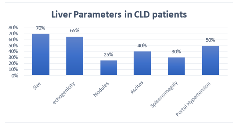
Figure 1: Liver Parameters in CLD patients Correlation with Laboratory Results
The study highlights the significant role of ultrasound in the assessment of chronic liver disease. Ultrasound is effective in detecting liver enlargement, echogenicity changes, and complications such as ascites and splenomegaly. The strong correlation between ultrasound findings and laboratory parameters emphasizes its utilityin monitoring diseaseprogression and guiding management. Ultrasound serves as a first-line imaging modality due to its non-invasive nature, availability, and ability to provide real-time assessment. However, it has limitations, including operator dependency and inability to provide histological information. Future studiescould explore the integration of ultrasound with elastography and other imaging modalities for a more comprehensive evaluation. This study underscores the criticalrole of ultrasound in the assessment of chronic liver disease (CLD) in a cohort of 100 patients. The findings demonstrate that ultrasound is not only effective in diagnosing liver abnormalities but also serves as a valuable tool for monitoring disease progression and guiding management decisions. The high prevalence of hepatomegaly (70%) and increased echogenicity (65%) in our cohort aligns with existing literature, whichindicates these are common ultrasonographic findings in patients with CLD. The association between echogenicity changes and serum liver enzymes (ALT and AST) suggests that ultrasound can reflect underlying liver pathology and hepatic inflammation.
The detection of liver nodulesin 25% of patients is noteworthy, as it raisesthe potential concern for hepatocellular carcinoma, particularly in individuals with chronic viral hepatitis and cirrhosis. This highlights the importance of regular surveillance using ultrasound for high-risk
populations, as recommended by various clinical guidelines (e.g., AASLD). The identification of ascites in 40% of patients and its correlation with elevated bilirubin levels emphasizes the role of ultrasound in evaluating complications associated with advancedliver disease, such as portal hypertension. Additionally, the finding of splenomegaly in 30% of patients furtherindicates portal hypertension, reinforcing the utilityof ultrasound in diagnosing and monitoring these complications. The significant correlations between ultrasound findingsand laboratory parameters support the use of ultrasound as a complementary tool in the clinical management of CLD. While ultrasound provides important morphological information, it should be integrated with laboratory assessments and clinical evaluations for a comprehensive understanding of the patient's condition. However, it is essential to acknowledge the limitations of ultrasound, including operator dependency and potential variability in interpretation. Future studies could explore the combination of ultrasound with elastography and other imagingmodalities, such as MRI or CT scans, to enhance diagnostic accuracy and provide a more comprehensive evaluation of liver disease.
Ultrasound is a valuable tool in the assessment of chronic liver disease, aiding in diagnosis, monitoring, and management. Its non-invasive nature and correlation with clinical findings make it indispensable in clinical practice. Further research is warranted to optimize its use and integrate new technologies in the management of CLD. In conclusion, ultrasound is a valuable, non-invasive imaging modality in the assessment of chronic liver disease. Its ability to detect liver abnormalities, evaluate complications, and correlate with clinical and laboratory findingsmakes it an indispensable tool in managingCLD. Regular use of ultrasound can aid in early detection of complications and guide therapeutic interventions, ultimately improving patient outcomes. As technology advances, integrating ultrasound with emerging techniques will likely enhance its utility in the clinical settings
Dear Editorial Team, Clinical Medical Reviews and Reports. My experience with the journal was highly positive. The peer-review process was rigorous, constructive, and completed in a timely manner. The reviewers provided valuable comments that helped improve the quality and clarity of our manuscript. The editorial office was professional, responsive, and supportive throughout all stages of the publication process. Communication was clear and efficient, and any questions were addressed promptly. Overall, I found the journal to maintain high scientific standards and an excellent publication workflow. I would be pleased to consider submitting future work to this journal. Best wishes from, Elena Popa.

It was my pleasure to submit my testimonial concerning the Reviewer Board of our Scientific Journal “Brain and Neurological Disorders”. The Reviewers focused on some modifications and their contribution was helpful. The ladies of our Editorial Office were also supported my efforts. It was my honor to have such a co-operation and I am looking forward for more collaboration.

Dear Grace Pierce, Editorial Coordinator of Journal of Clinical Research and Reports, Thank you for the speedy and efficient peer review process. I appreciate the fact that your peer reviewers do not take months to respond like with some other journals. I would also like to thank the editorial office for responding quickly to my questions. It is an excellent journal. I plan to submit more manuscripts in the future. Best wishes from, Robert W. McGee

Dear Grace Pierce, Editorial Coordinator of Journal of Clinical Research and Reports, Working with you and your team on our recent publication in JCRR has been a truly wonderful and enjoyable experience. The responses were prompt, and the reviewers were patient, constructive, and highly professional. One reviewer in particular gave me the feeling that a professor was carefully reading and commenting on my coursework, which was deeply touching. The entire process was straightforward and hassle‑free, with no tedious online forms to complete. I highly recommend this journal. Best wishes from, DR Aibing Rao, Head of R&D

I Appreciate the Opportunity to Share my Experience with the Journal of Clinical Research and Reports. The peer review process was timely and constructive, and the feedback provided helped improve the quality of our manuscript. The editorial office was professional, responsive, and supportive throughout the process, ensuring smooth communication and efficient handling of the submission. Overall, it was a positive experience collaborating with your team.

Dear Mercy Grace, Editorial Coordinator of Obstetrics Gynecology and Reproductive Sciences, We would like to express our gratitude for your help at all stages of publishing and editing the article. The editors of the magazine answer all the necessary questions and help at every stage. We will definitely continue to cooperate and publish other works in the Obstetrics Gynecology and Reproductive Sciences! Best wishes from, Alla Konstantinovna Politova,
