
Research Article | DOI: https://doi.org/10.31579/2690-4861/082
1* College of Science, Department of Biomedical Sciences, Cihan University-Erbil, Kurdistan Region, Iraq.
2 College of Dentistry, Babylon University, Iraq.
3 College of Science, Department of General Biology, Cihan University-Erbil, Kurdistan Region, Iraq.
*Corresponding Author: Homady, M. H, 1College of Science, Department of Biomedical Sciences, Cihan University-Erbil, Kurdistan Region, Iraq.
Citation: Homady M. H, ALquraishi, L, Ubeid, M. H, Juma, A. S. M. (2021) Ultra structural Studies of Mouse Liver in Castrated Subjects Treated with Grape Juice. International Journal of Clinical Case Reports and Reviews. 6(3); DOI: 10.31579/2690-4861/082
Copyright: © 2021 Homady M. H, This is an open-access article distributed under the terms of the Creative Commons Attribution License, which permits unrestricted use, distribution, and reproduction in any medium, provided the original author and source are credited.
Received: 07 December 2020 | Accepted: 25 January 2021 | Published: 27 January 2021
Keywords: liver; castration; grape juice; kupffer cells; scanning electron microscope
The present study was designed to evaluate the alterations in the liver tissues especially ITO and Kupffer cells of both castrated and castrated mice treated with10 µl/g of grape juice. The present study was conducted on forty five healthy males of Swiss albino mice, which were divided into 3 groups (N= 15 mice per each group). The first group was intact (control); the second one was castrated and the third group was castrated treated with 10 µl/g of grape juice. The ultra-structure sections of liver tissue from intact (control) group showed normal structure of both hepatocytes and sinusoids, with smooth external surfaces. Whereas ultra-sections from castrated group resulted in: degeneration of hepatocytes, deterioration of sinusoids; rough external surfaces, with aggregations of lipid droplets and white blood cells as compared with the control group. However treatment of castrated subject's with10µl/g of grape juice resulted in activation the regeneration processes in both hepatocytes and sinusoids, with smooth appearance of the external surfaces as compared with control group.
The present study concluded that surgical castration aggravated increased hepatic steatosis and increased inflammatory response by increased activation of Kupffer and Ito cells, these effects could be reserved by aggravated increased grape juice administration.
The mammalian liver is the largest internal digestive organ, which is indispensable in many essential physiologic processes and vulnerable to be impaired by a wide variety of factors, such as toxins, microorganisms, metabolic products, circulatory materials and metabolism of carbohydrates, lipids and proteins. [1, 2, 3].
Nonalcoholic fatty liver disease (NAFLD) is the most common chronic liver disease in the world. It is present in 30% of the general adult population. In reality, NAFLD comprise as a spectrum of hepatic abnormalities that are observable in liver histological slides, from a simple intrahepatic accumulation of fat (steatosis or nonalcoholic fatty liver, NAFL) to various degrees of necrotic inflammation (NASH nonalcoholic steatohepatitis, NASH) [ 4, 5, 6]. Kupffer cells, the hepatic resident macrophages, represent the largest group of macrophages in the body and account for about 20–25% of non-parenchymal cells in the liver (intrasinusoidal cells) [7, 8]. As the critical component of the innate immune system, Kupffer cells can be activated by various endogenous and exogenous stimuli, and play a key role in regulating the phenotype and function of neighboring parenchymal and non-parenchymal cells [9]. Hepatic stellate cells (HSCs) also called Ito cells, fat-storing cells, lipocytes, perisinusoidal cells and vitamin A-storing cells located in the space of Disse between hepatocytes and sinusoidal endothelial cells. These cells constitute approximately 10%of the total number of liver cells [10, 11, 12 and 13], and their cytoplasm is especially rich in lipid droplets and show long branched cytoplasmic processes that may reduce the lumen of sinusoid capillaries, in such a way modulating the liver sinusoidal blood flow. When the liver is damaged, the hepatic stellate cells change their shape and transform via a process named “activation” into the myofibroblast [14, 15 and 16]. All stages of NAFLD including cirrhosis and hepatocellular carcinomaassociated with hepatocyte steatosis [17]. Castration is a way of studying the consequences of extreme testosterone deficiency in animal models [18]. Castration promotes progression to steatohepatitis through activation of the ER (endoplasmic reticulum) stress pathway and enhancement of macrovesicular droplet.
Some medicinal plants may exert promising pharmacological properties and improve the effectiveness of conventional medications as complementary agents [19]. Vitis vinifera (Grape) are one of the most consumed fruits globally. It possesses a wide range of pharmacological activities due to its rich polyphenol ingredients most of which have been demonstrated to have therapeutic or health promoting properties [20, 21], among them, flavonoids are the most abundant and widely studied, recent studies have shown that the beneficial health effects promoted by consumption of grape and grape products are attributed to the unique mix of polyphenolic compounds. As the largest group of grape polyphenols, flavonoids are the main candidates considered to have biological properties, including but not limited to antioxidant, anti-inflammatory, anti-cancer antimicrobial, antiviral, cardioprotective, neuroprotective and hepatoprotective [22, 23, and 24]. Therefore, the aim of the present study was to determinethe effects of castration, and treatment of castrated subjects by using grape juice on both Ito and Kupffer cells by using Scanning Electron Microscope (SEM).
Materials and Methods
Swiss albino male mice weighting between (14-17) g., and aged (3weeks) were used in the present study, the mice were obtained from the Animal House, Faculty of Science/ University of Kufa. Animals were kept in ventilated cages, with a temperature of (25±2Cº) at 12:12 h light, dark cycle was used balanced, rodent food pellet and water were provided ad libitum [25, 26 and 27]. All experimental protocols using live animals were first reviewed, approved and accepted according to guidelines for the care and use of laboratory animals in biomedical research [28, 29 and 30]. A total number of 45 Swiss albino mice were used in the present study. Animals were divided into 5 groups (N=15), and the treatment was started at the age of 21 days for 6 weeks as:
Group I: Intact male mice received tap-water as control.
Group II: Castrated male mice received tap-water as (positive group).
Group III: Castrated male mice treated daily with (10µl/g) of grape juice,
The surgical castration method was done according to [31, 32]. Black grape (Vitis vinifera) obtained from local market (Baghdad, Iraq) 100g of grape was blundered by using a commercial blender without separating the seeds, and then it was filtered to remove the residue. The resulting extract (10 mls was stored in the refrigerator at 4˚C, and used after one hour. The extract was prepared according to [33]. A previous study documented that 10µl/g/day of grape juice extract was effective dose [34, 35, 36]. For this protocol, we used in the present study 10µl /g/day and was given daily as orally administered for six weeks.
Animals were sacrificed at the end of the experiments, with using ketamine and xylazine as anesthetic drugs to anesthetize the mice. The preparation procedures for Scanning Electron Microscopy has been described by [33]. Number of both Ito and Kupffer cells in cytoplasmic color in form of brown scale intensity was done according to [37].
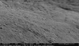
The present findings of electron micrograph of liver tissue from intact male mice (control) showed a normal structure ofboth hepatocytes and sinusoids with normal smooth external surface(Figures, 1, 2).
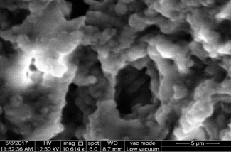
However the ultrastructure of liver tissue from castrated male mice
Showed degeneration processes of hepatocytes with deterioration of sinusoid (Figure 3) as well as aggregation of lipid droplets with rough external surfaces (Figure 4). Moreover, castration was also resulted in aggregations of white blood cells (Figure 5).
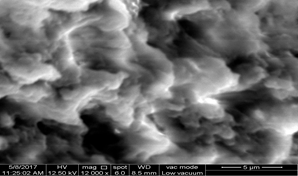
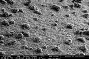
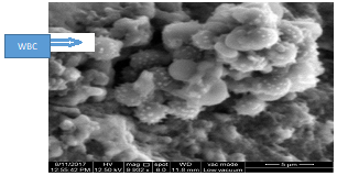

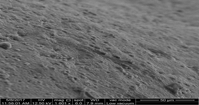
Scanning Electron Microscopical Results: The present results of the electron micrograph of liver tissue from intact (control) group showed a normal structure of hepatic lobules with smooth external surface. Whereas the results of castratedsubjects revealed, degeneration processes of hepatocytes and deterioration of sinusoid, with rough external surfaces, as well as aggregations of lipid droplets with concomitant increase in activation of immunity through aggregation of white blood cells. The interpretation of these findings may be attributed to hepatic changes as a results of castration that caused damaged or/may reductions in sinusoidal perfusion arise initially from the effects of hypertrophied hepatic parenchymal cells, swollen with accumulation of lipids withinand on their external surfaces. The extension in parenchymal cell plates caused narrowing and distortion the lumen of sinusoids as well as the intrasinusoidal lumen. These results are in agreement with the findings of [38, 39, 40], who stated that, hepatic steatosis altering the architecture of the sinusoidal network. Studied by [41, 42], demonstrated that the simple steatosis fatty livers may progress to steatohepatitis, and then to fibrosing steatohepatitis with the initiation of capillarization of the sinusoids. The present results are in concomitant with the findings of [43, 44], who concluded that the adhesion of leukocytes to the sinusoidal endothelium is followed by leukocyte infiltration into the hepatic parenchyma to form inflammatory foci. Many studied demonstrated that the deposition of collagen in the space of Diss accentuates the narrowing and distortion of the sinusoidal lumen, further restricting microvascular blood flow [45, 46, and 47]. Electron micrograph of liver tissue from castrated group treated with grape juice showed regeneration processes in both hepatocytes and sinusoids with smooth appearance of external surfaces. Such effects in the present study may be attributed to the protective effects of polyphenolic compounds which have multiple biological effects, including antioxidant, inhibiting lipid peroxidation activity, reduce the lipid load in the liver and reduce the swelling of hepatocytes that resulted in the dilation of spaces between them.
Conclusions: The present study demonstrated that impaired metabolic process related with surgical castration enhanced immune and inflammatory response, represented by activation of Kupffer and Ito cells which lead to hepatic apoptosis and contribute to increase nonalcoholic fatty liver disease. On the other hand the intra gastric application of Grape juice has dramatic effects to restore liver structure in castrated animals.
Dear Editorial Team, Clinical Medical Reviews and Reports. My experience with the journal was highly positive. The peer-review process was rigorous, constructive, and completed in a timely manner. The reviewers provided valuable comments that helped improve the quality and clarity of our manuscript. The editorial office was professional, responsive, and supportive throughout all stages of the publication process. Communication was clear and efficient, and any questions were addressed promptly. Overall, I found the journal to maintain high scientific standards and an excellent publication workflow. I would be pleased to consider submitting future work to this journal. Best wishes from, Elena Popa.

It was my pleasure to submit my testimonial concerning the Reviewer Board of our Scientific Journal “Brain and Neurological Disorders”. The Reviewers focused on some modifications and their contribution was helpful. The ladies of our Editorial Office were also supported my efforts. It was my honor to have such a co-operation and I am looking forward for more collaboration.

Dear Grace Pierce, Editorial Coordinator of Journal of Clinical Research and Reports, Thank you for the speedy and efficient peer review process. I appreciate the fact that your peer reviewers do not take months to respond like with some other journals. I would also like to thank the editorial office for responding quickly to my questions. It is an excellent journal. I plan to submit more manuscripts in the future. Best wishes from, Robert W. McGee

Dear Grace Pierce, Editorial Coordinator of Journal of Clinical Research and Reports, Working with you and your team on our recent publication in JCRR has been a truly wonderful and enjoyable experience. The responses were prompt, and the reviewers were patient, constructive, and highly professional. One reviewer in particular gave me the feeling that a professor was carefully reading and commenting on my coursework, which was deeply touching. The entire process was straightforward and hassle‑free, with no tedious online forms to complete. I highly recommend this journal. Best wishes from, DR Aibing Rao, Head of R&D

I Appreciate the Opportunity to Share my Experience with the Journal of Clinical Research and Reports. The peer review process was timely and constructive, and the feedback provided helped improve the quality of our manuscript. The editorial office was professional, responsive, and supportive throughout the process, ensuring smooth communication and efficient handling of the submission. Overall, it was a positive experience collaborating with your team.

Dear Mercy Grace, Editorial Coordinator of Obstetrics Gynecology and Reproductive Sciences, We would like to express our gratitude for your help at all stages of publishing and editing the article. The editors of the magazine answer all the necessary questions and help at every stage. We will definitely continue to cooperate and publish other works in the Obstetrics Gynecology and Reproductive Sciences! Best wishes from, Alla Konstantinovna Politova,
