
Review Article | DOI: https://doi.org/10.31579/2692-9406/155
International Higher School of Medicine, Bishkek and National Center of Cardiology and Internal Medicine, Bishkek, Kyrgyzstan.
*Corresponding Author: Abyt Ibraimov, International Higher School of Medicine, Bishkek and National Center of Cardiology and Internal Medicine, Bishkek, Kyrgyzstan.
Citation: Abyt Ibraimov (2023), Twenty Years of the Cell Thermoregulation Hypothesis, J. Biomedical Research and Clinical Reviews. 8(3); DOI:10.31579/2692-9406/155
Copyright: © 2023, Abyt Ibraimov. this is an open-access article distributed under the terms of the Creative Commons Attribution License, which permits unrestricted use, distribution, and reproduction in any medium, provided the original author and source are credited.
Received: 08 June 2023 | Accepted: 15 June 2023 | Published: 22 June 2023
Keywords: cell thermoregulation; condensed chromatin; chromosomal heterochromatin regions; human body heat conductivity; obesity; alcoholism; drug addiction
Thermoregulation at organism level is the well-established fact. The question on possibility of thermoregulation at the cell level remains opened. Based on study of distribution of chromosomal heterochromatin regions (HRs) in the genome of various human populations, in norm and at some forms of pathology the hypothesis about thermoregulation existence at the cell level has been presented. The essence of hypothesis of cell thermoregulation (СТ) is elimination of the temperature difference between the nucleus and cytoplasm when the nucleus temperature becomes higher than in the cytoplasm. The higher eukaryotes use a dense layer of peripheral condensed chromatin (CC) as heat conductor for a more efficient elimination of the temperature difference between the nucleus and cytoplasm. The СС localized between a nucleus and cytoplasm is made of chromosomal HRs. Since individuals in the population differ in the quantitative content of chromosomal HRs in the genome, then this circumstance is reflected in the rate of elimination of the temperature difference between the nucleus and cytoplasm. The phenotypic manifestation of CT is the level of body heat conductivity of the individuals in the population with all the ensuing consequences for the organism.
Thermoregulation at organism level is the well-established fact. The question on possibility of thermoregulation at the cell level remains opened. Based on study of distribution of chromosomal heterochromatin regions (HRs) in the genome of various human populations living in different climatic and geographical conditions of Africa and Eurasia, in norm and at some forms of human pathology the hypothesis about thermoregulation existence at the cell level has been presented [1]. The essence of hypothesis of cell thermoregulation (СТ) is elimination of the temperature difference between the nucleus and cytoplasm when the nucleus temperature becomes higher than in the cytoplasm. The nucleus, in contrast to the cytoplasm, cannot conduct heat directly in the extracellular space, from where the heat is taken by the circulating flow of blood and lymph. Thus, the nucleus can transfer surplus heat only in the cytoplasm. With this, the nucleus has two options: either by increasing its volume or increasing the heat conductivity of the nuclear envelope. As the first option is limited, and the second one is hampered because of thickness of the cell membranes, apparently the higher eukaryotes took advantage of the opportunity of a dense layer of peripheral condensed chromatin (CC) as heat conductor for a more efficient elimination of the temperature difference between the nucleus and cytoplasm. The CC localized between a nucleus and cytoplasm is made of chromosomal HRs, which are one of the forms of higher organization of non-coding, so-called "excess" DNA in the genome of higher eukaryotes. Despite the fact that the CT hypothesis is already 20 years old, it has not attracted the attention of researchers. The reasons that support the existence of CT on the example of a man are discussed below.
Why is the possibility of thermoregulation at the level of individual cells not being paid due attention?
The lack of interest in the possibility of the existence of cell thermoregulation has quite objective reasons. Recognized internal sources of heat (cellular metabolism, muscle contraction, ion pump, in rare cases brown fat burning) in the body are localized in the cytoplasm. Organisms receive energy from the environment in the form of potential energy contained in the chemical bonds of molecules of fats, carbohydrates and proteins. Energy metabolism of cells (formation, transfer and transformation) occurs mainly in cytoplasm. Apparently, it is taken for granted that if the temperature in the cytoplasm rises above the optimal level for the body, then it will be freely excreted into the intercellular space. The cell nucleus is usually not considered as one of the important internal sources of heat, despite the fact that very active biochemical processes take place there (reparation, recombination, rearrangement, modification, restriction, replication, transcription and other processes associated with DNA).
However, the question of whether thermoregulation can also exist at the level of individual cells remains open. Apparently, it is taken for granted that the level of intracellular temperature cannot rise to dangerous physiological values (e.g., >37 ⁰C in homoeothermic organisms) due to the small size of cells individually. If this happens, then the presence of interstitial fluid in the intercellular space and the circulation system (blood and lymph circulation) will not allow the temperature in the cells to rise above the optimal temperature for the body of this organism. Therefore, the level of thermal energy released in the process of cellular metabolism cannot be so important that in the process of evolution appeared some special mechanisms for thermoregulation at the level of individual cells. Obviously, for the same reasons, it is considered that the nucleus and cytoplasm use the same mechanisms in removing excess thermal energy. It is considered that diffusion and possibly convection are the primary means to passively remove the heat generated inside the cell [2]. However, there are facts and observations, seemingly unrelated, but indirectly indicating the possibility of thermoregulation at the cellular level. Below we will discuss some of them.
Why should due attention be paid to the possibility of thermoregulation at the level of individual cells?
Based on the study of distribution of chromosomal Q-heterochromatin regions (Q-HRs) in the genome of various human populations living in different climatic and geographical conditions of Africa and Eurasia, in norm and at some forms of human pathology, the hypothesis was put forward about the possibility of thermoregulation at the level of individual cells [1].
Initially, the CT hypothesis was proposed as an attempt to explain the following facts obtained on human populations [1]:
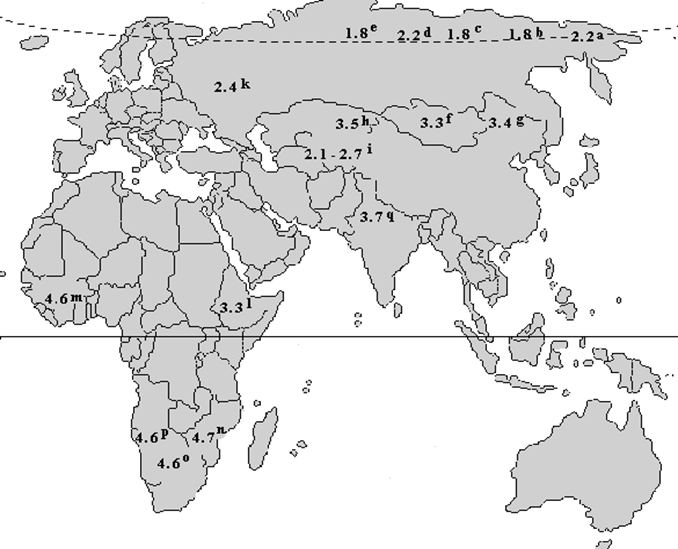
Figure 1:The values of the mean number of chromosomal Q-HRs, calculated per individual, in populations permanently living in different climatic and geographical zones of the Old World (a-Chukchi, b-Nenets, c-Selkups, d-Khanty, e-Yakuts, f-Mongols, g-Chinese, h-Kazakhs, i-Kyrgyz (highlanders of Pamir and Tien Shan), k-Russians, l-Ethiopians (Ethiopian Highlands), m-Negroes from Guinea-Bissau, n-Negroes from Mozambique, o-Negroes from Zimbabwe, p-Negroes from Angola, q-Indians from northern India [1].
2) these differences proved to be related to features of the ecological environment of the place of permanent residence, and not to racial and ethnic composition of the population;
3) the amount of Q-HRs in the genome tend to decrease from southern geographical latitudes to northern ones, and from low-altitude to high-altitude ones;
4) different age groups have different amount of Q-HRs, the greatest number of Q-HRs is characteristic of neonates, while the lowest – of elderly subjects;
5) individuals capable of successfully adapting themselves to the extreme high-altitude climate (e.g. mountaineers) and of the Far North (e.g. oil industry workers of polar Eastern Siberia) are characterized by extremely low amounts of Q-HRs in their genome;
6) individuals with a lower amount of Q-HR in their genome proved to be prone to alcoholism and obesity, while those with a greater amount of Q-HR – to drug addiction [3-21].
However, over time it became conceivable that some other facts and observations that still have no explanation could be partially explained within the framework of the CT hypothesis, in particular:
a) the existence of "excess" DNA in the genome of higher eukaryotes in general and why they consist mainly of non-coding, repeating sequences of nucleotides;
b) the organization of a part of the "excess" DNA into the so-called chromosomal HRs, which make up from 10% to 60% of the DNA of the genome in higher eukaryotes (in humans, HRs make up 15%-20%);
c) formation of chromosomal HRs in interphase cells in the form of a dense layer of peripheral condensed chromatin (CC) around the nucleus. The location of heterochromatin on the periphery of the nucleus is usually associated with its genetic inertness, which suggests that active biochemical processes occur closer to the center of the nucleus;
d) the role that chromosomal HRs play is still essentially unknown. This is also reflected in the variety of hypotheses. These range from the idea that heterochromatin has no function, consisting of ‘selfish’ DNA, to the assumption that it has an important role in development and evolution. However, none of them backed up by solid evidence when it comes to demonstrating their phenotypic manifestation;
e) the origin of homoeothermic animals;
f) the emergence of nucleolus and chromocenters in interphase nucleus;
g) the role chromosomal bands (C, G, Q, and T) in development and evolution. It has been shown that G-, Q- and R-bands are absent in plants and are always present in the chromosomes of higher vertebrates (reptiles, birds and mammals). In the case of invertebrates, fish and amphibians, it is difficult to identify these bands. Some chromosomal bands in some insects are equivalent to C-segments, and G-, Q- and R-bands appear to be absent. With the help of differential staining of chromosomes in plants, it is possible to identify mainly C-bands. However, chromosomal bands are best detected in the karyotype of higher primates, especially in humans;
h) the meaning of the presence only in the karyotype of three higher primates (Homo sapiens, Pan troglodytes and Gorilla gorilla) a new type of constitutive heterochromatin - Q-heterochromatin;
i) the existence of a broad quantitative variability of chromosomal Q-HRs only in the genome of human populations despite the fact that they present in the genome of two other higher primates (chimpanzee and gorilla);
j) the origin in the karyotype of modern man of 46 instead of 48 chromosomes, characteristic of higher primates;
k) high physiological plasticity of a man, which allowed him to adapt to climatic conditions other than Africa and to master the whole land of the Earth.
l) a man is able to run continuously for such long distances that no homoeothermic mammalian can do;
Of course, this is an incomplete list of questions that could be explained within the framework of CT hypothesis. This list may still expand if it turns out that CT really exists in Nature.
What is the cell thermoregulation?
To better present our understanding of cell thermoregulation (CT), it is necessary to briefly recall some fundamental facts about chromosomal HRs in the genome of higher eukaryotes. To-date two types of constitutive heterochromatin are recognized: C– and Q-heterochromatin [22,23,24]. There are several significant differences between them: C-heterochromatin is found in the chromosomes of all the higher eukaryotes, while Q-heterochromatin - only in man, the chimpanzee and gorilla [25-27]. C-heterochromatin regions (C-HRs) are known to be invariably present in all the chromosomes of man, varying mainly in size and location (inversion) (Figure. 2).
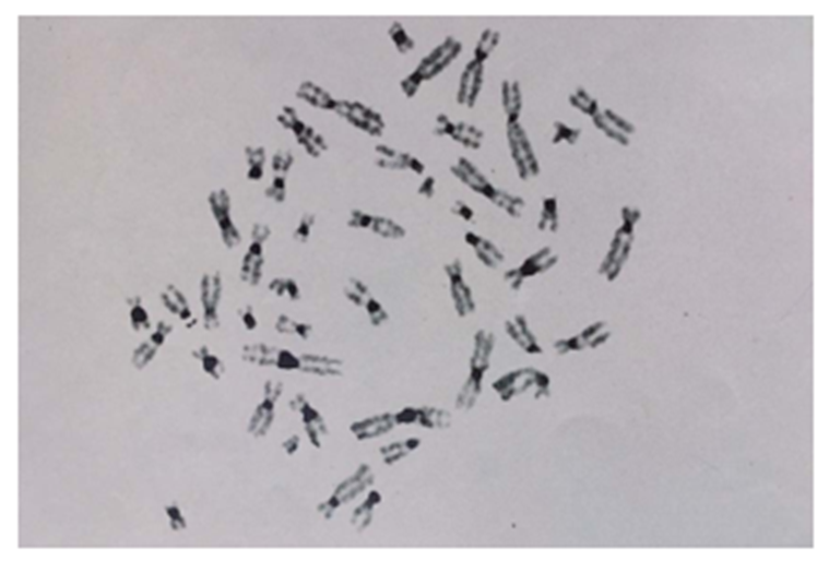
Figure 2: Human chromosomal C-heterochromatic regions (C-HRs) after C- staining. C-HRs of chromosomes (dark-colored areas) are localized on all chromosomes in the human karyotype without exception [28].
Q-heterochromatin regions (Q-HRs) may be completely absent on any of the Q-polymorphic chromosomes without any noticeable pathological or other phenotypic consequences for the carrier, while the complete absence of C-HRs even on one chromosome is an extremely rare phenomenon [24].
Only seven pairs of autosomes (3, 4, 13-15, 21 and 22) and the Y chromosome can have Q-HRs in the human karyotype. The main morphological expression of the phenomenon of wide variability of Q-HRs
of human chromosomes is that individuals in populations differ in the number, localization, size and intensity of fluorescence. It should be emphasized that there is no individual in human populations who would have Q-HRs simultaneously on all 25 potentially polymorphic loci of 46 chromosomes in his karyotype. Usually, the number of Q-variants in the human karyotype in populations ranges from 0 to 10 (Figure. 3) [14].

Figure 3: Q-heterochromatin regions localized on the Q-polymorphic loci of three autosomes (3cen, 3cen, 13p11, 13p11, 13p13, and 21p13) and on the q12 locus of chromosome Y [29].
Here it is necessary to emphasize once again that: a) the broad quantitative variability of chromosomal Q-HRs is present only in the genome of human populations, despite the fact that they occur in the genome of two more higher primates; b) human populations do not differ from each other in the quantitative content of chromosomal C-HRs [10,30].
Now about what we mean by the cell thermoregulation (CT). CT refers to the process of equalizing the temperature difference between the cytoplasm and the nucleus. The inner surface of the nuclear membrane is occupied by heterochromatin in the form of a dense layer of condensed chromatin (CC) and lamina (Figure. 4).
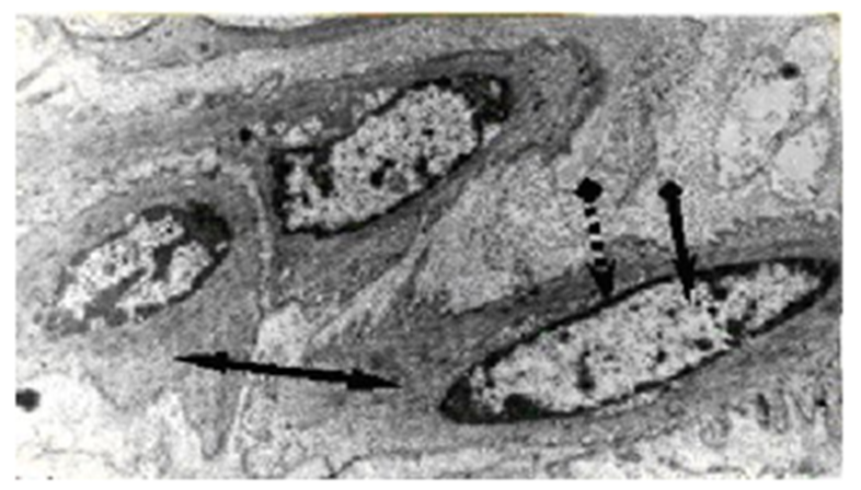
Figure 4: Electron micrograph of smooth muscle cells of a bull (x 8 000). Condensed (heterochromatin) and decondensed chromatin (euchromatin); ∙∙∙∙> peripheric layer of condensed chromatin; → euchromatin; and ↔ cytoplasm can be seen in the nuclei [28].
We believe that the structural basis of CT is the peripheral layer of CC. The transfer of thermal energy between the cytoplasm and the nucleus is carried out through this dense layer of peripheral CC, located inside the nuclear envelope [1].
Temperature homeostasis in the nucleus is necessary, first of all, for the optimal flow of complex molecular processes associated with the metabolism of DNA and RNA, which are important for the synthesis of normal proteins and enzymes. Therefore, the hypothesis postulates that the cell strives to maintain a constant temperature. As is known, chromosomes have internal (reparation, recombination, rearrangement, modification, restriction) and external (replication, transcription, packaging, organized movement) metabolic activities, which are accompanied, among other things, by the release of thermal energy. If, for some reason, the temperature in the nucleus begins to exceed that in the cytoplasm, there is a need for dissipation of excess heat outside the nucleus.
We believe that unlike the cytoplasm the cell nucleus is in the most difficult position in maintaining intracellular temperature homeostasis. Indeed, in order to get rid of excess thermal energy that is produced in the metabolic process, the nucleus has two choices: increase in size and/or increase the density of the nuclear envelope to speed up the transfer of heat to the cytoplasm. The first way to dissipate excess thermal energy из ядра is difficult due to the fact that the cytoskeleton and intracellular organelles do not allow this. The second variant is possible if the nucleus can increase the density of the CC layer inside the nuclear membrane in order to efficiently remove excess heat.
It is obvious that the thickness and density of the peripheral CC layer around the cell nucleus depends on the total number of chromosomal C-HRs in the genome. However, in the genome of three higher primates, unlike other animals, in addition to C-HRs, there are also Q-HRs, which, as we believe, further increase the packing density (compactization) of CC. In other words, CC in three higher primates (which contains chromosomal C- and Q-HRs), being the most densely packed material, should have the highest heat conductivity in the interphase cell with all the ensuing consequences [1,29,31-41].
But this does not mean that CT is exclusively a product of chromosomal HRs. In the chromosomes of higher eukaryotes in addition to HRs there are other tightly packed regular areas, like G+ and Q+ bands, which make up the bulk of the bodies of metaphase chromosomes. In the composition of the chromosomal G+ and Q+ bands there are heterochromatin materials (intercalary heterochromatin). As it is known the telomeric regions of chromosomes also contains heterochromatin.
We also believe that in the removal of heat surplus from a nucleus a nucleolus is actively involved. Thus, some of the chromosomes have nucleolar organizer regions (NORs), which contain ribosomal cystrons. In humans, NORs are localized on acrocentric chromosomes (13-15, 21 and 22). As it is known, NORs, together with chromosomal HRs form the nucleolus bodies. Since chromosomal HRs are in the body and around the nucleolus, there is nothing surprising in the assumption that they due to its high density can promote removal of excess heat from the nucleolus and further to CC around the nucleus [42,43].
It is old-established fact that in interphase nucleus chromosomal HRs coalescent into a structure called chromocenters. However, their biological role in the life of cells is still unclear. Existing hypotheses link the possible biological role of chromocenters with the behavior of chromosomal HRs in the cell cycle. We believe that the nuclear areas where excess heat is generated form temporary structures in the form of chromocenters and nucleoli, connecting them with a peripheral layer of СС around the nucleus. The essence of the proposed hypothesis is the assumption that the chromocenters, along with the nucleolus and the dense layer of peripheral СС participate in СТ. Namely, chromocenters are involved in the removal of excess heat from the "hot" areas of the interphase nucleus through СС in the cytoplasm [42-44].
We also believe that the genetic inertness of chromosomal HRs may not be the only reason for their localization and compaction on the nuclear periphery. It seems highly probable to us that the dense packing of chromosomal HRs on the nuclear periphery is caused by two reasons: a) the need for cell thermoregulation to effectively remove (dissipate) excess heat from the nucleus; and b) the risk of damage to the thin structure of the nuclear membrane from exposure to high temperature, emanating from the biochemically highly active interphase nucleus. The first reason, apparently, does not need any additional arguments, since the CC layer located on the nuclear periphery is the densest and, accordingly, the most highly heat-conducting structure in the interphase cell, with all the ensuing consequences for the CT. The second reason is related to the features of the cell membrane. As it is known, cell membranes are very sensitive to temperature fluctuations: they become too solid at low temperatures, and they become too liquid to perform their functions normally at high temperatures. Of course, to remove excess heat from the nucleus, a layer of cell membrane would be ideal due to their thickness. However, the high vulnerability of cell membranes to temperature fluctuations apparently “forced” Nature to use a dense CC layer of HRs, lamina, nucleolus, chromocenters and cytoskeleton to protect their functional integrity.
The lamina layer is located between the CC and the nuclear envelope. For what reason? It is generally accepted that the lamina exists just below the inner nuclear membrane to give its strength and shape to the nucleus. But, in principle, the lamina could be located outside the nuclear membrane, like a cell wall on the plasma membrane in plant cells or in prokaryotes, if the task of the lamina is limited only to strengthening and shaping the nucleus. It is possible that there is a deep biological meaning in the localization of the lamina directly under the inner nuclear membrane - to protect the nuclear envelope from the harmful effects of high temperature emanating from the biochemically highly active nucleus by effectively removing and dissipating excess thermal energy.
Despite the fact that the nucleoli and chromocenters were discovered more than a hundred years ago, their biological meaning is still not completely clear. We believe that in order to protect the nuclear membrane from the deleterious effects of high temperature emanating from the nucleus, nucleoli and chromocenters are formed, and in some cases Nature uses the services of B-chromosomes. The essence of our hypothesis lies in the assumption that the nucleoli, chromocenters and B-chromosomes, consisting of constitutive heterochromatin, are involved: a) in the localization of “hot” areas in the interphase nucleus; b) in the direction of the “flow” of thermal energy to very limited areas of the nuclear membrane, in order to protect the functional integrity of the entire nuclear membrane. In other words, if heat is removed evenly around the entire circumference of the nucleus, then there may be a real danger of damage to the entire nuclear envelope [28,35,42-44].
The observations that human chromosomes 1, 9 and 16, which do not contain ribosomal cistrons but have large blocks of chromosomal HRs and are in contact with the nucleolus [46], still have no rational explanation. The predominant spatial proximity of the sex chromosomes to the nucleoli in interphase nuclei is in the same situation [47]. As it seems to us, a rational explanation of these facts is the assumption of their participation in the "localization" of excess heat in the nucleolus, and then contacting with CC to dissipate it into the cytoplasm [42,44].
Filaments of the cytoskeleton can extend from one end of the cell to the other, covering tens or even hundreds of micrometers. It is known that plectin and other plakins can interact with protein complexes that connect the cytoskeleton to the interior of the nucleus. These complexes are composed of the SUN proteins of the inner nuclear membrane and the KASH proteins (also called nesprins) of the outer nuclear membrane. The SUN and KASH proteins bind to each other in the lumen of the nuclear bilayer, forming a bridge connecting the nuclear and cytoplasmic cytoskeletons. The SUN proteins within the nucleus bind to the nuclear lamina or chromosomes, while the KASH proteins in the cytoplasm can bind directly to actin filaments and indirectly to microtubules and intermediate filaments through association with motor proteins and plakins, respectively. This link serves to mechanically connect the nucleus to the cytoskeleton and is involved in many cellular functions, including the movement of chromosomes within the nucleus during meiosis, the positioning of nuclei and centrosomes, nuclear migration, and the global organization of the cytoskeleton [Alberts 2008]. In this regard, we believe that excess thermal energy is removed from the nucleus into the interstitial fluid of the intercellular space, mainly with the help of the cytoskeleton, and not due to diffusion and convection, as is commonly believed [2]. And, finally, there is no known mechanism for a cell to actively dissipate excessive thermal energy. It is considered that diffusion and possibly convection are the primary means to passively remove the heat generated inside the cell. This explanation is strongly objected to. Matter of fact, ‘Inside the cell the molecules are mostly associated with polymeric structures (cytoskeletal polymers or membranes) and thus exist in very heterogeneous, solid-state environments that alter their behavior dramatically compared to free molecules in test tubes’ [45]. As such, highly localized heat sources are expected to create a subcellular temperature gradient. In other words, the interacting molecules in the cell do not float freely, as in a test tube with a water solution. Therefore, diffusion and convection cannot be the primary means to remove the heat generated inside the cell. Consequently, it is necessary to look for other additional mechanisms for removing surplus heat from the cell, and especially from its largest organelle - the nucleus.
Experimental verification of the hypothesis of cell thermoregulation.
The CT hypothesis has not yet been directly experimentally confirmed. Of course, it would be ideal if someone were able to show the following in vivo: the rate of heat transfer from the nucleus to the cytoplasm depends on the number of chromosomal HRs in the genome and the packing density (compactization) of CC in the interphase cell. No one has done such an experiment yet. However, the ultimate goal of our study is to experimentally test the hypothesis of CT, which, as we believe, is the main biological role (effect) of chromosomal HRs [1].
In essence, the idea of CT refers to the process of equalizing the temperature difference between the cytoplasm and the nucleus and then the entire cellular part of the body. Ultimately, we are talking about the body heat conductivity (BHC). The simplest idea of the BHC can be represented on the basis of the known laws of physics. It follows from the second law of thermodynamics that heat transfers from a hot body to a cold one until the temperature difference disappears. It is also obvious from general theoretical considerations that the same substance, depending on its density, should have different heat conductivity. The higher the density, the higher the heat conductivity, that is, the higher the rate of transfer of heat. CC is the densest area in the interphase cell, therefore, all other things being equal, it should have the highest heat conductivity.
Heat conductivity (HC) due to the transfer of energy is one of the three transfer phenomena existing in nature (heat conductivity, diffusion and internal friction or viscosity). All substances have HC: gases, liquids and solids. Unlike gases and liquids, convection is not possible in solids, therefore, the thermal transfer is carried out only by heat conductivity.
In thermal physics, the measurement of HC of solids (for example: metal) is carried out by determining the coefficient of thermal conductivity by the calorimetric method. The thermal transfer takes place through a metal rod, the ends of which are placed in a calorimeter with water taken at temperatures Т1 and T2 (T1 > T2). It is necessary to experimentally determine the amount of heat transferred and the time to measure the coefficient of HC of a given metal rod.
It is obvious that the direct transfer of the HC measurement method used in thermal physics is not acceptable for the human body, both for technical and ethical reasons. However, we tried to approach the solution of this problem indirectly, by assessing the HC of only a part of the human body. To do this, we had to modify the generally accepted technique of physicists so that it was acceptable to humans.
It is obvious that the living body has some initial heat conductivity. Nevertheless, as an important physical characteristic of a living organism, it has not yet attracted the attention of scientists. We failed to find not only a special method, but even no attempts to evaluate the body heat conductivity (BHC) of living organisms in vivo in the literature. This is not surprising, because it turned out that even experts in the physiology of thermoregulation never raised this issue [48,49]. Apparently, they do not a priori allow for the possibility of the existence of hereditary variability at the BHC in animals, including humans. However, all this can be understood, because the animal body is not a homogeneous physical mass. Therefore, we were forced to develop a methodological approach for assessing the BHC for humans.
In 2007, the idea was developed that CT, if it really exists in Nature, should also manifest itself at the level of the whole body in the form of different BHC of individuals in a population [32]. Since direct in vivo measurement of the temperature of different parts of cells is still not possible, then it was decided to somehow indirectly estimate the heat conductivity of the whole body.
Perhaps the most difficult methodological problem in determining the human BHC of a man was the creation of a temperature gradient between the human body and the source of thermal load individually for each person. It is known that people differ from each other in the temperature of the palm, and we decided to take advantage of this circumstance. Our empirical experience has shown that in order to create a temperature gradient between the human body and the source of thermal load, an acceptable value by which it is possible to painlessly increase the temperature of “hot” water in a water bath (relative to the temperature of the palm) is ~9 °C. Of course, this number does not hide any fundamental physical characteristic of the human body. But, it is also not desirable to increase the temperature of “hot” water by more than 9 °C. In this case, we could encounter, in addition to unpleasant sensations, the denaturation of proteins in cells in individuals whose temperature of the palm is close to core temperature.
It has been found that the temperature of the palm itself, and especially when taken into account in close connection with the temperature of the forehead surface can give very informative results about the human BHC [41]. It is by studying the distribution of the temperature difference between palm and forehead surface we managed to identify the existence of significant differences in gender and racial and ethnic origin at the population level [28,41].
The main difficulty in searching for a possible relationship between the number of chromosomal HRs in the genome and the human BHC was the complete absence of any methodological approach in this regard. If the determination of the number of chromosomal HRs in the human karyotype is already a well-established methodological procedure, then this cannot be said when it comes to measuring the human BHC due to the complete lack of any experience in this matter. Therefore, it is too early to say that we can measure the human BHC with the same accuracy as physicists do when measuring the thermal conductivity of nonliving bodies, such as metals. At best, we can only evaluate the human BHC as high, medium, or low, and apparently, it is still far from an accurate measurement in numbers [28,32,41].
Over the years, we have tested many methods of evaluating of the human BHC. By trial and error, we managed to significantly improve and simplify the technique of measurement of human BHC with the involvement of some technical tools and approaches. In particular, we began to use a water bath with a thermostatic device (water bath WTB6 from MEMMERT GmbH + Co., Germany) and pyrometers for non-contact thermometry of the surface of the human body.
The measurement of the human BHC is carried out indoors at room temperature (20 °С-24 °С). At first, the temperatures of the surface of the forehead and the palm of the hands were measured in the studied individuals. To prepare “hot” water for a given individual the number 9 added to the temperatures of the surface of the left palm. For example, if the temperature of the palm of the individual's left hand is 31.0 °C, then the temperature of the “hot” water should be 40.0 °C. The studied individual sat on a chair with a back, the torso was straightened, the head was raised, the arms hung naturally at the sides, and the muscles were relaxed. Then the individual slowly immersed the left hand up to the wrist in a water bath filled with “hot” water. Every minute, the temperature of the surface of the right palm of the studied individual was measured with a pyrometer to the nearest tenth of degrees Celsius (t °С). The experiment continues (thermal load of the human body through the left hand) until the temperature increase in the surface of the right palm reaches its maximum value (“temperature peak”) and begins to decrease (for details see [28,41]).
How do we interpret these data? We believe that the time of the onset of the temperature peak on the surface of the right palm reflects the rate of heat transfer in the human. In other words, we believe that a human with high BHC is able to: a) conduct heat through his body faster than other individuals in the population; b) equalize the temperature difference in different parts of the body faster; and c) remove (dissipate) excess thermal energy outside of his body faster in order to maintain the required temperature level in the body. If the temperature peak on the surface of the right palm occurs within the first 5 minutes from the start of the thermal load, then such an individual is considered as a human with a high BHC, from 6-10 minutes, as an average one, from 11 minutes and above, as a low BHC (for details see [41]).
In the process of testing the method, it also turned out that there is a statistically significant relationship between the level of human BHC and the magnitude of the temperature difference between distant parts of his body: e.g., forehead and palm or armpit and palm surface temperature. We believe that the smaller temperature difference between different parts of the human body reflects the high heat-conducting capacity of this organism in the sense that: a) such an organism equalizes the temperature differences between different parts of the body more efficiently; b) removes (dissipates) excess heat energy outside of the body faster. As it turned out, the temperature of the surface of the palm at rest also reflects the BHC level; individuals with a high palm temperature have a high BHC, and vice versa [41].
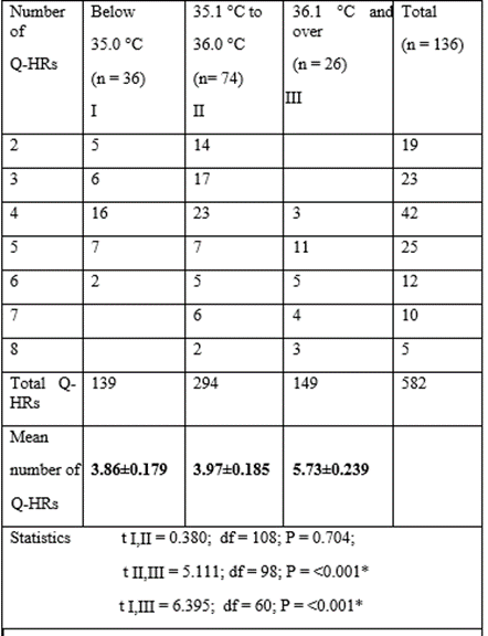
Table 1 shows the relationship between the number of chromosomal Q-HRs in the human genome and the rate of reaction of the body to the controlled thermal load, which was determined by the time (in minutes) to the occurrence of peak temperature on the surface of the right palm.
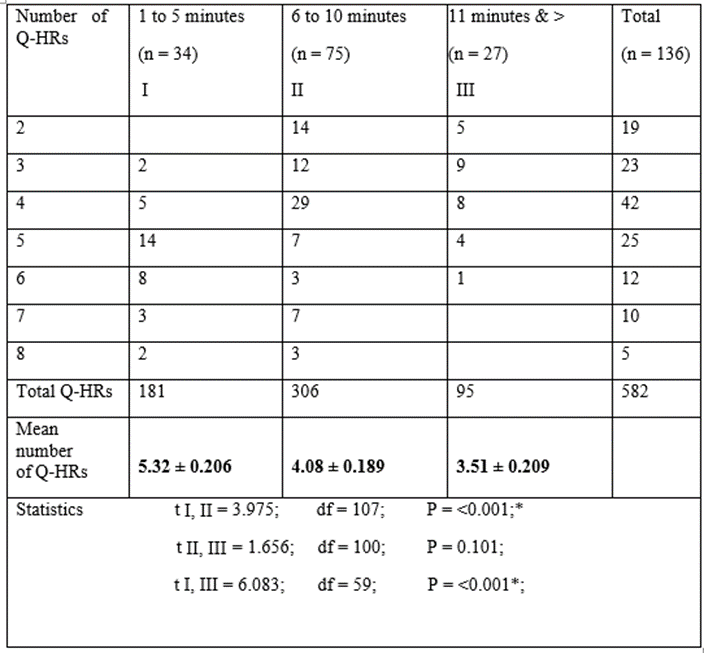
Table 1: The distribution and mean numbers of chromosomal Q-HRs and time to occurrence of temperature peak on the surface of the right palm.
As can be seen in this Table, there is a statistically significant relationship between the number of chromosomal Q-HRs in the human genome and the reaction of the body to the controlled thermal load. In individuals whose genome contains more chromosomal Q-HRs than the population average, the peak temperature occurs in the first five minutes of the thermal load, and vice versa. The relationship between the quantity of chromosomal Q-HRs and the temperature difference between the surface of the right palm and the forehead at rest is shown in Table 2.

*These differences are statistically significant.
Table 2: The distribution and mean numbers of chromosomal Q-HRs and the temperature difference between the surface of the right palm and the forehead.
As can be seen in Table 2 the more chromosomal Q-HRs in the human genome, the smaller the temperature (T) difference between the forehead surface and the right palm, and vice versa. Table 3 shows a different pattern: the greater the number of chromosomal Q-HRs in the human genome, the higher the temperature of the surface of the right palm at rest, and vice versa.
*These differences are statistically significant.
Table 3: The distribution and mean number of chromosomal Q-HRs and the temperature of the surfaceof the right palm.
How do we interpret these data? We believe that the time to occurrence of peak temperature (T) on the right palm reflects the rate of heat conductivity, while the value of T of the right palm’s surface at that moment seems to reflect the quantity of thermal energy in the individual’s body. If the peak T on the surface of the palm occurs in the first five minutes after the thermal load is applied, then such an individual is considered to have a high BHC, and vice versa. In other words, we believe that a human with a high BHC conducts heat through the body quicker and eliminates any excessive through the body shell quicker to better maintain inner body temperature at a constant level.
A statistically significant relationship between the number of chromosomal Q-HRs in the genome and the T difference between the forehead surface and the right palm at rest may also characterize the heat conducting ability of the human body — the smaller the T difference, the higher the BHC, and vice versa. We believe that a smaller T difference between the forehead surface and the palm reflects the high heat conductivity of the body, so that such an organism equalizes the T difference between the different parts of the body more effectively, thereby successfully avoiding overheating in high temperature conditions. The temperature of the right palm at rest, presumably, also reflects the level of BHC: individuals with a high T at the palm may have higher BHC, and vice versa.
What does the hypothesis offer to explain the facts indicating the possibility of the existence of cell thermoregulation?
From our point of view, the cell thermoregulation (CT) hypothesis can explain the above facts, provided that: a) there really is a problem with the removal of excess metabolic heat from the cell nucleus; b) the peripheral CC layer consisting of chromosomal HRs is a heat conductor between the nucleus and the cytoplasm; c) the packing density (compactification) of the CC depends on from the total number of chromosomal HRs; d) individuals in the population really differ from each other in their BHC level and it depends on the number of chromosomal Q-HRs in their genome; e) the mechanisms of human physiological thermoregulation are the same, but they are implemented under different physical conditions due to the fact that the bodies of individuals in the population differ in their level of heat conductivity with all the consequences that follow from this.
Let's start by analyzing: a) the consistent intra- and inter-population differences in the quantitative content of chromosomal Q-HRs in the genome of human populations living in different ecological environments; b) the tendency to decrease the number of Q-HRs in the human genome from southern geographical latitudes to northern and from low to high altitude ones; c) the number of Q-HRs in elderly groups, in people who are able to successfully adapt to the extreme high-altitude climate and the Far North, in individuals with alcoholism, obesity and drug addiction.
We want to assert that the individuals with a high BHC equalize better and faster temperature differences between different parts of the body, and vice versa. If that is the case, then, for example, the well-known resistance of the indigenous inhabitants of the southern latitudes to high ambient temperatures would find a rational explanation. Namely, southerners, due to the high heat conductivity of their bodies, more effectively equalize temperature differences in different parts of the body and quickly remove (dissipate) excess heat into the environment. In this case, the natives of the Far North and high altitudes can better and longer retain metabolic heat in the organism due to the low heat conductivity of their bodies, with all the ensuing consequences for their adaptation (for more details, see [10-15,28,50]).
The facts of the low number of chromosomal Q-HRs in the genome of older people compared to newborns and children [5,51,52] can be explained within the framework of our hypothesis. We believe that the decrease in the amount of Q-HRs in older age groups is associated with the negative selection of children in the first days, months and years of life, in which the levels of BHC are high for a given ecological environment. It is known that the small weight and relatively large surface of the child’s body lead to a discrepancy between heat production and heat removal. The younger the child, the greater the body area per unit of body weight and, consequently, the greater its heat loss. At the same time, 0.069 m2 of body surface falls on 1 kg of a child's weight, while in an adult - 0.025 m2, i.e., the cooling surface of a child is more than twice that of an adult per 1 kg of weight. Moreover, in a newborn, 2/3 of the entire circulating blood is in the skin. This is also largely due to the huge surface of the child's skin in relation to body weight. The predominance of a vasodilating skin response to both warm and cold stimuli reflects a biological adaptation to prevent body overheating. At the same time, we should not forget about the sensitivity of the child's body to cold irritants and the danger of colds associated with this. Since it is known that in the first years of life, children die in the absence of timely and effective medical care, mainly from respiratory diseases, as well as infections due to hyperthermia. The same probably occurs in subsequent years of life, since it is known that during ontogenesis the number of chromosomal Q-HRs does not change.
Let us briefly consider the situation with alimentary obesity. We found that individuals suffering from alimentary obesity have a significantly lower BHC compared to the control sample [28,33.35]. The main question remains open – why are some individuals thin and others fat even in a relatively homogeneous environment? It is believed that the answer to this question will be obtained by identifying the genes responsible for obesity in humans. With respect to obesity, we believe the following: in patients with alimentary obesity with a low content of Q-HRs in the genome, under favorable living conditions (temperature comfort) and material standard of living, part of the calories in a low heat-conducting body accumulates in the form of body fat due to poor removal (dissipation) of excess metabolic heat from the body.
We would answer the notorious question: “Why are some individuals thin and others very fat?” - instead of existing points of view that believe that obesity is the result of a lack of internal discipline in food ingestion, or the presence in the genome of structural genes responsible for obesity in humans, that obesity is not just a personal failure or the result of a malfunction of any structural genes (here we mean only alimentary obesity). We believe that, apparently, there is a very wide diversity in the human population in the functioning of energy and temperature homeostasis, and this diversity is associated with the BHC level of the individual. In individuals with low BHC, even when consuming the same amount of food as people with normal weight, under comfortable living conditions, more fat will be deposited due to a smaller number of chromosomal Q-HRs, which, as we believe, are involved in human thermoregulation as part of CC in the cell nucleus [1,31-33].
Alcoholism and drug addiction are exclusively human pathologies. Our data show that the number of chromosomal Q-HRs in the genome seems to have something to do with the pathogenesis of alcoholism and drug addiction. As we have shown, in the genome of individuals who abuse strong alcoholic beverages, the number of chromosomal Q-HRs is extremely small, while at the same time, people with drug addiction, on the contrary, have a lot of Q-HRs [34,35].
The possible role of BHC in the development of alcoholism appears to us as follows; the frequency of consuming strong alcoholic beverages tends to increase with increasing latitude (from low to high) and altitude. At the same time, the number of Q-HRs in the genome in a population tends to decrease as the geographical latitude increases and the height of the place of permanent residence above sea level increases. [1,9,14,28].
Let us consider the simplest example. Often life and the harsh climate in the Far North or the high-altitudes predispose, in a certain sense, to the intake of strong alcoholic beverages just to obtain a feeling of thermal comfort. But at the same time, as we believe, the same dose of alcohol for people with different BHC can lead to different consequences. So, a feeling of thermal comfort in individuals with low BHC occurs after in taking a relatively large amount of alcohol in one feast due to delayed “warming up” and equalization of the temperature difference in different parts of the body, which eventually leads to more severe intoxication with a hangover syndrome than in individuals with normal or high BHC. In other words, the lower the BHC of an individual, the more strong alcohol is required due to the slow warming of the whole body necessary for the onset of a feeling of thermal comfort in the whole body.
Addiction to drugs in drug addicts, that is, individuals with high BHC also occurs due to the desire to get a sense of thermal comfort., but this “pleasure” specifically comes from the “narcotic cooling” of the body, with the resulting emotional or other sufferings. We believe that the psycho-emotional effects of alcohol and drugs on the body are determined by the depth of violation of temperature homeostasis at the cellular level, but manifested in diametrically opposite directions, that is, ethanol causes alcohol intoxication, raising body temperature (the oxidation of 1 g of ethanol produces 7 kcal), and drugs, on the contrary, lower it, causing a state of narcotic stupor.
The natural human desire in a hot climate to enjoy the “deep coolness” or thermal comfort in the North or the highlands would be a completely justified desire if they were not satisfied with narcotic stupor or alcohol intoxication. However, the notorious propensity of southerners to take drugs, and northerners or highlanders to strong alcoholic beverages, could be partly explained by the different content of Q-HRs in their genome [6,11-13,16-18] and, accordingly, associate them with the human BHC [28,33,34].
Regarding the origin of homoeothermic animals, let us recall once again that temperature has a fundamental influence in all chemical and biochemical reactions. Maintaining the relative constancy of the internal temperature is a necessary condition for normal life. Some living beings maintain temperature homeostasis in the body due to external sources of energy (poikilothermy), others due to the energy of food consumption (homoeothermy). However, it is unknown the origin of homoeothermic organisms. Despite the fundamental similarity of the mechanisms of the central organ-based physiological thermoregulation, even among the higher vertebrates exists poikilothermy and homoeothermic animals. We believe that homoeothermy is not the result of the evolution of physiological mechanisms of thermoregulation. Homoeothermy is the result of the evolution of high repetitive DNAs in the genome, some of which formed the chromosomal HRs. Chromosomal HRs constitutes the material basis of cell thermoregulation, which is responsible for the removal of excess thermal energy from the nucleus into the cytoplasm. Homoeothermic organisms, unlike poikilotherms have chromosomal G-, Q- and R-bands and capable of faster and more efficient leveling of temperature difference between the nucleus and the cytoplasm with all the ensuing consequences [28,39 40,43].
It has been shown that chromosomal G-, Q- and R-bands are absent in plants and are always present in the karyotypes of higher vertebrates (reptiles, birds and mammals). In the case of invertebrates, fish and amphibians, it is difficult to identify these bands. With the help of differential staining of chromosomes in plants, it is possible to identify mainly C-bands. Chromosome bands (CBs) are dense regions of the chromosomes of higher eukaryotes. Based on study of CBs the hypothesis about their participation in the CT has been presented. The essence of hypothesis is that the CBs as part of peripheral layer of CC serves as heat conductor for a more efficient elimination of the temperature difference between the nucleus and cytoplasm [43,44].
We do not know the cause of origin only in the karyotype at the ancestors of three higher primates (Homo sapiens, Pan troglodytes and Gorilla gorilla) a new type of constitutional heterochromatin - Q-heterochromatin. However, the existence of a broad quantitative variability of chromosomal Q-HRs has been established only in the genome of populations of modern humans, despite the fact that they occur in the genome of two other higher primates. In addition, the origin of 46 instead of 48 chromosomes in the karyotype of modern man, characteristic of higher primates, is still not the subject of special research.
However, cause and effect of such chromosome rearrangement is unknown. A hypothesis has been proposed that natural selection caused merger of two pairs of autosomes into one chromosome. In the changed climate of the East Africa individuals with less amount of chromosomal Q-HRs in genome were the most adapted. Two pairs of acrocentrics in the genome the ancestor of modern man, which merged into a single chromosome, apparently, carried on their short arms of Q-HRs with a very high frequency, preventing the birth of individuals with a low number Q-heterochromatin. With the merger of these two pairs of acrocentrics into one, the number of autosomes bearing the Q-HRs reduced from nine to seven pairs, as in the modern human. Such chromosome rearrangement resulted in two important consequences: а) chromosomal Q-HRs distributed into seven Q-polymorphic autosomes, so that it was possible to give birth to the individuals with different, including the low, number of Q-heterochromatin; b) in the population individuals with low number of Q-HRs appeared, able to adapt to new, harsher climatic conditions. With the lapse of time, these individuals formed a new population in the new territory, where individuals with a number of chromosomal Q-HRs like the modern man, and with the number of 46 chromosomes in the genome began to dominate. Thus, the cause of the origin of the 46 chromosome karyotype from an ancestral 48 chromosome line was natural selection, and an effect was adaptation, i.e. individuals with different, including the low number of Q-HRs, got the advantage to open up and to colonize new ecological zones of the East Africa (for details see [36,40].
Speaking about the high physiological plasticity of a human, which allowed him to adapt to climatic conditions other than East Africa and to master the whole land of the Earth, we mean the following. In fact, there is nothing special behind the figure 46. There are many animals and plants with this number of chromosomes. Here, in our opinion, the localization, composition, and especially wide quantitative variability of chromosomal HRs in the human karyotype are important. The uniqueness of the human karyotype is as follows: 1) unlike other higher eukaryotes, only humans and two other higher primates have both types of constitutive heterochromatin - C- and Q-heterochromatin; 2) among higher primates, the largest number of chromosomal C-HRs is in the human karyotype, and they are localized on its three autosomes (1, 9 and 16) and on the Y chromosome. A human owes the highest heat conductivity of his body to this very circumstance; 3) in contrast to chimpanzees and gorillas, the number of chromosomal Q-HRs in individuals in a population varies and ranges from 0 to 10; 4) such a wide quantitative variability is associated with an uneven distribution of the number chromosomal Q-HRs on seven Q-polymorphic autosomes; 5) the phenotypic manifestation of such hereditary variability is the differences between individuals in a population from each other in terms of BHC levels with all the ensuing consequences for the organism.
As is known, a man is able to run continuously for such long distances that no homoeothermic mammalian can do (e.g., corral hunting, when a primitive hunter in the conditions of African heat can drive an animal to death). In short, from our point of view this curious circumstance is due to the uniqueness of the karyotype of modern man. Namely, only man has the highest BHC among mammals, since in his karyotype, in addition to Q-HRs, there are autosomes (1, 9 and 16) that carry the largest blocks of C-HRs among higher primates.
The organ-based (organismal) thermoregulation system includes the hypothalamus in the brain, as well as the sweat glands, skin, and circulatory system. At the same time, the question remains little studied, how did the organismal system of thermoregulation arise in the process of evolution? We have not been able to find special studies devoted to the origin of the circulation system (CS) in the process of evolution, despite the fact that its anatomy and physiology are one of the most studied areas of biology and medicine. The fact is that CS is the first and main link in maintaining temperature homeostasis in higher eukaryotes, without which the organ-based thermoregulation system cannot function. We believe that cell thermoregulation, which appeared before the development of the organismic level of maintaining temperature homeostasis, apparently played a decisive role in the emergence of the CS (for details see [53].
Within the framework of the CT hypothesis, other well-known facts could be explained. For example, more and more countries located in low geographical latitudes began to take part in international sports competitions. It is noteworthy that athletes living in these regions achieve great success in sports that require (in addition to other factors) efficient heat removal from the body (e.g. football, professional boxing and marathon running). In contrast, athletes from higher geographic latitudes predominate in water sports, winter sports, and alpinism [16,17,29,38]. It has been established that natives of low geographical latitudes have more chromosomal Q-HRs in their genome [6,11-14,18,20]. Because southerners' bodies have relatively high BHC level [32], it is not surprising that they are successful in sports requiring efficient heat dissipation. Indeed, an athlete with a high BHC level cannot make much progress in water and winter sports due to the fact that his body will cool too quickly. However, this same athlete is likely to be more successful in sports that require efficient heat dissipation from the body. In the same way, it would be possible to explain why men tolerate heat stress better than women, and the latter are more resistant to cold than men, since men have a total number of chromosomal HRs on average twice as much as women (for more details, see [28]).
The biological role of “excess DNA” in eukaryotes, which consists of short repeating sequences of nucleotides and does not code for proteins and enzymes remains unclear. The part of this DNA in the interphase cell is complexed with proteins into highly compacted structures, referred to as CC. The purpose of the CT hypothesis is to provide information, that it is possible that in higher eukaryotes the biological role of non-coding DNA, which form a dense layer of CC around the cell nucleus in the interphase cell, is their participation in maintaining intracellular temperature homeostasis. Although the issue of thermoregulation at the cellular level is not yet on the agenda of scientists, nevertheless, an attempt has been made to collect data indicating the possibility of the existence of such a phenomenon in higher eukaryotes. Since direct in vivo measurements of temperature changes in different parts of the cell are not yet technically possible, indirect methods for studying the supposed phenomenon (cell thermoregulation) by studying the heat conductivity of individual parts of the human body and the number of heterochromatic regions of chromosomes in his genome are proposed. It is assumed that the phenotypic manifestation of chromosomal HRs at the level of the human body is the level of heat conductivity of his body, which role is to be assessed in normal and pathological conditions. Since the number of chromosomal HRs in the genome does not change in ontogenesis, then it is possible that the BHC level may turn out to be the same constitutional feature as skin color, eye, body types, height and other congenital physical human features.
Acknowledgements
I express my sincere gratitude to the colleagues who worked with me over the years – E.I. Aksenrod, G.U. Kurmanova, G.O. Karagulova, A.A. Akanov, T.S. Meimanaliev, M.T. Sultanmuratov, I.K. Moldotashev and A.K. Kazakova.
I apologize to those authors, whose works were not cited, or were cited only through reviews, owing to space limitations.
Conflicts of Interest: none.
Funding has not been received for the study.
Statement of Consent/Ethical approval: Not required.
Dear Editorial Team, Clinical Medical Reviews and Reports. My experience with the journal was highly positive. The peer-review process was rigorous, constructive, and completed in a timely manner. The reviewers provided valuable comments that helped improve the quality and clarity of our manuscript. The editorial office was professional, responsive, and supportive throughout all stages of the publication process. Communication was clear and efficient, and any questions were addressed promptly. Overall, I found the journal to maintain high scientific standards and an excellent publication workflow. I would be pleased to consider submitting future work to this journal. Best wishes from, Elena Popa.

It was my pleasure to submit my testimonial concerning the Reviewer Board of our Scientific Journal “Brain and Neurological Disorders”. The Reviewers focused on some modifications and their contribution was helpful. The ladies of our Editorial Office were also supported my efforts. It was my honor to have such a co-operation and I am looking forward for more collaboration.

Dear Grace Pierce, Editorial Coordinator of Journal of Clinical Research and Reports, Thank you for the speedy and efficient peer review process. I appreciate the fact that your peer reviewers do not take months to respond like with some other journals. I would also like to thank the editorial office for responding quickly to my questions. It is an excellent journal. I plan to submit more manuscripts in the future. Best wishes from, Robert W. McGee

Dear Grace Pierce, Editorial Coordinator of Journal of Clinical Research and Reports, Working with you and your team on our recent publication in JCRR has been a truly wonderful and enjoyable experience. The responses were prompt, and the reviewers were patient, constructive, and highly professional. One reviewer in particular gave me the feeling that a professor was carefully reading and commenting on my coursework, which was deeply touching. The entire process was straightforward and hassle‑free, with no tedious online forms to complete. I highly recommend this journal. Best wishes from, DR Aibing Rao, Head of R&D

I Appreciate the Opportunity to Share my Experience with the Journal of Clinical Research and Reports. The peer review process was timely and constructive, and the feedback provided helped improve the quality of our manuscript. The editorial office was professional, responsive, and supportive throughout the process, ensuring smooth communication and efficient handling of the submission. Overall, it was a positive experience collaborating with your team.

Dear Mercy Grace, Editorial Coordinator of Obstetrics Gynecology and Reproductive Sciences, We would like to express our gratitude for your help at all stages of publishing and editing the article. The editors of the magazine answer all the necessary questions and help at every stage. We will definitely continue to cooperate and publish other works in the Obstetrics Gynecology and Reproductive Sciences! Best wishes from, Alla Konstantinovna Politova,
