
Research Article | DOI: https://doi.org/10.31579/2693-2156/041
Department for Cardiothoracic Surgery, University Hospital Augsburg, Germany.
*Corresponding Author: Sebastian Reindl, Department for Cardiothoracic Surgery, University Hospital Augsburg, Germany.
Citation: Reindl S., Jawny P.,Girdauskas E., Raab S. (2022). Thoracic trauma with suspected cardiac injury on admission: how often is a cardiothoracic surgeon required, J. Thoracic Disease and Cardiothoracic Surgery, 3(2) DOI: 10.31579/2693-2156/041
Copyright: © 2022 Sebastian Reindl. This is an open-access article distributed under the terms of The Creative Commons Attribution License, which permits unrestricted use, distribution, and reproduction in any medium, provided the original author and source are credited.
Received: 05 July 2022 | Accepted: 12 July 2022 | Published: 19 August 2022
Keywords: chest wall; blunt thoracic trauma; penetrating thoracic trauma; cardiac injury; ventricular injury; hemopericardium
Introduction: Cardiac involvement in the setting of thoracic trauma is possible with both blunt and penetrating mechanisms. Overall, structural cardiac injury is rare, but when it occurs, it requires immediate diagnosis. We evaluated our process in decision-taking and further surgical procedures if necessary. The aim of this retrospective study is (1) the analysis of cardiac injury patterns and their therapeutic approaches, (2) in how many of these cases a cardiothoracic surgeon is needed in trauma room care and (3) in how many patients cardiothoracic surgery was indicated.
Patients and Methods: We analyzed all blunt and penetrating trauma patients with suspected cardiac injury at the time of admission between 7/2016 and 7/2021. Sonography, cardiac enzymes, and electrocardiography were obtained obligatorily. Computed tomography (CT) was carried out in hemodynamically stable patients. Trauma room protocols were analyzed including available Injury Severy Scores (ISS). Data on cardiac injuries, operations, access routes, outcomes and hospitalization were evaluated.
Results: In total, 43 patients with cardiac injury at the time of admission could be identified. Contusio cordis was detected in 27 patients (63%), in whom conservative therapy was performed. We indicated surgical intervention in 16 patients (37.2%): nine patients (21%) after blunt chest trauma with cardiac or pericardial injuries, mean ISS 37.3 (9.7). These included tricuspid regurgitation after traumatic rupture of chordae tendineae and rupture of the left atrial appendage. Seven patients (16%) underwent surgery for penetrating chest trauma: six for cardiac knife injury, one patient for iatrogenic drainage dislocation in the left ventricle. Mortality was 22% in surgically treated blunt trauma patients, whereas no mortality was observed in penetrating trauma.
Discussion: The most common cardiac injury after blunt thoracic trauma is contusio cordis. In these patients’ surgical treatment is usually not necessary. In contrast, thoracic trauma with structural cardiac injury needs a coordinated and interdisciplinary management in a center with cardiothoracic surgery. In addition to anamnesis and clinical examination, sonography with echocardiography and CT in particular play a decisive role in a rapid diagnosis. In the patients presented in this study, more than every third case required cardiothoracic surgery (16 out of 43 patients, 37.2%). Thus, cardiothoracic expertise should be present for all trauma room patients with suspected cardiac injury.
In trauma room care, the trauma leader – usually a trauma surgeon – decides on necessary specialists for the respective patient and injury patterns. Thoracic trauma plays a key role in the care of severely injured patients (Abbreviated Injury Scale, AIS, injury severity ≥ 2). In the distribution of injuries according to the AIS, thoracic injuries hold a share of 45% in all patients [1]. Blunt (most often traffic accidents or falls from different heights) in comparison to penetrating trauma mechanisms play a major role in terms of numbers (96.3% vs. 3.7%) [2–4]. Currently available figures from the TraumaRegister DGU® (German Trauma Society) put heart injuries at about 1.8% of blunt and about 2.6% of penetrating trauma [5]. The proportion of cardiac contusion is up to 76% in the context of blunt trauma mechanisms, with arrhythmias and disorders of the conduction system as typical findings [6]. Clinically, cardiac trauma manifests asymptomatically to highly acute with signs of infarction due to coronary involvement [7]. Due to the immediate retrosternal location, the right heart is affected more often. In case of highly acute symptoms and hemodynamic instability, ECLS (extracorporeal life support) is also a treatment option [8–10].
The initial diagnostics in the trauma room is of vital importance in cardiac trauma. Sonography (eFAST) and computed tomography (CT-thorax, angio-CT of the thorax or trauma room spiral) are integrated in the ATLS algorithm and allow a fast and reliable diagnosis of cardiac and / or pericardial injuries [11,12]. The establishment of a pleural drainage is of major importance and in many cases already represents the definitive care in preclinical and clinical settings [5,6]. By this, an emergency thoracotomy can be quickly decided for or against. [13,14]. The diagnostic standards also include the measurement of cardiac enzymes, whereby in terms of sensitivity cardiac troponin I or T has largely replaced creatine kinase (CK) or myocardial proportion (CK-MB) [15]. The 12-lead electrocardiography (ECG) can rule out cardiac involvement at an early stage or indicate myocardial damage. Both parameters are suitable for follow-up in the case of cardiac.
Consequently, an interdisciplinary therapeutic approach is of significant importance. If there is a suspicion of cardiac involvement, it is advisable to consult a cardiothoracic surgeon at an early stage. In this context, an interdisciplinary decision on acute life-threatening injuries to be treated primarily is indispensable. Thus, cardiac injuries in the triage of injury patterns are sometimes not to be treated immediately, but in the timely course of polytrauma care. However, there are currently no comprehensive guidelines for cardiac injury patterns.
In order to elucidate how decision-taking for further diagnostics and therapy takes place in a level one trauma center, we selected patients in whom a cardiac injury was either obvious or very probable, e. g. penetrating injury in the cardiac box. In addition, patients were selected after a blunt thoracic trauma for whom cardiac injury had already been mentioned as the admission diagnosis. Therefore, the aim of this study is
Patient selection
For the present study, all patients with blunt or penetrating chest trauma in the period 07/2016 to 07/2021 at the University Hospital Augsburg were retrospectively analyzed. We identified a group of patients with cardiac injury as the leading diagnosis either in the prehospital or in the trauma room setting. Cardiac injuries as a secondary diagnosis and traumatic aortic injuries were not considered.
Trauma Room Management
Procedure and diagnostics for trauma room patients were carried out according to a standardized protocol. First, there was a structured handover from the emergency physician to the trauma leader. He has also determined beforehand which surgical expertise, i. e. a cardiacthoracic surgeon, had to be present. The patient was then taken to the trauma room. Accordingly, a decision is made whether immediate surgical intervention such as emergency thoracotomy was necessary, or whether the patient's cardiovascular system was stable enough for further diagnostics. These included physical examination, sonography of the abdominal and intrathoracic organs according to the eFAST algorithm and blood sampling including troponin I and ECG. Here again the decision is made whether surgical intervention such as pleural drainage or thoracotomy is necessary, or whether a CT scan is performed. With the knowledge of these findings, the decision on the further procedure was then made. Subsequently, it was analyzed which surgical therapy was carried out in which urgency and time interval after hospital admission.
Statistical Analysis
For the patients identified, trauma room protocols were analyzed including available Injury Severy Scores (ISS). Data on cardiac injuries, operations, access routes, outcomes and hospitalization were collected and evaluated. Statistical calculations were performed using IBM SPSS Statistics, release 28 (IBM Inc, Armonk, New York, USA). For qualitative data, absolute and relative frequencies were calculated. Quantitative results are presented by mean (standard deviation) (range). To compare two groups, student’s t-test was used. Because of the rather small sample sizes, exact tests were performed. The result of a statistical test was considered statistically significant if the p value was less than 0.05.
Patient Characteristics
A total of 44 patients with cardiac injuries at the time of admission were identified. One patient with suspicion on hemopericardium, in which a serous pericardial effusion was shown intraoperatively, was excluded. The 43 patients were 28 men (65.1%) and 15 women (34.9%). The mean age at admission was 45.5 (30.0) years. Within our patient collective, blunt thoracic trauma was significantly more common than penetrating: 36 vs. 7 (83.7 vs. 16.3%).
In total, we indicated surgery in 16 patients (37.2%): nine procedures for blunt trauma, seven procedures for penetrating trauma. A significant age difference was not found in patients with indication for surgery: 49.9 (18.9) vs. 44.4 (24.1) years (p = 0.585).
The detailed characteristics of patients with cardiac injuries after blunt and penetrating thoracic trauma are listed in table 1.
Blunt Cardiac Injuries
As the most common cardiac injury cardiac contusion (contusio cordis) could be identified in 27 cases (75.0% of blunt traumas). These patients were treated conservatively regarding the cardiac involvement but were closely monitored on intensive care units. For this purpose, laboratory controls of troponin I as well as echocardiographic follow-up checks were carried out to assess regional wall movement disorders, a persistent or progressive reduction of the ejection fraction as well as pericardial effusion. ECG controls were also used to detect indications of newly occurring ischemia at an early stage. These patients did not require interventional or surgical therapy. The average length of stay was 10.0 (11.3) days. Among this group, an 85-year-old patient (i. e. 3,7% of patients with cardiac contusion) died on the day of her admission due to severe thoracic trauma with higher-grade dislocated rib fractures C1 to C9, undislocated sternal fracture and progressive bradycardia due to cardiac contusion.
We indicated surgical treatment of a cardiac injury in a total of n = 9 patients (25.0 % of blunt trauma) based on the following diagnoses:
The average Injury Severity Score (ISS) calculated in this subgroup was 37.3 (9.7). Surgery took place 5.0 (5.1) days after the accident event. The surgical access was chosen according to the injury pattern present: median sternotomy (n = 1), anterolateral thoracotomy (n = 7), video-assisted thoracoscopy VATS (n = 1).
Severe pericardial rupture with luxatio cordis had been confirmed in two patients. Due to the serious injuries, thoracic surgery in both patients could only be performed after 8 and 12 days following the accident (deceleration trauma due to traffic accidents in both patients). Via anterolateral thoracotomy, the rupture was reconstructed by pericardial mesh plasty, and concomitant intra-thoracic injuries were treated (see figure 1).
A. Shown is a pre-operative chest X-ray. Heart and mediastinum are clearly shifted towards the left thoracic wall.
B. Postoperative chest X-ray after pericardial mesh plasty via anterolateral thoracotomy. Heart and mediastinum are centered.
C. Trauma room CT spiral. Ventral rupture of the anterior thoracic wall with affection of the adherent pericardium (arrow). The heart is shifted towards the left side. Massive lung contusion can be seen dorsal.
D. Intraoperative videoscopic photograph after pericardial mesh graft plasty.
Figure 1: Casuistry 1: 61-year-old male patient after car accident. Intubation at accident site. The chest tube was also inserted preclinically.

A. Spiral CT showed left sided rib fractures with dislocated endings affecting the pericardium.
B. Via contrast enhancement active bleeding of a pericardial vessel can be seen. Intraoperatively, arrosion of the A. pericardiophrenica with massive bleeding was diagnosed.
C. Postoperative chest X-ray after ligature of the vessel and osteosynthesis of corresponding dislocated rib fractures. Unstable vertebral injuries were treated within the same operating procedure. The patient remained paraplegic.
Figure 2: Casuistry 2: 45-year-old patient after motorbike accident. Patient presented with fracture-related arrosion of the A. pericardiophrenica and a fulminant intrathoracic hemorrhage. After relief of the hemothorax this could be treated by a vascular ligature with subsequent osteosynthesis of the intrapleural dislocated fracture endings of the ribs.

A. TEE shows a high-grade valve regurgitation, vena contracta 0,757 cm.
B. Postoperative chest X-ray after tricuspid valve repair.
Figure 3: Casuistry 3: 17-year-old male after fall accident. Transesophageal echocardiography revealed a clearly visible rupture of the septal papillary muscle of the tricuspid valve, which prolapsed into the right atrium and led to high-grade valve regurgitation. In addition, there was a traumatic subarachnoid hemorrhage, a subdural hematoma, and a rupture of the pelvic ring. Initially, neuro- and pelvic surgery was provided. After 13 days, the tricuspid valve was reconstructed via a median sternotomy in extracorporeal circulation by transmural refixation of the septal cusp and implantation of a 32 mm annuloplasty ring.
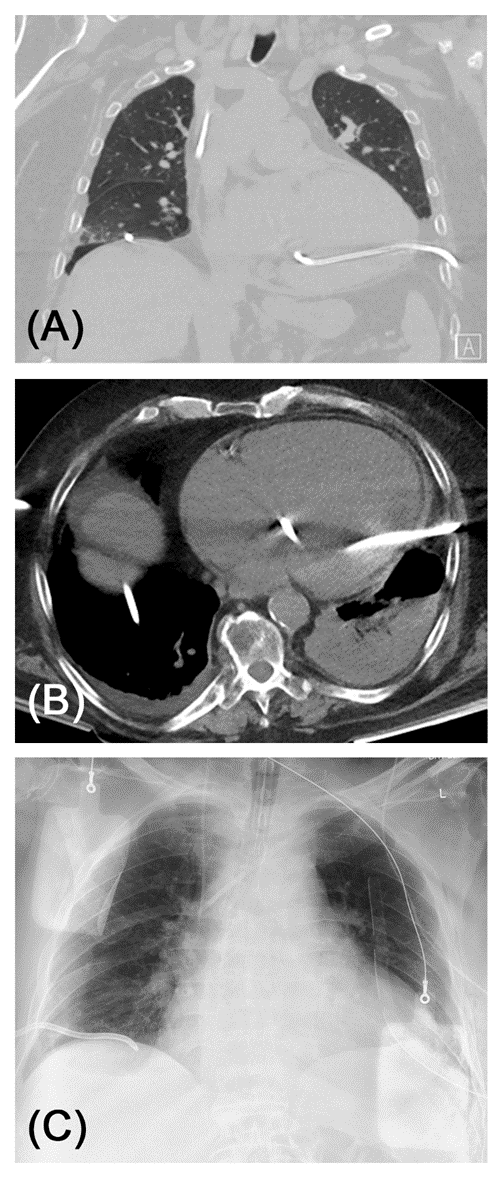
A. Pre-operative CT of the thorax (coronary reconstruction, lung window) clearly shows malpositioning of the tube inside the left ventricle.
B. The tip of the tube reaches the basal portion of the aortic valve apparatus. No aortic regurgitation was found in echocardiography.
C. Postoperative chest X-ray after removal of the tube via anterolateral thoracotomy and direct suture of the left-ventricular perforation. A new pleural drainage was inserted. External defibrillation paddles can be seen.
Figure 4: Casuistry 4: 87-year-old patient with cardiomyopathy and severely impaired left ventricular contractility. As part of cardiac decompensation, bilateral pleural effusions had developed. To relieve progressive dyspnea, a pleural drainage had been applied on both sides, left-sided it had come to a misalignment of the drainage via falsa into the left ventricle. The surgical therapy could be carried out here without a median sternotomy via anterolateral thoracotomy.
Both patients died after 14 and 18 days on intensive care therapy, respectively.
Apart from patients with luxatio cordis, there were no deaths, so that a mortality rate of 22.2 % is recorded in this group of patients. The average
intensive care treatment was 13.0 (5.6) days with a total hospitalization of 18.2 (6.2) days. One patient required long-term ventilation with tracheotomy (11.1%).
Penetrating Cardiac Injuries
Cardiac injury due to penetrating chest trauma was present in a total of n = 7 cases (i. e. 16.3% of all patients): exclusively male, age 44.4 (24.1), six thoracic knife stab injuries, one iatrogenic perforation of the left ventricle by mispositioning of a pleural drainage. No ISS was calculated for these patients. Except for the iatrogenic injury, the foreign bodies were no longer in situ. All patients received surgical care, six patients immediately after admission, one patient two days after the injury; mean 0.2 (0,7) days.
In five patients, an injury to the right ventricle was found intraoperatively. Ventricular suture was performed via anterolateral thoracotomy. Therefore, the pericardial lesion was expanded to a pericardial window and the hemopericardium was relieved. The cardiac injury then was sutured with monofilament, non-absorbable polypropylene threads (thickness 2-0 or 3-0) with a Teflon felt as an abutment. The course of the coronary arteries was strictly observed. Intrapericardial and intrapleural drainages were placed. A left ventricular injury was shown in one patient. In this case, median sternotomy was chosen as surgical access to treat the injury under optimal exposure. A coronary injury was not found in any patient.
There were no deaths in this group of patients. The average intensive care treatment was 5.0 (2.6) days with a total hospitalization of 6.4 (3.5) days. No patient required long-term ventilation or tracheotomy. Patients after suicide attempt remained in intensive care units for monitoring until they were transferred to a psychiatric follow-up.
Clinical Relevance of Cardiac Injuries in Blunt Thoracic Trauma
The focus of this study is on cardiac injuries, which were already mentioned as the main diagnosis during admission. Here we were interested in the extent to which they already require cardiosurgical expertise in the emergency room and how quickly they need to be treated. Most cardiac injuries in chest trauma are associated with compression and deceleration mechanisms. In these patients the most common cardiac injury is cardiac contusion. This is congruent with the data presented in this study: 75.0% of blunt trauma showed this type of cardiac injury. These patients usually do not require any further therapy and can be treated in centers without cardiothoracic surgery expertise [16]. They have a particularly good prognosis. The rarer group of patients with structural heart injuries (25% in our cohort) require completely different care. Because of the vital threat to patients, a chest trauma should always be evaluated to determine whether there is a structural heart injury [8,9].
Cardiologic expertise for arrhythmias and non-perforating coronary injuries is necessary. We did not diagnose these rare complications during our observation period. However, we have certainly observed perforation of the pericardium with and without dislocations. These were treated at a later stage (after 5.0 (5.1) days), since other injuries required more urgent therapy - provided hemodynamic stability was present. Pericardial injuries are the most common entity that led to surgical intervention in our study. Cardiac dislocation occurs rarely but is then associated with high mortality in view of the typically severe polytrauma (ISS 37.3 (9.7) in our cohort). The critical injuries in blunt trauma patients require significant longer intensive care and hospitalization in comparison to penetrating cardiac injuries: ICU stay in blunt trauma 13.0 (5.6) days vs. 5.0 (1.6) in penetrating trauma (p=0.026); hospital stay in blunt trauma 18.2 (6.2) days vs. 6.4 (3.4) in penetrating trauma (p=0.003) (see also table 1).
| blunt trauma | penetrating trauma | p | |
| m:f | 2:1 | 7:0 | |
| Age [years] | 49.9 (18.9) (17-74) | 44.4 (24.2) (19-87)) | 0.585 |
| accident | deceleration 6 fall 2 entrapment 1 | knife stab wound 6 iatrogenic LV perforation 1 | |
| ISS | 37.3 (9.7) | n.a. | n.a. |
| cardiac injury | luxation cordis 2 pericardial injury 4 arrosion of pericardiophrenic 1 tricuspid rupture 1 LAA rupture 1 | RV injury 5 LV injury 1 LV perforation 1 | |
access route thoracotomy median sternotomy VATS | 7 (77%) 1 (11%) 1 (11%) | 5 (71%) 1 (14%) 1 (14%) | |
| operation | pericardial plasty 2 pericardial drain 4 vessel ligature 1 LAA clip 1 tricuspid reconstruction 1 | RV suture 4 RV hemostasis 1 LV suture 2 | |
outcome survived TT deceased | 7 (77%) 1 (11%) 2 (22%) | 7 (100%) 0 (0%) 0 (0%) | |
| ICU stay [days] | 13.0 (5.6) (3-22) | 5,0 (2.6) (1-9) | 0.026 |
| hospital stay [days] | 18.2 (6.2) (12-29) | 6.4 (3.4) (1-11) | 0.003 |
| time to surgery [days] | 5.0 (5.1) (0-13) | 0.3 (0.7) (0-2) | 0.021 |
Tables: legend: f = female, m = male, VATS = video-assisted thoracoscopy, LAA = left atrial appendage, TT = tracheotomy, ICU = intensive care unit
Laceration on the pericardium is usually accompanied by hemorrhagic pericardial effusion, but tamponade does occur rarely in these cases, as relief in the pleural cavity is possible. A reconstruction of the pericardium with relief of the effusion by a pericardial and pleural drainage is the therapy of choice. Care was always taken how to position the patient and operations were carried out with the patient ready for immediate thoracotomy.
Structural injuries to the heart, such as ruptures of the myocardial wall or the valve apparatus, are clinically rare [17–19] whereby a relevant preclinical mortality is pointed out here. A partial amputation of the left atrial appendage could be treated without median sternotomy and use of extracorporeal circulation: a left atrial appendage (LAA) clip was used here, which is usually used for occlusion of the atrial appendage in rhythm surgery [20]. If necessary, the immediate preoperative –"prophylactic" – puncture of the inguinal vessels is also useful to establish a veno-arterial ECLS in acute deterioration of the vital parameters without loss of time. Injuries to the valve apparatus are rare and then require conventional surgical repair.
Clinical Relevance of Cardiac Injuries in Penetrating Thoracic Trauma
In our experience, penetrating cardiac injuries need often be surgically explored or treated. Penetrating injuries that only affect the anterior mediastinum are explicitly excluded here, as they can also be treated conservatively. Most often, our analysis found knife stab wounds, which more often affect the right ventricle due to the anatomical location behind the ventral chest wall. After relieving the pericardial effusion, a direct suture is usually possible via an anterolateral thoracotomy, whereby attention must be paid to the course of the coronary arteries. Iatrogenic perforations of cardiac structures are possible, but very rare.
In the patients shown in this study it was always possible to perform computed tomography to determine the exact location of the injury. This also meant that the access route was not always a median sternotomy, but also a lateral thoracotomy. A thoracoscopy should continue to be viewed critically here, since it was sufficient only once in the case of a laceration of the ventricle. A second time a conversion to thoracotomy was needed due to bleeding. With the involvement of the left ventricle, the median sternotomy provides the best exposure and at the same time allows the rapid commissioning of extracorporeal circulation if it is necessary for the treatment of myocardial injury.
A distinction must be made between trauma room thoracotomy as ultima ratio for critical trauma patients. It is associated with a very poor prognosis in both penetrating and blunt trauma (21,22). In the patients presented here, an emergency thoracotomy in the operating room was required several times, but never a trauma room thoracotomy.
In the patients presented in this study, more than every third case required cardiothoracic surgery (16 out of 43 patients, 37.2%). Thus, cardiothoracic expertise should be present for all trauma room patients with suspected cardiac injury. From this retrospective analysis, we derive the following recommendations for suspected structural cardiac injuries:
Like most publications on this topic, the present study includes retrospective data analysis and must be critically discussed from this point of view. However, randomized controlled trials are difficult to implement for understandable reasons. Retrospective data collection is based on a relevant selection bias in sense of high prehospital mortality of cardiac injuries, in particular due to pericardial ruptures with cardiac dislocation and cardiac or coronary ruptures with consecutively fulminant pericardial tamponade. The presented data thus include pre-selected patients who reach the hospital alive with the aforementioned injury patterns. In literature, mortality from penetrating injuries to the heart is up to 86 % and from vascular injuries of the thorax up to 92 % (15,23). In autopsy studies, the detection of cardiac involvement in fatal thoracic trauma is described up to 96 % [24].
Nevertheless, the patient data analyzed here reflect the spectrum of cardiac injuries associated with blunt and penetrating in a maximum care hospital. If the patient reaches the hospital primarily alive or can be stabilized in the trauma room, a high survival rate can be achieved by the cardiothoracic surgeon. In our case series, mortality for blunt injuries to the heart was 22% of the patients receiving surgical care, while no mortality was recorded for penetrating injuries.
The authors declare that the research was conducted in the absence of any commercial or financial relationships that could be construed as a potential conflict of interest.
Sebastian Reindl prepared the manuscript and figures. Philipp Jawny performed data collection and did statistical analysis and testing. Stephan Raab corrected the manuscript and checked for scientific relevance and correctness.
None.
None.
Dear Editorial Team, Clinical Medical Reviews and Reports. My experience with the journal was highly positive. The peer-review process was rigorous, constructive, and completed in a timely manner. The reviewers provided valuable comments that helped improve the quality and clarity of our manuscript. The editorial office was professional, responsive, and supportive throughout all stages of the publication process. Communication was clear and efficient, and any questions were addressed promptly. Overall, I found the journal to maintain high scientific standards and an excellent publication workflow. I would be pleased to consider submitting future work to this journal. Best wishes from, Elena Popa.

It was my pleasure to submit my testimonial concerning the Reviewer Board of our Scientific Journal “Brain and Neurological Disorders”. The Reviewers focused on some modifications and their contribution was helpful. The ladies of our Editorial Office were also supported my efforts. It was my honor to have such a co-operation and I am looking forward for more collaboration.

Dear Grace Pierce, Editorial Coordinator of Journal of Clinical Research and Reports, Thank you for the speedy and efficient peer review process. I appreciate the fact that your peer reviewers do not take months to respond like with some other journals. I would also like to thank the editorial office for responding quickly to my questions. It is an excellent journal. I plan to submit more manuscripts in the future. Best wishes from, Robert W. McGee

Dear Grace Pierce, Editorial Coordinator of Journal of Clinical Research and Reports, Working with you and your team on our recent publication in JCRR has been a truly wonderful and enjoyable experience. The responses were prompt, and the reviewers were patient, constructive, and highly professional. One reviewer in particular gave me the feeling that a professor was carefully reading and commenting on my coursework, which was deeply touching. The entire process was straightforward and hassle‑free, with no tedious online forms to complete. I highly recommend this journal. Best wishes from, DR Aibing Rao, Head of R&D

I Appreciate the Opportunity to Share my Experience with the Journal of Clinical Research and Reports. The peer review process was timely and constructive, and the feedback provided helped improve the quality of our manuscript. The editorial office was professional, responsive, and supportive throughout the process, ensuring smooth communication and efficient handling of the submission. Overall, it was a positive experience collaborating with your team.

Dear Mercy Grace, Editorial Coordinator of Obstetrics Gynecology and Reproductive Sciences, We would like to express our gratitude for your help at all stages of publishing and editing the article. The editors of the magazine answer all the necessary questions and help at every stage. We will definitely continue to cooperate and publish other works in the Obstetrics Gynecology and Reproductive Sciences! Best wishes from, Alla Konstantinovna Politova,
