
Review Article | DOI: https://doi.org/10.31579/2690-1919/157
*Corresponding Author: Rolf Bambauer, Formerly, Institute for Blood Purification, Frankenstrasse 4, 66424 Homburg, Germany.
Citation: Rolf Bambauer, Ralf Schiel (2021) Therapeutic Apheresis and Immunosuppression in Renal Diseases. J Clinical Research and Reports, 7(3); DOI:10.31579/2690-1919/157
Copyright: © 2021 Rolf Bambauer, This is an open access article distributed under the Creative Commons Attribution License, which permits unrestricted use, distribution, and reproduction in any medium, provided the original work is properly cited.
Received: 22 March 2021 | Accepted: 31 March 2021 | Published: 09 April 2021
Keywords: therapeutic apheresis; rapidly progressive glomerulonephritis; anti-basement membrane antibody glomerulonephritis; immune complex nephritis; nephrotic syndrome; acute kidney injury; myoglobulinemic renal failure; hemolytic uremic syndrome
Therapeutic plasma exchange (TPE) with hollow fiber modules is used in different severe diseases since more than 40 years. The authors try to give an overview of therapeutic apheresis (TA) in renal diseases. The updated information on immunology and molecular biology of different renal diseases are discussed in relation to the rationale for apheresis therapy and its place in combination with other modern treatments. The different renal diseases can be treated by various apheresis methods such as TPE with substitution solution, or with online plasma or blood purification using adsorption columns, which contain biological or non-biological agents. The following diseases are discussed: rapidly progressive glomerulonephritis (RPGN) including anti-glomerular basement membrane antibody glomerulonephritis (anti-GBM RPGN), RPGN with or without glomerular deposition (ANCA-ab), pauci-immune RPGN, immune complex nephritis (ICN), and various glomerulonephritis with nephrotic syndrome (NS), acute kidney injury (AKI), myoglobulinemic renal failure, hemolytic-uremic syndrome (HUS), and kidney transplant rejection. For the renal diseases, which can be treated with TA, the guidelines of the Apheresis Applications Committee (AAC) of the American Society for Apheresis (ASFA) are shown.
Therapeutic apheresis (TA) summarizes different extracorporeal blood purification techniques that removes inflammatory mediators, antibodies and other toxic substances, which are pathogenic in various diseases and is used in many autoimmune disorders [1]. TA has proved itself in a series of immunological-, metabolic diseases, and intoxications. More selective plasma separation and immunoadsorption (IA) with immobilized monoclonal or polyclonal antibodies etc. have been introduced in clinical routine.
With the hollow fiber modules in TA, a complete separation of the corpuscular components from the plasma is reached and due to increases blood flow rate and higher efficacy [2]. There is no advantage that TA using centrifuges has shorter treatment times such as TA using hollow fibers shown by Hafer et al. [3]. More important is to keep the blood levels with antibodies, and/or pathogenic substances on a very low level over long time during the treatment. However, the substances that should be eliminated could invade into the intravascular space and be eliminated by the membrane separators. Furthermore, cell damage - especially to thrombocytes – occurs less using membranes than centrifuges for all cell separation. The adsorption technologies allow the most selective separation of plasma components without the use of any substitution solution [2]. Membrane techniques are simple and safe to apply and can be competitive to other plasma separation and treatment technologies [4].
The hollow fiber modules in therapeutic plasma exchange (TPE) are mostly used in nephrology, as many of these membranes can be used with currently available dialysis equipment. Nephrologists have an extensive training in the management of blood purification treatments including vascular access, anticoagulation, volume management and prescription for solute clearance [5]. The renal indications for TPE expand the clinical practice of nephrologists [6].
Only a few prospective controlled trials are available that are of adequate statistical power to allow definitive conclusions to be reached regarding the therapeutic value of plasma exchange. This drawback reflects, in part, the relative rarity of most of the disorders under investigation. To compensate, many investigators have understandably grouped heterogenous diseases together, often retrospectively, and used historical controls. The latter design is potentially hazardous, given that earlier diagnosis, recognition of milder cases, and improved general care over time may be lost as a benefit of plasma exchange [7]. Most histories of many diseases commonly treated by TA (e.g., cryoglobulinemia, SLE) are characterized by episodes of exacerbation and remission, further underscoring the importance of adequate concurrent controls:
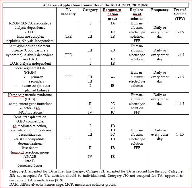
For those diseases for which the use of TA is discussed, the guidelines on the use of TA from the Apheresis Applications Committee (AAC) of the American Society for Apheresis (ASFA) are cited [8, 9]. Especially the categorization and indications of different diseases of the AAC are mentioned (Table 1). Bambauer et al. discuss the TA methods such as TPE and different semi- or selective plasma exchange methods [10].
Rapidly Progressive Glomerulonephritis (RPGN)
RPGN is a diffuse glomerulonephritis that frequently begins acutely. RPGN is a histologic diagnosis, and can occur from a number of etiologies, including anti-basement membrane antibody glomerulonephritis (ABM-ab-GN), which is very rare, antineutrophil cytoplasma antibodies (ANCA), and even IgA nephritis. The histological characteristics are usually capillary emboli with necrosis of the capillary walls and semi-lunar formation, and deposition of IgG and C3 along the glomerular basement membrane. Most cases are simultaneously accompanied by acute kidney injury [11]. More than 90 percent of patients with RPGN due to Goodpasture´s / anti-GBM RPGN have anti-GBM antibodies in their circulation.
RPGN consists of rapid loss of renal function with the histologic finding of crescent formation in over 50 % of glomeruli [11]. Histologically is observed a proliferation of cells within Bowman´s space of the glomerulus due to the extravasations of proteins into the space. These cells consist of proliferating parietal epithelial cells as well as infiltrating macrophages and monocytes. RPGN is not a single disease entity but is a clinical syndrome with a different number of etiologies. Histologic classification divides RPGN into three subtypes based on the immunofluorescence pattern on renal biopsy [8].
The incidence is 0.85 per 100.000/year. Importantly, when discussing RPGN, a number of entities are frequently included in case series and trials, thus confounding results [11]. Therapy consists of high-dose corticosteroid (e.g., methylprednisolone) and cytotoxic immunosuppressive drug (e.g., cyclophosphamide or azathioprine) [9]. Other drugs have been used include leflunomide, deoxyspergualin, tumor necrosis factor blockers, calcineurin inhibitors, and antibodies against T cells, or human monoclonal antibodies (HMA).
The rationale for therapeutic apheresis is that RPGN with dialysis dependence (Cr > 6 mg/dL) and RPGN with diffuse alveolar hemorrhage have the Category I with the recommendation grade (RG) 1A and 1C. RPGN dialysis independent has the Category III with the recommendation grade 2C [8]. Because of the benefit of plasma exchange in the crescentic GN of anti-GBM, plasma exchange was applied to all causes of RPGN [9]. The role of TPE has been examined in some trials in pauci-immune and immune complex GNs and in the treatment of pauci-immune GN. Results of other trials indicate that TPE may be beneficial for dialysis-dependent patients presenting with severe renal dysfunction; however, is no therapeutic benefit over immunosuppression in milder disease. The predominance of pauci-immune GN cases in these series may account for these results [9]. Immunoadsorption is the extracorporeal method that most effectively removes pathogenic immune complexes and antibodies [12]. The frequency of TA is every other day. The volume treated is 1 – 1.5 total plasma volume and the substitution solution could be a human-albumin-electrolyte solution. Treatment is for 1 – 2 week followed by tapering with less frequent treatments. The duration of therapy is not well defined in the literature. Some trials have stopped TA if there is no response after 4 weeks of therapy.
PEXIVAs, an international randomized controlled study comparing TPE versus no TPE and standard versus reduced dose steroid regimen on the primary composite outcome of end stage renal disease (ESRD) or death in patients with ANCA-associated vasculitis (AAV) represents the largest study on the role of TPE in AAV [13]. In the patients under TPE was not significantly associated with their risk of primary outcomes, mortality, and side effects. Further, it was suggested that TPE might be effective in suppressing ESRD in the early stages of treatment [14]. The PEXIVAs study did not show the addition of TPE to standard therapy conferred benefits in patients with severe ANCA-associated vasculitis, but it did show that a reduced-dose regimen of oral glucocorticoids was noninferior to a standard-dose regimen [15].
Anti-Basement Membrane Antibody Glomerulonephritis
(Goodpasture Syndrome, ABM-ab-GN)
In anti-basement membrane antibody glomerulonephritis, antibodies appear which that are directed against a peptide component of one of the two non-collagen parts of type IV collagen. However, type IV collagen is found not only in the kidney, but also in the vessels of other organs, such as the lung [16]. The mechanisms responsible for the production of antibodies against the antigens are still not clear.
A large number of diseases have been associated with Goodpasture syndrome based on different cases; however, the most consistently reported associations are with membranous nephropathy and anti-neutrophil cytoplasmatic - associated vasculitis. Only a small part of ANCA GN have anti-GBM ab, mostly it has thought to be an environmental or infectious exposure that triggers onset of these diseases. It is reasonable to speculate that for both membranous and ANCA-positive vasculitis damage to the kidney elicits an immune response against the GBM, leading to the production of antibodies, which may or may not contribute to disease progression [16]. ANCA GN responds to TPE even when patient on dialysis and anti-GBM GN does not.
The formation of anti-basement membrane antibodies is frequently limited in duration. The autoantibodies cause severe disturbances in the permeability in the lung with significant deterioration in diffusion capacity and hemoptysis. The renal deposition of this autoantibody frequently leads to rapid deterioration in renal functioning, which expresses itself histologically in a necrotizing glomerulonephritis in part. Linear deposits of IgG can be immunohistologically detected both at the basement membrane of the lung, as well as of the kidney [17, 18]. An antigen with a probable size of 26,000 – 28,000 daltons is considered responsible for these deposits, its immunogenic epitopes being located on the stable glomerular domain NC1 of collagen IV [18]. The antigen is primarily present in a hexamers form and forms monomers and dimers [18, 19]. Antigen determinants are exposed after dissociation and can thus bind specific antibodies. This molecule seems to be present in all basal membranes, in particular in those of the glomeruli, renal tubuli, the Bowman capsule, the lung, and the plexus chorioideus, in the placenta, but also in those of the aorta and the small intestine.
De Lind van Wijngaarden et al. observed that chronic and acute tubulointerstitial lesions predict the glomerular filtration rate (GRF) at 12 months, yet it was the use of TPE and the number of normal glomeruli on biopsy that remained positive predictors of dialysis independence in the same time interval [18]. This is important because it suggests that unaffected glomeruli determine long-term renal outcome at 1 year. In a second study, the same group extended their work in determining the rate of renal recovery [19]. In the MEPEX study, 69 dialysis-dependent patients who were part of the TPE trial, plasma exchange was superior to pulse methylprednisolone with respect to the change of coming of dialysis. The outcome measure depended on the relative number of normal glomeruli.
Treatment with TA also provides the possibility of improvement in cases of pulmonary bleeding, which based on the same immunological process, even when renal function is already irreversibly impaired [20, 21]. A final long-term prognosis for patients whose condition improved after TA cannot be made. As basement membrane antibody formation often ceases during treatment, recovery, or at least partial recovery is possible.
The rationale for TA is that RPGN with dialysis dependence (Cr > 6 mg/dL) and RPGN with diffuse alveolar hemorrhage have the category I with the RG 1A and 1C. RPGN dialysis independent has the Category III with the RG 2C (Table 1) [8]. Because of the benefit of TPE in the crescentic GN of anti-GBM, TPE was applied to all causes of RPGN. The frequency of TA is every or every other day until anti-glomerular basement membrane antibodies are detectable. The volume treated is 1-1.5 total plasma volume, and the substitution solution could be a 5 % human-albumin-electrolyte solution or fresh frozen plasma (FFP). Treatment is for 1-2 week followed by tapering with less frequent treatments. The duration of therapy is not well defined in the literature. Some trial have stopped TA if there is no response after 4 weeks of therapy. TA should be continued until antibodies fall to undetectable levels in patients with active disease and anti-GNM antibodies present [8].
Immune Complex Nephritis (ICN)
Many types of glomerulonephritis are initiated by the deposition of immune complexes, which induce tissue injury via either engagement of Fc receptors on effector cells or via complement activation [22]. The pathogenic consequences of systemic autoimmune disease is thought to trigger by the generation of antibody and subsequent tissue deposition of immune complexes (IC). Modulation of the autoantibody response disrupts pathogenesis by preventing the formation of ICs; however, uncoupling IC formation from subsequent inflammatory response seems unlikely because of the apparent complexity of the IC-triggered inflammatory cascade [23].
In idiopathic symptomatic RPGN, which is frequently caused by an immune complex nephritis, the therapeutic concept is not as clear-cut as with anti-glomerular basement membrane antibody nephritis. Sieberth et al. demonstrated in a study that a combined therapy of TPE and immunosuppression is superior to immunosuppressive therapy alone [24]. An improvement in renal function is possible in more than 60 percent of cases, if either pulse therapy (high dose therapy with corticosteroids) or TPE is administered. In view of the devastating pathophysiologic consequences of interaction between circulation immune complexes and the basement membrane was found, that TPE in combination with immunosuppression should be carried out as quickly as possible [25]. Pusey et al. recommended TPE for severe cases of immune complex nephritis [26].
Combination of corticosteroid and cyclophosphamide or rituximab, and/or MMF, and TAC has been recommended for remission induction of ANCA-associated vasculitis [27]. This is the first report demonstrating the efficacy of a multitarget therapy of corticosteroid, mycophenolate mofetil (MMF), and tacrolimus (TAC) for remission-induction of intractable ANCA-associated glomerulonephritis developed independently of systemic lupus erythematosus (SLE)
RPGN with or without Glomerular Deposition (ANCA ab) Pauci – Immune RPGN
Approximately 60 percent of patients with RPGN present with crescentic glomerulonephritis characterized by few or absent immune deposits, the so-called pauci-immune RPGN. Patients with this disease have either Wegner´s granulomatosis; ANCA-ab associated vasculitis, polyarthritis nodosa, or “renal-limited” pauci-immune GN (Table 1) [28]. These diagnoses may represent a spectrum of manifestations of a single disease, because there is marked overlap of clinical and histopathologic features, and several patients have anti-neutrophil cytoplasmatic antibodies (ANCA) in their blood which are more common that anti-GBM. The concentration of circulating ANCA correlate with the disease activity in some patients, and ANCA may contribute to the pathophysiology of pauci-immune RPGN through reactivity with neutrophils or endothelial cells, and other inflammatory mechanisms [11, 28, 29].
The prognosis of pauci-immune RPGN in general has been poor. Precise therapy therapeutic recommendations are difficult to obtain from the literature, because most series comprise patients with different types of RPGN. However, available data suggest that 80 percent of such patients’ progress to ESRD without therapy with high dose immunosuppression or cytotoxic drugs. Some trials have evaluated the efficacy of TA as an adjunct to conventional immunosuppressive in patients with pauci- immune RPGN [28, 30-32].
In milder forms of pauci–immune RPGN, the generation of antibody and subsequent tissue deposition of immune complexes (IC), the results of the randomized trials argue against the role for TA, however, suggest a potential benefit when TA is used as an adjunct to conventional immunosuppressive therapy in patients with severe disease. This relative lack of efficacy probably reflects the efficiency of conventional immunosuppressive agents in halting inflammation and preserving renal function in most patients. These conclusions are supported by the results of uncontrolled trials, suggesting a response rate of 70 percent in patients with RPGN treated with TA, similar to that of patients treated with immunosuppressive therapy with a response rate of 60 percent. In most cases of RPGN, a treatment of TA in the early phase of the disease seems to be necessary.
A combination of cytapheresis and standard immunsosuppressive therapy of prednisolone and cyclophosmide was reported as a successful treatment. In five patients with a myeloperoxidase antineutrophil cytoplasmatic antibody associated vasculitis, the renal function improved and the pulmonary hemorrhage disappeared [29]. Others reported of successful treatments with immunoadsorption and immunosuppressive therapy [32, 33]. In the above mentioned MEPEX study, de Lind van Wijngaarden et al showed that in patients with dialysis-dependent, ANCA-associated vasculitis, the chances of recovery differ depending on the type of adjunctive treatment, the percentage of normal glomeruli and glomerulosclerosis, the extent of tubular atrophy, and the presence of arteriosclerosis. Even with an ominous biopsy at diagnosis in combination with dialysis dependence, the chance of renal recovery exceeds the chance of therapy-related death when the patient is treated with plasma exchange as adjunctive therapy [34]. The PEXIVAS trial did not show that the addition of TPE to standard therapy conferred benefits in patients with severe ANCA-associated vasculitis, but it did show that a reduced-dose regimen of oral glucocorticoids was noninferior to a standard-dose regimen [15].
Therapy Recommendations for RPGN
RPGN therapy possibilities were extended in recent years to include TA. Antigens, antigen-antibody complexes, and immune complexes can be eliminated from the blood with the aid of plasma exchange. A corresponding therapy enables immunomodulation through suppression or stimulation of antibody formation, as well as a temporary remission of the inflammation through inhibition of the mediators. TPE combined with immunosuppression therapy seems to us to be advisable, particularly in view of the unfavorable prognosis for RPGN, with its complex causes.
The therapy recommendation is based on the few uncontrolled and controlled studies available [25, 28, 31, 35, 36]. TPE is indicated in combination with an immunosuppressive therapy with prednisolone (intravenous pulse therapy, or oral therapy), cyclophosphamide (intravenous pulse therapy or oral therapy), or azathioprine in indicated in the following cases:
Preparation for kidney transplant with anti-basement membrane antibodies still detectable in the serum.
With high titers of circulating immune complexes or other antibodies, which could damage the kidney and other organs, IA with protein-A, or sheep polyclonal antibodies can be more effective than the TPE procedure.
TA used for renal indications, even in elderly patients is relatively safe. Trends towards death in elderly patients may be multi-factorials and not necessary related to TA [36]. TA may be decrease end of end-stage renal disease or death in patients with RPGN [37]. The combination of TA with immunosuppressive therapies including biologics seems to be more effective as TA alone, but additional trials are required. However, other authors prefer in cyclophosphamide-resisistent ANCA-associated GN a multitarget therapy, a combination of corticosteroids, MMF, and TAC, but additional trials are required [27].
Glomerulonephritis with Nephrotic Syndrome (NS)
Classification is classified morphologically, and thus does not provide a uniform description of the disease. Differing etiologies can result in considerable variations in the clinical features, as well as course and prognosis. Therefore, it is difficult to establish generally applicable therapeutic concepts and customized treatment for the individual patient is the norm [38]. The variable clinical courses of this heterogenous disease group render it almost impossible to carry out controlled therapy studies. The clinical successes and failures are to be found, as are therapy-produced complications, e.g., infections, sterility, loss of hair, and others. The benefits of immunosuppressive therapy must be weighed against these complications. The aim of therapy for glomerulonephritis is to prevent terminal renal insufficiency and the risks of nephrotic syndrome, therefore some therapeutic possibilities are discussed here.
The cause of nephrotic syndrome lies in changes in the electrophysiological characteristics of the filtration barriers and of the plasmaproteins. The anionic charge on albumin is retained by the negative charge of the glomerular filter - including the basement membrane and the epithelium - obviously play a decisive role [7]. Hemodynamic changes, such as increase in venous pressure, can favour the filtration of proteins.
Nephrotic syndrome of various GN often reacts to corticosteroids in varying doses, administered over a period of 4 - 8 weeks. Patients with frequent relapses are also treated with 2 - 3 mg/kg BW/day cyclophosphamide [11]. Cyclosporin A has also been successfully applied in nephrotic syndrome [7]. High doses of immunoglobulin (IgG) for nephrotic syndrome, administered 0.4 g/kg BW IgG on three successive days were reported, and repeated every 21 days over a period of one year. Other therapeutic measures for nephrotic syndrome are anticoagulants, thrombocyte inhibitors, ACE inhibitors, immunosuppressive drugs, lipid reducers, biologics, and diets [39-41].
The prognosis for focal sclerosing glomerulosclerosis (FSGS), usually accompanied by nephrotic syndrome, is considerably less favourable. Cases with nephrotic syndrome are recorded as having a survival rate of 70 percent after six years. Without nephrotic syndrome, this rate reaches 85 percent. Patients with this form of glomerulonephritis are comprised of steroid – sensitive and a steroid - non-sensitive groups, and an appropriate therapy must be selected. Non-reaction to steroids is an indication for a therapy with cyclophosphamide, chlorambucil, or cyclosporin or other immunosuppressive therapy [42]. FSGS is caused by a variety of factors, however, one type that recurs after transplantation and has been associated with circulating factors, can be treated with TPE.
In resistance to medication or severe progression of the disease, additional TA therapy should be considered, as a continuing treatment given once a week, or every two weeks, or once a month. After transplantation, as many as 40 percent of patients with nephrotic syndrome have recurrences. The glomerular abnormalities in patients with established disease include focal and segmental glomerulosclerosis and hyalinosis, although fusion of epithelial-cell foot processes may be the only abnormality early in the course of disease [7]. Some patients with recurrent focal glomerulosclerosis have a response to treatment with TPE, LDL apheresis and IA there may be different circulating factors that alter the glomerular barrier to protein filtration [43].
In the guidelines on the use of TA in clinical practice-evidence-based approach from the AAC of the ASFA has the primary and secondary FSGS the Category III with the RG 1C, and for the FSGS recurrent the category I with the recommendation grade 1B (Table 1) [8, 9].
The treatment in native kidneys with FSGS is primarily with corticosteroids for at least 6 months prior to trying second-line agents such as cyclophosphamide, chlorambucil, or azathioprine. For resistant cases, TPE is being currently an option. Several investigators worldwide have used TPE in the management of patients with FSGS in transplanted organs, in an attempt to save the graft. Although there is no standardized treatment for recurrent FSGS post-transplant, the majority of regimens use a combination of an immunosuppressant such as cyclophosphamide, biologics, and TPE. Other therapeutic options include high-dose cyclosporine, angiotensin converting enzyme inhibitors, and indomethacin and/or tacrolimus. Another approach to prevent recurrent FSGS is several sessions of pre-emptive TPE immediately prior to and following the transplant [7]. More recently, rituximab and mycophenolate mofetil have also been used in conjunction with diagnosed in order to halt the process and maintain renal function [8, 9].
In certain FSGS patients appears to contain an ill-defined “permeability factor”, probably a glycoprotein of molecular weight of 30 – 50 kDa that includes profound leakage of albumin when incubated with isolated rat glomeruli. Such factor is removed by TPE and the decrease in serum concentration coincides with improvement in proteinuria. The immediate onset of proteinuria following transplant is mediated by this factor, prophylactic TPE may be instituted in high-risk patients. Some reports describe the use of Staphylococcal protein-A columns in recurrent FSGS. The duration of the procedure is to begin with three daily exchanges followed by at least six more TPE in the subsequent 2 weeks, for minimum of nine procedures. Tapering should be decided on a case-by-case basis and is guided by the degree of proteinuria. Timing of clinical response is quite variable and control of proteinuria may take several weeks to months. Some patients have received long-term monthly exchanges as maintenance therapy [8, 9].
The nephrotic syndrome consisting of massive proteinuria, hypoalbuminemia, edema, and hyperlipidemia, is a common complication of glomerular disease in children and adults. The annual incidence of nephrotic syndrome ranges from 2 – 7 per 100,000 children, and prevalence from 12 – 16 per 100,000. There is epidemiological evidence of a higher incidence of NS in children aged below 10 years from South ASIA [40]. The primary cause of NS is idiopathic. There is evidence pointing to a role of the immune system in pediatric minimal change glomerulonephritis (MCGN). Another hypothesis has described an association between allergy and MCGN in children. Relapses in this of syndrome are triggered commonly by minor infections and occasionally by reactions to be stings or poisoning. Abnormalities of both humoral and cellular immunity have been described. Finally, the induction of remissions by corticosteroid, alkylating agents, or cyclosporine therapy provides indirect evidence for an immune etiology [8].
Although they are massively proteinuria, patients with MCGN, do not have a generalized glomerular leak to macromolecules. The clearance of neutral macromolecules in MCGN is actually less than normal over a range of molecular radii. In contrast, the clearance of anionic macromolecules is significantly increased. This and several other lines of evidence suggest that proteinuria results from a loss of fixed negative charges of anionic glycosaminoglycan’s in the glomerular capillary wall [8]. The mechanisms through which these charges are lost are unknown. The traditional view is that massive albuminuria, in NS causes a decrease in intravascular oncotic pressure, which allows extravasation of fluid and hypovolemia, increased aldosterone and antidiuretic hormone secretion, and renal salt and water retention. An alternative explanation for retention of salt and water in NS is a decreased glomerular filtration rate, with a decreased filtration fraction.
Minimal change glomerulonephritis usually takes a benign course and can be well treated with customary therapy measures. In severe cases, therapy with prednisolone and cyclophosphamide over a period of 8 to 12 weeks is indicated [44-45]. Cyclosporin has shown some efficacy in steroid-resistant NS [46]. A significantly rapid faster relief from steroid–resistant NS by using LDL apheresis than from steroid monotherapy is reported [46]. A rapid improvement of hypercholesterolemia by LDL apheresis in steroid–resistant NS will provide more rapid relief from NS than from steroid therapy alone. Others recommended in steroid-resistant NS intravenous steroids in high dose with alkylating agents, cyclophosphamide oral or pulse cyclophosphamide and mycophenolate mofetil [47].
Membranoproliferative glomerulonephritis (MPGN) usually occurs in combination with nephrotic syndrome and hypertension. The occurrence of nephrotic syndrome signifies a poorer prognosis. The effectiveness of medication with corticosteroids or pulse therapy, cyclophosphamides, anticoagulants, and intravenous immunoglobulins has not yet been established [48]. Experience with TA, especially with protein-A immunoadsorption has been presented [49, 50]. A successful treatment with protein-A IA in patients with relapsing nephrotic syndrome was reported. MPGN from cryoglobulinemia could be an indication for TA, too.
Nephrotic syndrome is the main symptom in perimembranous glomerulonephritis. In the case of acute nephrotic syndrome, it is advisable to undertake therapy with high doses of prednisolone as a pulse therapy over a period of 3 to 5 days or with 2 mg/kg BW in decreasing dosage for 2 to 3 months. A combination with TPE should be considered especially with the more selective procedures like cascade filtration, IA, and LDL-apheresis [46, 49, 51].
The symptoms in mesangioproliferative glomerulonephritis are not usually homogeneous. The prognosis is poorer if the condition is accompanied by nephrotic syndrome and hypertension. Here also, there are varying opinions exist with regard to corticosteroid and cytostatic therapy. Nephrotic syndrome justifies a trial therapy with cyclophosphamide. Although TA is indicated in severe cases of various types of glomerulonephritis. In severe, drug therapy-resistant cases, a combined TA and immunosuppression therapy is recommended, regardless of the degree of renal insufficiency [52].
Acute nephrotic syndrome in particular seems to be favourably influenced by regular TA treatment, for first, dysproteinemias and thus the edema can be improved, and second human albumin can be administered in larger doses. TA is theoretically a way of achieving an improved effect on the basal membrane. The elimination of cholesterol, LDL, and triglycerides might also reduce the atherogenic risk for these patients and thus prevent progression. TA should be considered as a useful therapeutic tool in the management of this disease [43]. The reports of the therapy of NS with more selective TA procedures like cascade filtration, IA, and LDL apheresis are very encouraging and show a possibility for treating severe cases of NS, if drug therapy fails. Renal diseases such as light chain nephropathy, dense deposit diseases and others as shown in Table 1 can be in severe cases and if the conservative therapy has failed, threated with TPE. As in the case of other renal diseases, controlled prospective studies are needed.
Acute Kidney Injury (AKI)
The renal diseases are concluded with some notes of acute kidney injury as an independent disease [53]. The variety of the causes that can trigger AKI justifies a close examination of this disease. Acute renal insufficiency means reversible renal damage with oligo-anuria, which is now generally called acute kidney injury. AKI presents unique, life threatening and organ threatening therapeutics challenges that require prompt accurate diagnosis and treatment. In some cases, AKI can also take a polyuria course. Damage to the kidneys varies depending on the degree and duration of pre-renal, renal or post-renal disorders (noxae). It is reversible only after the elimination of the noxae; in the case of structural disorders only after its repair; or it can remain irreversible.
AKI is also defined as an acute over hours or days developing renal function damage, which is measured by the glomerular filtration rate (GFR). Other renal functions are changed and decreased in AKI, such as the excretion of metabolic products and drugs, the reabsorption of filtrated substances, the regulation in acid-base and electrolyte disorders, and different endocrinological functions. The incidence of AKI is 2–5 percent in inpatients and up to 10-30 percent with intensive medical. The mortality has essentially remained unchanged in the last four decades and at 30 to 80 percent is very high. AKI is the most frequent and expensive renal disease with the highest course of morbidity and mortality in hospitals [54, 55]
As the causative noxae of AKI must be eliminated at the time of insult to the kidney and before it has destroyed, they can be influenced primarily during the period in which they act on the kidney. Thereafter, further measures against the noxae are no longer effective; all that then remains is life-long dialysis and/or kidney transplantation [55]. Given the new treatment possibilities, the customary classification of these factors in pre-renal, post-renal, and renal disorders is simplistic from a therapeutic point of view [53].
The treatment of plasma disorders in particular, TPE opens up a new approach. Conventional therapeutic methods are to be applied to blood flow and post renal disorders. Of course, in AKI it is important to eliminate or to influence all factors which can lead to AKI and to ensure sufficient administration of parenteral calories, including amino acids, glucose, and fatty solutions [55]. There is no guideline available for the therapy of AKI. In especially severe or therapy resistant course of AKI, the following TA methods could be discussed and implemented:
Despite the intensive and costly therapeutic modalities in AKI, the mortality of the AKI is always high, at 30 – 80 percent, and this has not essentially changed in the last four decades [55]. Approximately twice as many patients with severe diseases, such as multi-organ failure and AKI, die in intensive care units when compared with patients without AKI. These patients die not because of AKI, but because of the different complications that follow AKI.
Considering these facts and the inevitable unfavorable prognosis of the AKI, it is possible to add the option of TA to the conservative and extracorporeal therapies [56]. More of controlled studies should be done to improve the clinical outcome and decrease the high costs of this therapeutic method. Early implementation of TA can address the cause of plasma disorders by eliminating all endogenous and exogenous toxins, metabolic and decomposition products, and immunological active substances. Consistent implementation of TPE combined with dialysis and other conventional techniques may help to improve the yet poor prognosis for AKI.
Myoglobulinemic Renal Failure
An acute myolysis, as example, can induce severe disturbances of the renal function. A normal functioning kidney can eliminate free myoglobin with a molecular weight of 17,800 Dalton rapidly. Massive increase of myoglobin and their derivate in the blood as in acute crushing injuries and mass trauma is highly nephrotoxicity and causes a decreased circulation in the kidney and/and a metabolic acidosis. Acute kidney injury can develop rapidly [57]. Observations showed that, by along with myoglobin, peroxide free radicals of can be released, and can induce a disseminated intravascular coagulation (DIC), and can damage thrombocytes, the endothelium of the vessels, and disturb the metabolism of prostaglandin synthesis [58].
Rhabdomyolysis is also a clinical syndrome in which the contents of injured muscle cells leak into the circulation. This leakage results in electrolyte abnormalities, acidosis, clotting disorders, hypovolemia, and acute kidney injury. Many conditions, both traumatic and non-traumatic, can lead to rhabdomyolysis. Intervention consists of early detection, treatment of the underlying cause, volume replacement, urinary alkalinisation, and aggressive diuresis or hemolysis. Patients with rhabdomyolysis often require intensive care [59].
Elimination of myoglobin from plasma may be enhanced by TPE in patients with AKI [60]. Endothelin, a vasoconstrictive peptide that includes 21 amino acids, has a strong vasoconstrictive effect in the glomeruli. It leads to hypoxia and hypotension of the endothelium, which causes the endothelium to an increased release of endothelin. Cornellisen et al. evaluated 4 patients with rhabdomyolysis and showed that a single 2 liter TPE has no beneficial effects in the treatment in rhabdomyolysis [61]. Only one TPE with only 2 liters was done; all other case reports have used multiple daily TPE. In three patients with severe myoglobulinemia sufficient to cause renal failure, hemodialysis treatment could be prevented when TPE was used in an early stage [62].
The effect of TPE in the case of three patients with myolysis was very impressive. After operative removal of an abdominal glioblastoma, a one-year-old girl with myoglobulinemic muscular dystrophy and acute kidney injury displayed rapid normalisation of renal function, after 3 TPE and four HD treatment sessions [2]. Similar normalization in renal functioning was observed in the case of a 77- year-old patient with myoglobulinemic AKI after one TPE session with three liters. In a 19-year-old female patient, who suffered from malignant hyperthermia and AKI after administration of an anaesthetic during tonsillectomy, was cured after three sessions of TPE. In particular, in the case of malignant hyperthermia, which is rare but reaches a mortality rate of 60 – 70 percent, TPE seems to improve the poor prognosis, if applied at an early stage.
In a case of rhabdomyolysis complicated with increased serum bezafibrat level, TPE was used. The advocated bezafibrate being highly protein bound is unlikely to be cleared by hemodialysis. TPE was safe and effective in addition to supportive care for rhabdomyolysis associated with bezafibrate [63]. Ronco reported that attempts to use TPE in myoglobulinemia have resulted in higher sieving coefficients, but notes limitations due to low volume exchanges [64]. An 82-years old male patient who developed rhabdomyolysis while taking a combination of simvastatin and gemfibrozil and was successfully treated with TPE [65]. Improvement in kidney function when it does occur does so slowly over months of supportive care and dialysis. However, there are only a few cases of rhabdomyolysis reported in literature. Most are case reports. Other authors showed that neither plasmapheresis nor hemodiafiltration has been successful in patients with myoglobulinemic renal failure. Fortunately, most patients eventually regain normal kidney function [66].
In the case of myoglobulinemic renal failure: TPE interrupts, stops, or eliminates the
Dialysis and other conventional techniques may help to improve the yet poor prognosis for AKI.
Hemolytic-Uremic Syndrome (HUS)
Hemolytic-uremic syndrome is a disease that can lead to AKI and often to other serious sequelae, including death. The disease is characterized by microangiopathic hemolytic anemia, thrombocytopenia and AKI. The etiology and pathogenesis of HUS are not completely understood, and the therapy of HUS is complicated. After introduction of therapeutic apheresis as a supportive therapy in HUS, several authors reported successful treatment using TA in HUS in more than 87 percent of treated patients. The supportive therapy is indicated in severe courses of HUS and is superior to available therapy interventions. The pathophysiologic aspects of the different pathogenic types of HUS are discussed by Bambauer et al. [67].
Most cases are associated with infections with enterohemorrhagic E. coli (EHEC). These bacteria can be transmitted through contaminated food, animal and person to person contact. HUS is one of the most severe complications of a potentially avoidable food-borne infection. Other causes of HUS described as “typical” have to be differentiated since other factors including genetic disorders are of importance. A minimum of three different pathogenetic types, which lead to HUS, are subdivided. HUS caused by infection, idiopathic HUS (non-Shiga toxin HUS), and HUS in systemic diseases and after toxin exposure [68].
There have been reports of spontaneous recovery from HUS. The various etiological and pathogenetic assumptions have produced diverse therapy concepts. However, the total lethality in HUS was first reduced to 20 percent with the introduction of dialysis (69). If the therapy is administered early enough, two-thirds of cases recover without any impairment. In 10 - 20 percent of cases, however, lasting renal damage occurs. Other authors reported successful in HUS using TPE and successful treatment in HUS using IA with protein-A [70-73]. A compilation of therapeutic concepts for HUS implemented up to 2009 showed the success of HUS therapy with TPE/HD or IA/HD [67].
A simple plasma infusion as sufficient it is not adequate, since various, pathophysiological mechanisms are observed that cannot be explained solely this theory [74, 75]. However, substitution of plasma or coagulation factors is often necessary due to the severe coagulation problems in HUS. TA might be more effective than infusions alone, as it removes potentially toxic substances from the circulation. TPE or IA should be considered first-line therapy in situations that limit the amount of plasma that can be infused, such as renal or heart failure. Plasma infusion treatment is contraindicated in S. pneumonia induced non-Stx-HUS. It may exacerbate the disease because adult plasma contains antibodies against Thomson-Friedenreich antigen [76].
Different randomized controlled trials showed that TPE and/or dialysis as supportive therapy are still the most effective treatments in HUS [77]. This was observed in randomized controlled trials of any intervention for hemolytic-uremic syndrome and thrombotic thrombocytopenic purpura between 1966 and 2006. The outcome was listed for HUS, all-cause mortality, chronic reduced kidney function, and persistent proteinuria or hypertension at last follow up. None of the evaluated interventions such as fresh frozen plasma transfusion or dipyridamole, Shiga toxin binding protein and steroids was superior to supportive therapy alone for any outcomes [77].
The advantage of TA over other therapeutic procedures is that it intervenes at an early stage in the pathogenetic processes by quickly removing immune complexes and toxins. TA eliminates fibrinogen, fibrinogen degradation products, and other high molecular complexes, all of which can both support and inhibit coagulation. All other toxins produced by bacteriae and viruses like Shiga-toxin, the pathogenic pathway which follows the activation of the complement system of the factor HF1 with a partial HF1 deficiency and all other toxic substances can be quickly removed by TA.
The TA methods, which are introduced in HUS as a supportive therapy, are TPE and immunoadsorption with protein-A columns. Both methods are described elsewhere [67, 71, 72]. The rationale for TA in HUS is discussed controversially because of the limited and or conflicting data available in the literature. The rationale is that TA can effectively remove antibody or mutated circulating complements regulators [10]. TA seems a reasonable option considering the poor prognosis of HUS in adults [67]. The role of TA is uncertain but this treatment may be appropriate as supportive therapy under certain circumstances and with a defined therapeutic endpoint because of the high mortality.
2013 and 2019 the AAC of the ASFA divided HUS in 3 groups for TPE: Group 1 (diarrhea associated HUS) is a HUS due to complement factor gene mutations has the category II with the recommendation grade (RG) 2C. Group 2 is a HUS due to autoantibody to factor H (atypical HUS), and has the category I with the RG 2C. Group 3 is the typical HUS < 18 years. Group 3 has the category IV with the RG 1C (Table 1) [8, 9]. Due to the various and very different causes, which can lead to a hemolytic-uremic syndrome, there are no exact guidelines available for the therapy of HUS. This will acknowledge that choosing evidence-based therapies are often limited by our incomplete understanding of the various pathogenic cascade.
In HUS, a supportive therapy is indicated which include control of fluid and electrolyte imbalance, use of dialysis if required, control of hypertension, blood and plasma transfusion as required. Antibiotic treatment of E. coli O157:H7 colitis may stimulate further verotoxin production and thereby increase the risk of HUS. The use of dialysis like hemodialysis or peritoneal dialysis as required daily. However, untreated HUS in adults and children may progress to end in organ damage [78]. Platelet transfusion may actually worsen outcome.
TPE or IA is generally performed daily until the platelet count is normal. In TPE, the replacement fluid consists of human albumin-electrolyte solution (5 %) in 30 to 70 percent and fresh frozen plasma (FFP) in the remainder. The exchange volume per treatment should be 1 – 1.5 total plasma volume depending on the severity of the HUS. TPE may reverse the ongoing platelet consumption. By using IA, no replacement fluid is necessary only between the treatments FFP or coagulation factors may be transfused if required. An exchange volume of 3 – 4 L plasma corresponding to whole blood is recommended. The hemodialysis treatment can be combined with the TA.
A large outbreak of diarrhea and the HUS caused by an unusual serotype of Shiga-toxin-producing Escherichia coli (O104:H4) was in Germany in May to July 2011 with 3,167 without HUS and 16 deaths in the patients, and 908 with HUS and 34 deaths [79]. 241 patients with HUS were treated with TPE and 193 patients with TPE and eculizumab. The treatment strategy was dependent on disease severity (80). TPE and eculizumab in combination seems to be prudent and necessary prior to establishing new treatment guidelines.
Kidney Transplant Rejection
In chronic renal failure, a kidney transplantation is the decisive alternative to permanent dialysis. Rejection of the transplanted kidney is a grave problem. Although various therapeutic interventions to delay or prevent rejection exist and use steroids, immunoglobulins, immunosuppressives, cyclosporine A, triple drug, OKT3, and other new developed immunosuppressive therapies. Infections and rejection reactions are the most frequent complications of modern transplantation [81, 82]. Thus, acute kidney transplant rejection is considered as an indication for plasmapheresis [83, 84]. TA is indicated in the management of rejection crisis due to preformed specific antibodies or a high degree of immunization [81].
Immunological problems like performed donor-specific antibodies or a high degree of immunization complicate the outcome of donor transplantation. Postoperatively the antibody-mediated rejection or drug-related side effects of the medication can limit the therapeutic success of transplantation. Acute allograft rejection is one of the important complications after renal transplantation, and it is a deleterious factor for long-term graft survival. Rejection is a complex pathophysiologic process, which has been explained by transcriptome and proteome in RNA transcripts and proteins level respectively [85]. Therefore, therapeutic strategies include a primary avoidance of immunization, careful patient selection, a meticulous immunological workup and a proper follow up and therapeutic apheresis as improved therapy [86, 87].
After the blood group barrier had been successfully crossed in Japan in the 1980s, different protocols were developed for ABO-incompatible kidney transplantation and the procedure has gained widespread acceptance and has been implemented in most transplant centres [84-86]. Immunosuppression consists of tacrolimus, mycophenolate and steroids together with induction therapy with an IL-2-receptor blocking agent. The isoagglutinine antibodies against the donor can be eliminated. Firstly, the CD 19/20-positive pre-B cells with a single infusion of rituximab four weeks prior to transplantation and in a second step, the already existing antibodies are depleted by using therapeutic apheresis such as TPE or IA. Novel sensitization and production of antibodies is thereby efficiently prevented [87, 88].
The disadvantage by using TPE is the elimination of physiological proteins, the limitation to 1 – 1.5 total plasma volume (TPV) as treating dose and the potential for infectious complications such as HIV or hepatitis B or C by using plasma as substitution solution. Therefore, various groups use the IA with unselective IgG columns. Patients with performed HLA-antibodies, i.e. a high percentage of panel reactive antibodies, accumulate on the waiting list for kidney transplantation and can experience a substantially longer waiting time [81, 87]. Therefore, center specific desensitization protocols were developed in order to transplant these highly immunized patients within a reasonable time frame.
The transplantation procedure is problematic with deceased donor organs as the time for pre-conditioning of the recipient is extremely limited and the accompanying procedures are difficult to perform in time. If transplantation from a living donor with DSA is planned, different protocols were published to desensitize the recipient. These strategies require an intensive procedure, mostly consisting of the administration of intravenous immunoglobulins (IVIG), of intensified immunosuppression, pre- and postoperative TPE or IA and carry a higher risk for antibody-mediated rejection [81, 89-91]. TA in all forms can be applied to remove DSA and multiple HLA antibodies. No selective secondary adsorbers exist, and available columns with a selectivity for immunoglobulins would be considered the best option. Some treatments are usually needed to deplete to recipient of the DSA- and/or anti-HLA titer.
Acute antibody rejection of organ allografts usually presents as severe dysfunction with a high risk of allografts loss. HLA antibodies are involved in AMR [92]. The renal biopsy often cannot rule out one cause or the other with sufficient certainty, leaving the physician with the decision how to treat vascular rejection that can be caused by antibodies or cellular infiltration [93]. TA accompanied by T cell depletion (ATG, ALG, or OKT3) conversion to a tacrolimus-based immunosuppression and pulsed steroids, are used to limit the interstitial and vascular damage [91]. The use of IA targeted against IgG has been used successfully. It is not possible, due to conflicting and limited data, to give general recommendations in regard to the treatment of TPE or IA, the number of apheresis sessions and the best immunosuppressive therapy [94]. A screening for donor-specific antibodies should be performed to monitor the antibody titer during treatment, until 10 sessions with daily treatments initially followed by apheresis every other day can be necessary in a patient with vascular rejection (Banff IIb-III or AMR) [81, 90].
Recurrence or de novo thrombotic microangiopathy (TMA) in the transient patient is observed rarely with the use of calcineurin inhibitors or mTOR inhibitors or acute vascular rejection. Infectious diseases such as HIV, CMV, paravirus B 19, an inhibited or decreased activity of the von Willebrand factor-cleaving metalloprotease ADAMTS13 or mutations in complement receptors may also trigger microangiopathy with either limited or systemic manifestations [81].
TA can be attempted to ameliorate the course of the disease and subsequent graft damage, if switching to a different immunosuppressive regimen or the treatment of an underlying infection does not lead to an improvement of the TMA [94]. The treatment regimen is comparable to TMA in non-transplanted patients. The treated volume is usually one TPV with human albumin and/or fresh frozen plasma as substitution fluid and anticoagulation with heparin on a daily basis until platelet count and lactate dehydrogenase have normalized. Up to 50 percent of patients demonstrate a prompt exacerbation if daily TA is stopped. Continuation of TA on an alternate day strategy for at least two additional treatments can reduce the recurrence rate. Nevertheless, TMA reduces graft survival both in recurring or de novo TMA and treatment might not alter the progression of the disease [81].
Goodpasture syndrome or anti-GBM disease can occur de novo in patients following transplantation or as a manifestation of underlying Alport disease, but is rare (e.g., 3 percent of transplanted male Alport patients) [95-97]. The recipient´s immune system is exposed to a collagen component carried by the transplanted organ that is lacking in Alport patients and, consequently, the patient might develop antibodies against this antigen in the glomerular basement membrane. These antibodies may then induce post-transplantation anti-GBM disease.
The treatment of this condition and of de novo disease is identical to the strategy applied to non-transplanted patients. TA is used in order to remove the causative antibody. Both TPE and IA have been shown to deplete the patient effectively of antibodies and halt disease progression [98, 99]. The TA should be a rapid removal of the antibodies with daily treatments. Treatment frequency should be tapered later to antibody titer measurements. TA is accompanied by an intensified immunosuppressive regimen to suppress further antibody formation [81, 100].
Only few information is available about long-term results of kidney transplantation in adults with focal segmental glomerulosclerosis. However, primary FSGS recurs with uncertain incidence after kidney transplantation (presumably 20 percent). A circulating factor is assumed to play a causative role and TA has been successfully applied in patients with recurrent FSGS. In patients treated with a protein-A adsorption column or TPE, a dramatic but usually transient reduction in proteinuria has been observed [101]. This effect was larger with the use of IA, but more prolonged remissions were reported with the use of TPE with or without combination with cyclophosphamide [81, 100].
Therapeutic apheresis in transplantation as an important part of different therapy strategies like for therapy of several conditions such as AMR or ABOi transplantation is accepted today. TA enables the physicians to develop strategies to provide the best organ replacement to patients with high degree of immunization or performed DSA thereby expanding the use of living donation. The standard method has been TPE but it is currently more and more replaced by the more selective methods provided by immunoadsorption. Due to the considerable costs of IA, the selection and application of an adsorber and device for IA should be preceded by a judicious effort to characterize and plan the treatment. The specific characteristics of the clinical problem, the capabilities of the choice available and the current evidence have to be known to avoid high costs or inadequate therapy.
The guidelines on the use of TA in clinical practice-evidence-based approach of the AAC of the ASFA describe the antibody-mediated rejection and HLA desensitization as follows and give for the AMR renal transplant recipients and desensitization living donor due to donor specific HLA antibody the category I with the RG 1B. The desensitization high PRA deceased donor has the category III with the RG 2C [8, 9].
AMR affects less than 10 percent of renal allografts. Recipients at increased risk include those with previous transplant and high panel-reactive antibodies [8]. New immunosuppressive drugs are continually being developed to prevent and treat acute allograft rejection. All transplant recipients are placed on immunosuppressive therapy but individuals with a high likelihood of acute rejection, including those with HLA antibodies and recipients of cadaveric organs, receive more intensive regimens. The optimal regimen has yet not to be defined but include the use of cyclosporine, tacrolimus, mycophenolate mofetil, azathioprine, and ant lymphocyte globulin [2, 6]. Other monoclonal antibodies are rituximab, bortezomib and eculizumab [8, 9].
The rationale for therapeutic apheresis is that AMR and DSA, which are generated after transplantation, can be removed with TPE, double filtration plasmapheresis, lymphoplasmapheresis, and IA [7]. TPE is used to lower antibody titer below a critical threshold. TPE has been included in preparatory regimes for ABOi renal transplantation in addition to other immunosuppressive / immunomodulatory drugs Therapies; this is likely due to improved anti-rejections, improved detection of DSA, and improved definition of AMR using the Banff criteria. Previously there was a high graft loss rate with acute vascular rejection, current regimens, which include plasma exchange, have a graft survival rate of 70 – 80 percent [7].
TA can also be used prior to transplant to remove HLA antibodies. TPE is used in combination with immunosuppressive drugs pre-transplant until cross-match is negative. TPE is usually continued post-operatively and re-initiated in cases where AMR occurs. The ability to obtain a negative cross-match depends on the DSA titer. Using approximately 5 TPE pre-operatively, will allow the titer of ≤ 32 to become negative. The risk of AMR is approximately 30 percent with a small number of graft losses. The desensitization protocols should be used only in highly selected patients [7].
Patients should be started on immunosuppressive drugs prior to initiate plasma exchange to limit antibody re-synthesis. For desensitization protocols, there appears to be a correlation between the number of TPE needed pre-operatively to obtain a negative cross-match and the antibody titer [8]. The exchange volume will be 1 – 1.5 TPV and the replacement fluid can be a human-albumin (5 percent) electrolyte solution or FFP. TPE is also performed post-operatively for a minimum of three procedures. Further treatment is determined by risk of AMR, DSA titers, or the occurrence of AMR [9].
Further investigations and more controlled studies will show the importance of TA in the therapy strategies, but the financial aspects of TA are matter of regional negotiation and preference. To simplify reimbursement, transplant centres should define their needs aim for a standard reimbursement and to try to limit price variations of this very expensive therapy [81].
Different renal diseases can be treated by various apheresis methods. However, there are only a few prospective controlled trials available to allow definitive conclusions. RPGN is a clinico-pathologic entity consisting of rapid loss of renal function, usually a 50 % decline in GFR within some months. Therefore, TA is indicated in RPGN (ANCA associated) with dialysis dependence (Cr > 6 mg/dL), and in RPGN with diffuse alveolar hemorrhage (anti-glomerular basement membrane disease). TA in RPGN with dialysis independence is only indicated in severe cases if the immunosuppressive therapy has failed. In approximately 60 % of patients with RPGN present with crescentic glomerulonephritis (pauci-immune RPGN) with few or absent deposits, some trials have evaluated the efficacy of TA as an adjunct to conventional immunosuppressive therapy. FSGN is caused by a variety of factors, however, one type that recurs after transplantation and has been with circulating factors, can be treated with TA. MPGN from cryoglobulinemia could be an indication for TA, too. Only in severe cases of myoglobulinemic renal failure TA can be indicated as a supportive therapy. In the inevitable unfavourable prognosis of the AKI, therapeutic apheresis can be added to the conservative and extracorporeal therapies, if this therapy failed. His rationale for TA in HUS is discussed controversially. The treatment strategy is dependent on disease severity. TA and biologic agents, such as eculizumab, in combination seems to be prudent. TA is indicated in renal transplantation in ABO compatible antibody mediated rejection, desensitization, living donor, and positive crossmatch due to donor specific HLA antibody. In renal transplantation, ABO incompatible, TA is indicated for desensitization live donors and in humoral rejection. Further studies are necessary to prove the benefit of TA in renal diseases.
Dear Editorial Team, Clinical Medical Reviews and Reports. My experience with the journal was highly positive. The peer-review process was rigorous, constructive, and completed in a timely manner. The reviewers provided valuable comments that helped improve the quality and clarity of our manuscript. The editorial office was professional, responsive, and supportive throughout all stages of the publication process. Communication was clear and efficient, and any questions were addressed promptly. Overall, I found the journal to maintain high scientific standards and an excellent publication workflow. I would be pleased to consider submitting future work to this journal. Best wishes from, Elena Popa.

It was my pleasure to submit my testimonial concerning the Reviewer Board of our Scientific Journal “Brain and Neurological Disorders”. The Reviewers focused on some modifications and their contribution was helpful. The ladies of our Editorial Office were also supported my efforts. It was my honor to have such a co-operation and I am looking forward for more collaboration.

Dear Grace Pierce, Editorial Coordinator of Journal of Clinical Research and Reports, Thank you for the speedy and efficient peer review process. I appreciate the fact that your peer reviewers do not take months to respond like with some other journals. I would also like to thank the editorial office for responding quickly to my questions. It is an excellent journal. I plan to submit more manuscripts in the future. Best wishes from, Robert W. McGee

Dear Grace Pierce, Editorial Coordinator of Journal of Clinical Research and Reports, Working with you and your team on our recent publication in JCRR has been a truly wonderful and enjoyable experience. The responses were prompt, and the reviewers were patient, constructive, and highly professional. One reviewer in particular gave me the feeling that a professor was carefully reading and commenting on my coursework, which was deeply touching. The entire process was straightforward and hassle‑free, with no tedious online forms to complete. I highly recommend this journal. Best wishes from, DR Aibing Rao, Head of R&D

I Appreciate the Opportunity to Share my Experience with the Journal of Clinical Research and Reports. The peer review process was timely and constructive, and the feedback provided helped improve the quality of our manuscript. The editorial office was professional, responsive, and supportive throughout the process, ensuring smooth communication and efficient handling of the submission. Overall, it was a positive experience collaborating with your team.

Dear Mercy Grace, Editorial Coordinator of Obstetrics Gynecology and Reproductive Sciences, We would like to express our gratitude for your help at all stages of publishing and editing the article. The editors of the magazine answer all the necessary questions and help at every stage. We will definitely continue to cooperate and publish other works in the Obstetrics Gynecology and Reproductive Sciences! Best wishes from, Alla Konstantinovna Politova,
