
Research Article | DOI: https://doi.org/10.31579/2692-9392/018
*Corresponding Author: Fathy Abdolmejed, Ophthalmology Department, Tobruk University, Tobruk,
Citation: Abdolmejed F., Almajri K., Ali G., (2020) The serious eye inflammation of post intraocular Avastin injection. J. Archives of Medical Case Reports and Case Study. 3(2); DOI:10.31579/2692-9392/018
Copyright: © 2020 Fathy Abdolmejed, This is an open access article distributed under the Creative Commons Attribution License, which permits unrestricted use, distribution, and reproduction in any medium, provided the original work is properly cited.
Received: 26 August 2020 | Accepted: 07 September 2020 | Published: 14 September 2020
Keywords: endophthalmitis; intra vitreal injection; tobruk-derna – Libya
Retrospective statistical study a collection of all patients with post intra vitreal injection (IVI) endophthalmitis in last 10 year period between 2010–2019 in eye department of Tobruk medical center -Tobruk and eye department of Alwahda hospital - Derna. We aim in this study to review the microbiology, clinical characteristics, diagnosis, and management strategies of post-intra vitreal injection endophthalmitis, by comparing our results with the different studies designs and treatment protocols. There were 9 patients of the after intra vitreal injections endophthalmitis cases. The age average is 70 years, 6 cases are females and 3 casees are male, all of them were practice post intravitreal Avastin injection endophthalmitis. There were 8 patients had Diabetes mellitus (DM), 2 patients had cardiovascular disease and 1 patient had hepatitis C as a systemic disease. The most common causative organism after intra vitreous injection endophthalmitis is Coagulase-negative staphylococci (CNS) 100% from the 56% positive causative organisms and 44% are no detected microorganisms, the outcome of cases which managed with pars-plana-vitrectomy (PPV) improved significantly about 60 Letters (> 3 Lines) and cases managed without PPV > 15 Letters (> 3 Lines). The cases of post intraocular endophthalmitis which diagnosed and treated early with PPV give a good result and can save the vision. The cases of acute POE in the first 3 day had significant better function (P value = 0.02).
Conclusion:
The post intraocular endophthalmitis is a serious complication, but if treated early with PPV give a good result and can save the vision.
Endophthalmitis is a severe intraocular inflammation, due to bacterial or fungal infection, including involvement of the vitreous and/or aqueous humors. It can causes permanent damage to retinal photoreceptors that may lead to partial or complete loss of vision, even with intensive medical and surgical management. For that reason, endophthalmitis is an emergency condition. Endophthalmitis can be caused by either exogenous (micro-organisms diffuse inside the eye through surgery or trauma) or endogenous (through blood from inside the body, through infection or sepsis), [Callegan, Engelbert, Parke and Gilmore 2002].
The post intraocular injection is a rare postoperative endophthalmitis. This is 0.03%-0.072% of all cases of anti-endothelial vascular growth factor (VEGF) injection [Hoevenaars et al., 2012; Park, Ramasay, Shaw, Prasad & Ling, 2014]. The most common causative organisms are CNS, Staphylococcus aurous and Streptococci viridians. The post anti VEGF injection endophthalmitis in general has a moderate visual outcome 6/60 [Dhoot, Kunjukuuju, et al 2013, Solborg Bjerrum S, Kiilgaard JF ,et al, 2013].
The most common cases post intra vitreal injection endophthalmitis occur in first (3-4) days after operation. In this type the bacterial endotoxins and other bacterial products appear to cause direct cellular injury or indirectly enhance the inflammatory effect. [Solborg Bjerrum S, Kiilgaard JF, et al. 2013].
After the introduction of bacteria into the posterior segment, endophthalmitis follows one of two following paths: mild inflammation with effective treatment, which lead recovery of infection, or severe inflammation, not controlled with treatment, which lead to vision loss. [Callegan, Engelbert, Parke and Gilmore 2002].
The most common causes of endophthalmitis are from the patient's conjunctival flora, contamination of sterilised instruments, disposable supplies, solutions, surgical field, or the intraocular lens. [Fang, Y. T., Chien 2006]
Most patients with post-operative endophthalmitis complain from pain and visual loss, and present with signs of conjunctival injection, fibrinous exudation, hypopyon and vitritis. [Rudnisky, Wan, et al. 2013]
There are some methods in management of postoperative endophthalmitis, including prophylaxis, medical and surgical.
Prophylaxis: Following are some examples of prophylaxis: Treatment of pre-existing infections: for example: blepharitis, conjunctivitis, of same eye or contralateral eye [Dancer SJ, Stewart M, et al. 2012 ].
Instillation of 5% povidone iodine into the conjunctival fornices, at least 3-5 minutes prior to operations [Ciulla, Starr, Masket 2002, Joseph S Bertino, 2009]. Scrupulous preparation of the surgical site [Schein OD. 2007, Beyea SC . 2004].
Prophylactic antibiotics: Pre-operative topical fluoroquinolone given in regimens 1 hour to 3 days before operation. But in some studies does not appear to protect against POE [Hollands H, Wong J, 2007], while other studies say it is protective if used postoperatively [Rudnisky C, Wan D, 2013], protective patch will be placed over the eye following surgery on the day of surgery [Behrens-Baumann, Frank, Neß, 2010, Wallin et al. 2005].
There are four methods to confirm the diagnosis; [1] taking aqueous samples and vitreous samples. [2] conjunctival swabs [3] ultrasound to confirm the posterior segment involvement in the case of the cloudy anterior segment. [Malhotra, Mandal,et al. 2008]; [4] polymerase chain reaction (PCR), which can detect a small amount of bacterial DNA. [Cornut, Boisset, et al. 2013, Barry, Cordovés, et al. 2013]. The use of this technique is not without disadvantages, as there is a false-positive rate of 5%, due to sample contamination [Beselga, Campos et al. 2013, Zenith, Rose, Chan. 2012].
Medical treatment of POE: The diagnosis of POE should be prompt and treatment must be initiated as early as possible [Yannuzzi, Patel, 2014]. Endophthalmitis cases can be treated successfully if properly managed and useful vision can be retained [Novosad and Callegan 2011].
Topical antibiotics; which are used to protect the wound from infection but have limited benefit [Lundström, Wejde, et al. 2007]. Topical dexamethasone; which can be used initially 2-hourly in the severe cases. Topical mydriatic; which can be used twice daily.
Systemic medication: Systemic antibiotics: although some studies suggested systemic antibiotic treatment had no effect on the course and outcome of endophthalmitis, however, most recent retrospective still advise to use two types of broad spectrum systemic antibiotics to cover both gram positive and gram-negative organisms, the most commonly used antibiotics, for example: vancomycin 2x 1g i.v. to cover gram-positive organisms, and a third-generation cephalosporin (ceftazidime) 3x 2g i.v. to cover gram-negative organisms, [Novosad and Callegan 2011, Bertino, 2009]. In case of alternative clindamycin 3x 600 mg oral plus ciprofloxacin 2x 750 mg oral or 3x 400 mg i.v. [Behrens-Baumann, Frank, 2010].
Systemic steroid must be used in severe cases of endophthalmitis to limit the destructive complications of the inflammatory process, but should excluding fungal infections.
Intravitreal antibiotics and steroids: [Silver standard]; An intra-vitreal injection of antibiotic should be immediately given to decrease the concentration of most organisms and quickly sterilize the eye. [Behrens-Baumann-2011 and Pham 2005].
The most commonly used antibiotics are ceftazidime and vancomycin [Beselga D, Campos A, Castro M, et al. 2013]. Intra-vitreal administration of dexamethasone may block of the immune response, and decrease progressive damage to the intraocular tissues [Rehak. Blatz. Meier 2008].
Surgical treatment: Pars-plana-vitrectomy (PPV) is used for surgical treatment: [Gold Standard]; If a vitreoretinal surgeon and equipped operating room are available. The best method of treatment is immediate PPV, to decrease the chance of bacterial toxins to attack the retinal photoreceptors. Clearing the posterior segment and maintenance of transparency and vitreal diffusion, leading to faster recovery of vision [Behrens-Baumann-2011 and Rehak, Blatz and Meier, 2007].
MATERIALS AND METHODS
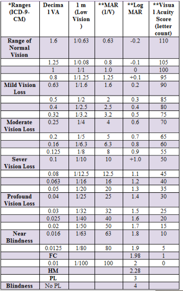
Retrospective study done through collection of the data from the Statistic office in Tobruk medical center-Tobruk and Alwahda hospital-Derna. The recorded data included clinical features of the most serious complication (endophthalmitis) of the post intra vitreal Avastin injection; the data was collected are visual acuity at diagnosis, age and sex of the patients, time of the disease, type, type of management. Additionally classified were operations protocol, type of organism that caused the disease, lab findings , systemic and local disease of affected patients, and the outcome for the patient through the VA findings. We depend on Ranges of vision loss and reading performance (Tab. No. 1) to a get the result of improvement of vision cases of post intraocular Avastin injection endophthalmitis (improvement of vision > 3 lines i.e > 15 letters on visual chart considered clinically a significant improvement). Statistical analysis was performed using Microsoft Office Excel program. Nominal and statistical significant variables were analysed using t- test for two samples. P value ≤ 0.05 or level (95%) was considered statistically significant.
Out of the 9 recorded patients of intravitreal injections endophthalmitis cases, there were 8 patients which receive the medication for treatment of diabetic macular edema, one case for treatment of age related macular degeneration AMD. The age average is 70 years, 6 cases were females and 3 cases were male, all of them were practice postoperative endophthalmitis. There were 8 patients had DM, 2 patients had heart disease and 1 patient had hepatitis C as a systemic disease.
There were 8 cases (89%) where complain from pain, redness and blurred of the vision and one case were pain non remarkable. There were 5 cases presented with synechia and hypopion in the anterior chamber and 4 cases without hypopion, all of the cases were with cells in anterior chamber and vitreous. All the cases after intravitreal medication injections where is acute i.e. the endophthalmitis occurs < 28 days, the most is in the first 10 days, and there are (66.7%) of cases occurs at first 3 days.
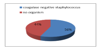
The most common causative organism after intra vitreous injection endophthalmitis is Coagulase-negative staphylococci (CNS) (100%) from the 56% positive causative organisms and 44% are no detected microorganisms, see the Figure.
There were 6 patients (66.7%) from all cases of after intravitreal injection were presented with near Blindness vision and two cases with sever blurred of vision and one case with mild blurred of vision. There were 7 patients managed with PPV + intra vitreous medication injection with washing the anterior chamber. The Log MAR Loss of Vision average at diagnosis is 1.98 and at discharge (after receive their management) is 1.3, the vision improved significantly after the management about 30 Letters i.e. improved (> 6 Lines). Although the cases which managed with PPV had worser visual acuity at time of diagnosis Log MAR 2.1 they had clinically better outcome after the management Log MAR 1.5 than the cases which managed without PPV (Log MAR at diagnosis 1.7 and 1.3 after the management), i.e. the outcome of cases of PPV improved significantly about 60 Letters (> 10 Lines) and cases managed without PPV >15 Letters (> 3 Lines).
Avastin is anti-vascular endothelial growth factor (anti-VEGF), these therapy has improved the quality of life for many patients with diabetic maculopathy, age-related macular degeneration, and other ocular diseases involving neovascularization and retinal edema. In these pathologies, the inhibition of intraocular VEGF is the only therapy that can preserve vision. [Semeraro et al., 2015].
Adverse events following intravitreal anti-VEGF injections has no relation to underlying ocular disease. The most common complications of intravitreal injection (IVI) are injection site discomfort, subconjunctival hemorrhage, vitreous reflux, transient increased intraocular pressure (IOP) and defect in the vision. The patient may also develop floaters, vitreous or retinal hemorrhage, and retinal detachment. [Frenkel MP, Haji SA & Frenkel RE, 2010]. The most serious complication of intravitreal injection is endophthalmitis and loss of vision. [Aiello et al., 2004].
In post intravitreal medication injection endophthalmitis, 44% of cases had positive organisms, all were Coagulase-negative staphylococci, making the most common causative organism in post- intravitreal medication injection endophthalmitis, was suggested with other studies. (Lyall et al. 2012 and Shah et al. 2011). The all cases of post- intravitreal medication were acute < 28, there were 6 patients (66.7%) occurred as acute endophthalmitis in the first 3 days after the injection. (Hoveenaars 2012 and Shah 2011).
Hoevenaars 2012 and Rudnisky 2013 reported the most cases of post IVI endophthalmitis cases treated with tap and injection of antibiotic. There were 7 patients (55.6%) had managed with PPV and intravitreal injection of antibiotic (vancomycin and dexamethasone), 2 patients had managed with only intravitral injection of antibiotic (vancomycin and dexamethasone) and there are 2 patients which had managed only with conservative treatment.
Peter Barry, Luis Cordovés, et al. 2013 and Zenith H. Y. Wu, Rose P. S. Chan, 2012, which advised immediately PPV make a significant outcome improvement cases of postoperative endophthalmitis, there were 7 patients managed with PPV + intra vitreous medication injection with washing the anterior chamber, the vision improved significantly after the management about 30 Letters i.e. improved (> 3 Lines). Although the cases which managed with PPV had worser visual acuity at time of diagnosis they had clinically better outcome after the management than the cases which managed without PPV, i.e. the outcome of cases of PPV improved significantly about (> 10 Lines) and cases managed without PPV Letters (> 3 Lines).
Although the postoperative endophthalmitis is sight threatening infection and destructive emergency disease, the appropriate prophylaxis is remain the best way of infection prevention. The delayed presentation, inappropriate management procedures, and severity of causative organism may account for the unfavorable visual outcome. In general post intravitreal endophthalmitis cases can be treatable condition especially when had diagnosed and managed early time with intravitreal antibiotics and early pars-plana-vitrectomy. Statistically we consist, the cases of acute intravitreal endophthalmitis in the first 3 day had significant better function outcome (P value = 0.024).
We would like to thank Mr. Hafez E.L Mansour (Lecturer at Tobruck University) for his help with data retrieval and statistical analysis.
CNV : Coagulase-negative-staphylococci
PPV : Pars-plana-vitrectomy
IVI : Intra vitreal injection
VEGF : Vascular endothelial growth factore
DM : Diabetes mellitus
CME : Cystoids macular edema
DMP : Diabetic maculopathy
AMD : Age related maculopathy
VA : Visual acuity
LP : Light perception
M : Hand movement
Dear Editorial Team, Clinical Medical Reviews and Reports. My experience with the journal was highly positive. The peer-review process was rigorous, constructive, and completed in a timely manner. The reviewers provided valuable comments that helped improve the quality and clarity of our manuscript. The editorial office was professional, responsive, and supportive throughout all stages of the publication process. Communication was clear and efficient, and any questions were addressed promptly. Overall, I found the journal to maintain high scientific standards and an excellent publication workflow. I would be pleased to consider submitting future work to this journal. Best wishes from, Elena Popa.

It was my pleasure to submit my testimonial concerning the Reviewer Board of our Scientific Journal “Brain and Neurological Disorders”. The Reviewers focused on some modifications and their contribution was helpful. The ladies of our Editorial Office were also supported my efforts. It was my honor to have such a co-operation and I am looking forward for more collaboration.

Dear Grace Pierce, Editorial Coordinator of Journal of Clinical Research and Reports, Thank you for the speedy and efficient peer review process. I appreciate the fact that your peer reviewers do not take months to respond like with some other journals. I would also like to thank the editorial office for responding quickly to my questions. It is an excellent journal. I plan to submit more manuscripts in the future. Best wishes from, Robert W. McGee

Dear Grace Pierce, Editorial Coordinator of Journal of Clinical Research and Reports, Working with you and your team on our recent publication in JCRR has been a truly wonderful and enjoyable experience. The responses were prompt, and the reviewers were patient, constructive, and highly professional. One reviewer in particular gave me the feeling that a professor was carefully reading and commenting on my coursework, which was deeply touching. The entire process was straightforward and hassle‑free, with no tedious online forms to complete. I highly recommend this journal. Best wishes from, DR Aibing Rao, Head of R&D

I Appreciate the Opportunity to Share my Experience with the Journal of Clinical Research and Reports. The peer review process was timely and constructive, and the feedback provided helped improve the quality of our manuscript. The editorial office was professional, responsive, and supportive throughout the process, ensuring smooth communication and efficient handling of the submission. Overall, it was a positive experience collaborating with your team.

Dear Mercy Grace, Editorial Coordinator of Obstetrics Gynecology and Reproductive Sciences, We would like to express our gratitude for your help at all stages of publishing and editing the article. The editors of the magazine answer all the necessary questions and help at every stage. We will definitely continue to cooperate and publish other works in the Obstetrics Gynecology and Reproductive Sciences! Best wishes from, Alla Konstantinovna Politova,
