
Research Article | DOI: https://doi.org/10.31579/2690-8794/032
*Corresponding Author: Osman Demirhan, Department of Medical Biology, Faculty of Medicine, Çukurova University, and 01330 Balcalı-Adana, Turkey, GMS: 05060229765.
Citation: Nesrin Çetinel, Osman Demirhan, Mustafa Demirtaş, Çağlar Emre Çağlıyan, Aslıgül Cüreoğlu, İnayet Nur Uslu, Yaşar Sertdemir; (2020) The Genotoxic Effect of Interventional Cardiac Radiologic Procedures on Human Chromosomes J, Clinical Medical Reviews and Reports. 2(5);DOI: 10.31579/2690-8794/032
Copyright: © 2020, Osman Demirhan:. This is an open access article distributed under the Creative Commons Attribution License, which permits unrestricted use, distribution, and reproduction in any medium, provided the original work is properly cited.
Received: 23 July 2020 | Accepted: 31 July 2020 | Published: 10 August 2020
Keywords: interventional radiology, angiography, coronary artery disease, cytogenetics, x-ray, contrast agents, chromosomal aberrations
In recent years, an important part of the ionizing radiation (IR) that human have been exposed for diagnostic purposes are interventional radiologic procedures. The X-rays and contrast media are used in angiography. The patients and staff members are exposed to X-ray during these procedures. While it is known that X-rays cause DNA damage and carcinogenesis, the effect of the contrast agent is still unknown. The aim of this study was to investigate the effect of X-rays and contrast agent on chromosomes of human patients. Peripheral blood samples were taken from 50 patients (30 males, 20 females, ages between 38-75 years). Chromosome analysis of peripheral lymphocytes in 50 patients were performed at 3 different periods; before the interventional radiologic procedure, 24 hours and 1-3 months after the procedure. Also, chromosome analysis was performed on 17 staff members working during interventional radiological procedures to investigate the effect of X-rays. Standard cytogenetic analysis techniques were used for this study. The frequency of chromosomal aberrations (CAs) was significantly higher in patients 24 hours after the interventional radiologic procedures than pretreatment (p=0,000). At the same time, CAs after 24 hours compared with those taken 1-3 months later, shown that the CAs were significantly reduced after 1-3 months (p=0,000). We also found that the frequency of CAs was also statistically higher in patients exposed to high radiation doses (p=0,042). Compared with the control group (n=30), CAs were found significantly higher in workers exposed to radiation. Our findings have shown that X-rays and contrast agents that used in interventional radiological cause chromosomal damage. For this reason, the dose of radiation to be given to the patient must be carefully selected. Due to the potentially high genetic damage of patients with coronary artery disease (CAD), the type and amount of medication to be given and the frequency of radiological diagnostic procedures to be performed should be meticulously adjusted.
Diagnostic ionizing radiation, such as X-rays are the largest man-made source of radiation exposure. The X-rays and iodinated contrast media are widely used for diagnostic radiology in angiography and in interventional radiology. Cardiac ionizing procedures expose both patients and medical staff to the highest radiation levels in diagnostic radiology. Exposure to IR may result in adverse health effect on patients and clinical cardiologists. There is a clear need to evaluate and establish biologic approaches for determining low-dose radiation effects in patients undergoing diagnostic X-ray procedures. Interventional cardiologists (ICs) are likely to receive high radiation exposure as a result of procedures they undertake. Unfortunately, many physicians are unfamiliar with radiation genotoxicity or the quantitative nature of the risks and IR risks are misunderstood. High radiation doses tend to kill cells, while low doses tend to damage or alter the genetic code of irradiated cells. Recently, as the number of diagnostic and interventional cardiac catheterisation procedures has greatly increased, serious radiation induced skin injuries and an excess of cataract development have been reported in exposed staff [1-3]. X-rays either acts directly on the DNA molecule or indirectly through the formation of reactive compounds that interact with the DNA molecule resulting in cytotoxicity of the cell [4]. IR can induce directly various forms of DNA damage, including the possibility of increasing the incidence of CAs and micronuclei (MN). CAs are the most fully developed biological indicator of IR exposure. It is widely accepted that there is an increased risk of cancer following exposure to IR; this is felt to be most likely due to damage to DNA strands during exposure [5]. However, a recent study has estimated that from 0.6% to 3% of all cancers are due to medical X-rays [6].
DNA double-strand breaks (DSBs) are the most relevant biologic damage induced by IR [7,8]. IR is uniquely energetic enough to overcome the binding energy of the electrons orbiting atoms and molecules; thus, these radiations can knock electrons out of their orbits, thereby creating ions. In biologic material exposed to x-rays, the most common scenario is the creation of hydroxyl radicals from x-ray interactions with water molecules; these radicals in turn interact with nearby DNA to cause strand breaks or base damage. The X-rays can also ionize DNA directly. Most radiation-induced damage is rapidly repaired by various systems within the cell, but DNA double-strand breaks are less easily repaired, and occasional misrepair can lead to induction of point mutations, chromosomal translocations, and gene fusions, all of which are linked to the induction of cancer [9]. IR can cause many types of DNA damage, in which DNA DSBs are most lethal, probably resulting in genomic instability and cause some diseases if unrepaired or misrepaired. Exposure of tissues and organs to IR can lead to cancer.
Despite all the above information, the aim of the present study is to investigate the effects of X-rays and contrast media on patients undergoing arteriography and interventional cardiologists.
The peripheral blood samples were collected by venipuncture from total 50 patients exposed to interventional radiological procedures (the X-ray and contrast medium) in the unit of angiography at the Department of Cardiology, Balcalı Hospital, Çukurova University, Adana, Turkey. The samples were taken: (1) before the procedures, (2) 24 hours after the examination and (3) 1-3 months after interventional radiological procedure in 25 patients. In the selected group of patients, 30 were males and 20 females, ranging in age from 38 to75 years (average age was 54, 31 years). In addition, the effects of the X-ray on chromosomes were investigated from 17 angiography workers (11 males and 6 females) occupationally exposed to the X-ray at the Balcalı Hospital, ranging in age 27 to 56 years and 30 individuals (17 males and 13 females) without any work-related exposure to ionizing radiation considered as control group whose ages ranged from 35 to 44 years. Informed written consent was obtained from all participants. The study was authorized by the Institutional Ethic Committee of Çukurova University.
Peripheral blood was collected into heparinised tubes from each subject for culture. Each sample was examined for expression of CAs in the Genetics Laboratory of the Department of Medical Biology and Genetics, Faculty of Medicine, Cukurova University. Lymphocyte cultures were set up by mixing 0.5 ml of whole blood samples, with 4.5 ml of PB-Max Karyotyping medium (Gibco). The cultures were incubated at 37oC for 72 h in an incubator, and colcemid (30 µl) was added 1, 5 h prior to harvesting. Standard cytogenetic techniques were used for harvesting and slide preparation. Three slides were prepared for each subject. The slides were prepared by trypsin GTG-banding and 50 metaphases/individuals were analysed on coded slides for structural CA, such as chromatid and chromosome breaks, deletions, acentric fragments, di-centric chromosomes, tetraploids, quadriradial exchange figures, and chromosomal exchanges. We conducted chromosome analysis using CytoVision software. Abberations were recorded according to the International System of Cytogenetic Nomenclature (ISCN).
SPSS 20.0 package program was used for statistical analysis of the data. Chi-square / Fisher Exact test was used to compare categorical measurements between groups. The Mann Whitney U test was used to compare the total irregularities without group distribution. The Wilcoxon Signed Rank test was used to compare pre-and post-treatment total irregularities without normal distribution. Statistical significance level was 0.05 in all tests. P values are given in tables with multiple comparison correction (Bonferroni).
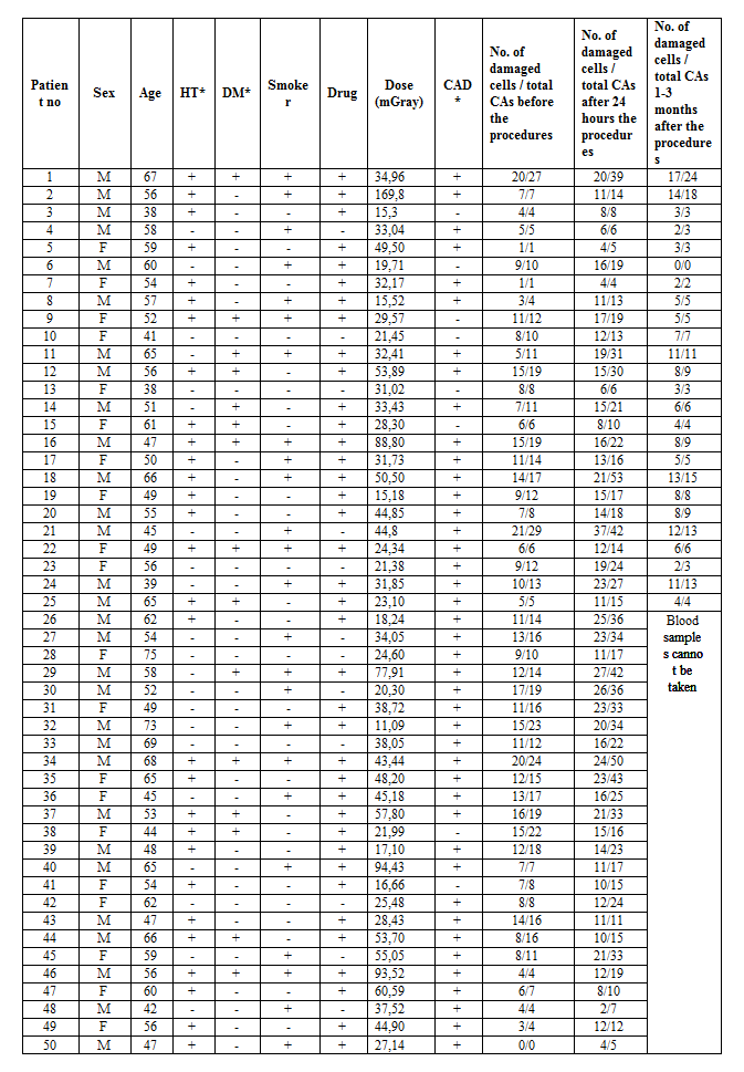
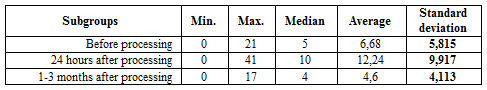
In our study; CAs occurred by analyzing 50 metaphase plates for each individual. Table 1 gives clinical, demographic characteristics and number of damaged cells for each patient.
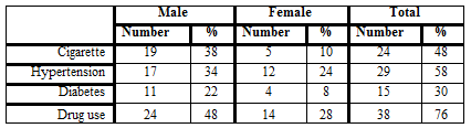
According to the clinical parameters (risk factors); 48% of the patients were smokers, 58% were hypertension, 30% had diabetes and 76% were using drugs (Table 2).
1. Chromosomal Abnormalities in Patients Before the Procedure
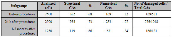
In this subgroup, 2500 cells were analyzed. One or more CAs were found in 459 of these cells. Total 531 structural (362, 68%) and numerical (169, 32%) CAs were observed (Table 3). Structural CAs were classified according to their significance; 27 (5,1%) deletions, 21 (4%) translocations, 2 (0,4%) duplications, 3 (0,6%) inversions, 20 (3,8%) chromatid breaks, 2 (0,4%) chromosome breaks, 8 (1,5%) dicentrics chromosomes, 2 (0,4%) neocentric structures, 238 (44,8%) fragile regions, 3 (0,6%) gaps and 36 (6,8%) 9qh+ chromosomes. Gonosomal chromosome mosaicisms; X chromosome loss was found in 3.5% (19 cases) and Y chromosome loss in 3.2% (17 cases) of the patients. XXXY was observed in one patient (0.2%), XXY in one patient (0.2%) and X chromosome in 4 patients (0.8%). Our study findings showed that X and Y losses mosaicisms were the most common chromosome aneuploidic mosaicisms in our patients. In addition, autosomal chromosome mosaicisms were observed in 3.9% (18 cases) of the patients. From these findings; autosomal monosomic mosaicisms (-20, -22, -18 and -21, respectively) were higher than autosomal trisomies (+21, +2, +8 and +18, respectively).
2. Chromosomal Abnormalities in Patients 24 hours after the procedure
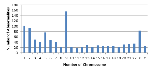
In this subgroup, 2500 metaphases were analyzed. A total of 1048 structural and numerical CAs were observed in 756 metaphases that including one or more various CAs (Table 3). A total of 1048 structural and numerical CAs were observed in 756 of 2500 cells examined (Table 3). These CAs were 73% structural and 27% numerical. Among the structural CAs; 6,8% (71) deletions, 3,7% (39) translocations, 0,3% (3) duplications, 0,7% (7) inversions, 0,2% (2) neocentric chromosomes, 0,4 (4) ascentric chromosomes, 0,1% (1) quadriradial chromosome, 4,1% (43) chromatid breaks, 0,4% (4) chromosome breaks, 1,1% (11) dicentrics chromosomes, 0,1% (1) isochromosome, 0,2% (2) ring chromosomes, 43,1% (476) fragile regions, 0,6% (6) gaps and 9,1% (95) 9qh+ chromosomes were found in the analyzed cells. In addition to observed structural CAs, 234 autosomal (22.3%) and 49 gonosomal (4.7%) numerical CAs were also detected. Among the gonosomal numerical CAs, 22 (2.1%) patients had X and 15 (1.4%) patients had Y chromosome loss, 2 (0.2%) patients had XXY and 10 (1%) patients had gonosomal chromosome mosaicisms such as +X [8] and +Y [2]. Autosomal monosomic mosaicisms (-22, -20, -21, -19 and -18, respectively) were higher than autosomal trisomic mosaicisms (+21). According to the findings obtained from the patients 24 h after the procedure the most affected chromosomes were 9, 1, 2, X, 5, 3, 6, 4 and 7, respectively (Figure 1).
3. Chromosome abnormalities at patients 1-3 months after the procedures
A total of 181 structural and numerical CAs were found in 166 defected metaphases of 1250 analyzed cells, at the subgroup 1-3 months after the procedure. The structural and numerical CAs were 119 (66%) and 62 (34%), respectively (Table 3). According to this findings, the proportions of fragilities, 9qh+, chromatid breaks, deletions, translocations and others were %42 (78), %8,8 (16), %6,6 (12), %3,8 (7), %2,7 (5) and %0,6 (1), respectively. Sixty-two (34%) numerical CAs were found of analyzed cells. Observed autosomal chromosome aneuploidic mosaicisms; -8 [4], -10 [1], -11 [1], -12 [1], -13 [2], -14 [1], -15 [5], -16 [2], -18 [3], -19 [2], -20 [6], -21 [1], -22 [6]; +20 [1] and +22 [1]. In addition, 1 marker chromosome was also observed. Gonosomal chromosome aneuploidic mosaicisms were; -Y [19], -X [4] and + X [1].
The mean values of CAs observed before the procedure, after 24 hours and after 1-3 months are shown in Table 4 and Figure 2.
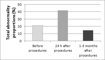
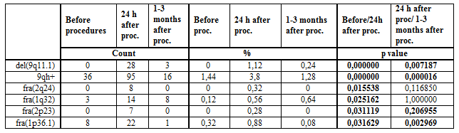
In the patient group, CAs (531/2500) observed before the processing were statistically significant (p=0,000) when compared with the abnormalities (1048/2500) of the subgroup 24 hours after the procedure (181/1250) (p=0,000). Furthermore, the chromosomal damages obtained 24 hours after the processing was compared with that after 1-3 months (181/1250), we found that the damage decreased statistically (p=0,000). Compared CAs of the before the processing and 24 hours after the processing, we found that abnormalities such as fra(1p36.1), fra(1q32), fra(2p23), fra(2q24), del(9q11.1) and 9qh+ (p=0.031629, 0.031119, 0.015538, 0.025162, 0.000000, 0.000000, respectively) were increased statistically (Table 5). This indicates that 1, 2 and 9 chromosomes are more affected than other chromosomes (Figure 1). Moreover, del(9q11.1), 9qh+ and fra(1p36.1) abnormalities were found significantly decreased when compared with 1-3 months after the processing (p=0,007187, 0,000016 and 0,002969) (Table 5).
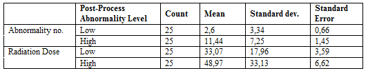
However, CAs were statistically higher (p = 0,000) in the patients before the processing (18.4%) than the control group (1.5%). For this reason, it was investigated whether patients were associated with CAs before processing with age, gender, smoking, drug use, DM, HT status and CAD. Accordingly, it was seen that there was no statistically significant relation between CAs and these factors (except sex) before the operation (page = 0.25, psmoker = 0.28, pHT = 0.089, pDM = 0.21, pdrug = 0.79, pCAD> 0.05). When CAs and sex of patients were compared before processing, it was statistically determined that CAs were higher in male patients (p = 0.042). It was found that the number of CAs was high in patients receiving high doses and this result was statistically significant (p = 0.042) (Table 6).
4. Findings from radiological workers and control groups
In this group, a total of 17 personnel (6 female and 11 male) working in angiography and radiological procedures and a total of 30 healthy (non-smokers and 17 women and 13 men) with no family history of cancer were compared in terms of CAs as a control group. The age range of the group of personnel was between 27-56, the overall age average was 41.23 ± 6.81 while the age range of the control group was 40-44, and the mean age was 37,13. 850 metaphase cells were analyzed from angiography workers occupationally exposed to the X-ray. Structural and numerical CAs were found in 134 (15.8%) of the cells. The 122 (14,4%) of these damages were structural CAs (5 translocations, 7 deletions, 2 inversions, 2 disentric chromosomes, 17 chromosomal local increases, 70 fragilites, 13 chromatid breaks, 1 chromosomal break, and 5 9qh+). In addition, 12 (1,4%) numerical gonosomal and autosomal aneuploidic mosaicisms were detected (the loss of X chromosome in 5 cells, the loss of Y chromosome in 2 cells, autosomal numerical deviations of -8 [1], -18 [1], -20 [1] and -21 [2]). In the control group, 22 (1.5%) structural CAs were detected in 1500 analyzed cells. All of these CAs were fragilities [fra(2p24), fra(5q31), fra(2q31), fra(6p21), fra(11q22), fra(6q25), fra(2q31), fra(2q13), fra(1q21), fra(1p36.1), fra(4q21), fra(12q24), fra(1q42), fra(1p32), fra(1q42), fra(3q27), fra(5q31), fra(6q23), fra(5q31), fra(2q31), fra(1q21) and fra(2q31)], and all of fragilities was found in 1/50 ratio.
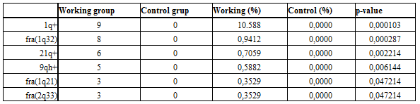
When the total chromosomal damages of the working and control groups were compared; statistically significant difference were found between the two groups (p = 0,000). According to this; the proportion of chromosomal damage in the working group was significantly higher than that of the control group. 1q+, fra(1q32), 21q+, 9qh+, fra(1q21) and fra(2q33) chromosome regions were found statistically higher in the working group (Table 7). Accordingly, it appears that the damages to these 6 chromosomal regions have increased significantly in the radiation exposed personnel.
Diagnostic X-ray procedures are the largest man-made source of IR exposure, and estimation of the risk is difficult. However, IR can be considered as a ‘two-edged sword’ in that it may lead to genetic modifications in exposed, surviving normal tissue. DNA damage is one of the detrimental effects of IR. But, the ability of angiography to produce DNA lesions has yet to be robustly demonstrated. Although many experimental in vitro studies have suggested DNA damage after exposure to X-rays and contrast media, we present in vivo results documenting that angiography scanning in daily clinical routine is associated with increased lymphocyte CAs in this study. But, this is not the first study to suggest that cardiac imaging may be able to cause CAs in peripheral blood lymphocytes tested before and after a IR and contrast media scan.
In the present study; CAs rate was compared with the pre-procedural patients (18.4%) and healthy control group (1,5%); the rate of chromosome damage was found to be very high in pre-procedural patients (p=0,000). The obtained CAs were found to be 68% structural and 32% numerical. The most common damage among these CAs is fragility and this is followed by deletion, translocation, chromatid and chromosome breaks, dicentric and other important structural damages. The fragility may be related to abnormalities in replication, resulting in single-strand DNA gaps, which, if not repaired, may lead to CAs such as deletions within the FS, or translocations or other rearrangements involving breakage at a FS [10]. These findings show that the CAs were significantly higher in cardiovascular patients compared to people who are not exposed to X-rays or healthy control group. Thus, our study found that the overall frequency of CAs was significantly higher in pre-angiography patients compared to the control group. What is the cause of these CAs observed before the patients are exposed to IR and contrast media?
NA damages have been reported to cause the development and progression of CAD [11]. The results of the present study have shown significant increase in genetic damage in lymphocytes of patients with CAD. It has often been reported that genetic damage may be caused by both, extrinsic and intrinsic environments [12]. The 58%, 30%, 48% and 76% of our patients with CAD had hypertension, diabetes, smoking and using drugs, respectively. Hypertension, obesity, hyperlipidemia, stress, smoking, age, sex, and family history are reported to increase risk for
CAD [13]. In our patients before exposed to X-rays, high CAs may be also associated with CAs in risk factors such as smoking, drug use, hypertension, diabetes, stress and age. Indeed, it has been reported that DNA damage in patients with CAD is higher than in controls [14,15]. It had documented that DSBs, oxidized pyrimidines and altered purines were significantly higher in leukocytes of patients with CAD [16]. Oxidative damage increases in aging and age related diseases [17, 18]. However, in some studies; excessive oxidative stress has been reported to play a role in the pathogenesis of cardiovascular disease. Increased production of oxidant free radicals was observed in patients with ischemic heart disease [19]. DNA and CAs can also directly or indirectly stimulate reactive oxygen species, thus providing the basis for cancer formation [20].
A progressive increase in spontaneous chromosome instability/chromosomal loss due to the ageing process is associated with the accumulation of DNA damage due to an age-related decline in DNA repair capacity [20]. Mammalian cells respond rapidly to DNA damage caused by external agents such as IR by rapidly activating the molecular machinery which aims at maintaining genomic integrity and thus preventing carcinogenic mutations. Some studies have found positive association of age with micronuclei acount [21, 22] while others have not [23]. Consequently, individual genetic differences in the ability to repair DNA damage may result in a different susceptibility towards the iodinated contrast agents and, therefore, increase the risk of developing exposure-related disease. The incidence of CAD and diabetes increases especially in people with metabolic syndrome including metabolic disorders such as obesity, hypertension and insulin resistance [17]. Therefore, genetic damages of drugs used in metabolic disorders cannot be ignored. In addition, cardiovascular drugs such as diuretics, beta blockers, calcium antagonists and ACE inhibitors have also been shown to stimulate cancer [24]. Studies have reported that aspirin causes CAs in Chinese hamster ovaries, lung fibroblast cells, and mouse germ cells [25, 26]. The 76% of our patients use drugs (coraspin, beloc, etc.). This suggests that the drugs used by patients may cause genotoxic effects and increase CAs. However, there was no statistical significance between pre-procedural damage and drug use in our study (p = 0.79). The CAs rate in our patients before and 24 hours after treatment was higher in male than in women and this difference was statistically significant (p=0.042). This suggests that X-rays and contrast media may cause more genetic damage in male patients. In one study, it was reported that DNA damage rates in chronic heart patients were not statistically different according to sex [27].
Contrast media is frequently used together with the X-ray in angiography and in interventional radiology. Previous studies have demonstrated that these compounds induce CAs. For this, cytogenetic analysis findings of diagnostic doses of the X rays and contrast media were investigated in experimental studies on cell cultures in vitro [28,29]. Parallel clinical investigations showed an increased genotoxicity in the peripheral blood lymphocytes of the patients undergoing angiography [29-31]. It is well recognized that iodinated contrast media have a cytotoxic effect, and this is felt to be one of the mechanisms responsible for contrast-induced nephrotoxicity [32]. In the present study, patients exposed to the X-rays and contrast media experienced CAs in 30.3% of the cells analyzed after 24 hours. It was found that 73% and 27% of these CAs were structural and numerical mosaicism, respectively. The 43.1% of the structural CAs were found to be fragile, and 18.7% were other significant damages (deletions, chromatid and chromosomal breaks, translocations, dentric, ascentric and marker chromosomes). When compared with the damage observed before the procedures; we found that this was statistically significant (p=0,000), and this significant difference is thought to originate from the X-ray and contrast media.
These findings show that CAs increase significantly in 24 hours after the procedure and that the X-rays and contrast media causes fragility, gaps and breaks on the chromosomes. In fact, it reported that interventional radiological procedures increased CAs in patients after the procedure. DSBs are the most relevant biologic damage induced by IR [34, 35]. The most prominent risk of iodinated contrast agents is nephrotoxicity, rare in patients without a history or symptoms of renal disease. The incidence of kidney injury was 1.3% after percutaneous coronary intervention [36]. Some studies [32, 37, 38] have suggested an association between contrast media and increased DSBs in individuals exposed to low-dose radiation but not necessarily high-dose radiation. The characterization of FS has demonstrated that they are associated with genes that relate to tumorigenesis and behavioural disorders [39, 40]. However, a study has estimated that from 0.6% to 3% of all cancers are due to medical X-rays [41]. It is widely accepted that there is an increased risk of cancer following exposure to IR; this is felt to be most likely due to damage to DNA strands during exposure. Damage to DNA strands can be demonstrated following exposure to X-rays, and new evidence is emerging that this effect may be compounded by administration of iodinated contrast agents. We conclude that application of iodinated contrast media leads to an increase in the extent of DNA damage following irradiation for diagnostic imaging purposes.
In the present study; CAs most frequently seen 24 hours after the procedure; the distribution of damages according to chromosomes appears to be mainly composed of chromosomes 9, 1, 2, X, 5, 3, 6, 4 and 7, respectively, and it was compared with those of the 6 regions on the 3 chromosomes (1p36.1, 1q32, 2p23, 2q24, 9q11.1-q13 and 9qh+) were statistically significant (p=0.031629, 0.031119, 0.015538, 0.025162, 0.000000, 0.000000, respectively). These findings show that chromosomes 1, 2, and 9 are significantly affected by the X-rays and contrast media. Since these chromosome-regions contain innumerable oncogenes, tumor suppressor genes, enzyme genes responsible for DNA repair mechanisms, genes responsible for apoptosis, or candidate genes, these damages may be considered as a risk factor for cancer risk. These overexpressed 1q32, 2q24 and 2p23 chromosome regions decreased significantly in 1-3 months after the procedure, but 1p36.1 region was still highly sensitive (p= 0.002969). This indicates that the damages occurred after 24 hours from the procedure was corrected/repaired at later times. However, it turns out that 1p36.1 region is still unrepairable and sensitive. Most radiation-induced damage is rapidly repaired by various systems within the cell, but DSBs are less easily repaired, and occasional misrepair can lead to induction of point mutations, chromosomal translocations, and gene fusions, all of which are linked to the induction of cancer [9]. It was found that p36.1 and q32 regions on chromosome 1 were the most affected, 24 hours after interventional cardiac procedure, and it was statistically significant (p=0,031629, p=0025,162) in our patients. The chromosome 1 has been reported to contain a large number of tumor suppressor genes in the short arm, leading to the formation of solid tumors. It has also been reported that the 1p36 band is a cancer breakpoint [42]. At the same time, the q24 and p23 regions of chromosome 2 were also significantly stimulated.
In the present study, the most affected chromosomes, by the X-rays, was chromosome 9. These structural chromosomal defects were observed as heterochromatin positive, chromatid and chromosomal breaks, deletions, translocations, inversions and isochromosome. Among these CAs, 9qh+ was the most common. Although this structure is considered polymorphism, clinical outcomes remain uncertain. Despite this uncertainty, our findings suggest that the X-rays and contrast media cause the increase of heteromaterial in the periscentromeric region of chromosome 9. Other structural damages on chromosome 9; 9p and 9q deletions were frequently observed in various malignancies [43-45]. It has been reported that deletions of the 9p are common in melanoma, glioma, leukemia and NSCLC cases [44]. NSCLC patients were reported to have undergone 90% deletion of 9p and breaks between 9q13-9p22 regions [46]. In patients with adenocarcinoma, it has been suggested that the 9q line contains multiple tumor suppressor genes [45]. Consequently, we can easily say that the deletion in the q11.1-q13 region, which is frequently repeated in one of our patients, will play an important role in cancer formation.
Numerous CAs are a common feature of some cancers, suggesting that this is a potent stimulus for tumor development by increasing genomic instability. Our patients have shown that autosomal and gonosomal mosaic CAs are high before and 24 hours after the procedure, and the X-rays and contrast media cause gonosomal mosaicisms (-X and -Y chromosomal monosomes). However, autosomal monozomic mosaicisms (-20, -22, -18, -21, -19) are more common than autosomal trisomic mosaicisms (+21, +2, +8, +18) among autosomal aneuploidic mosaicisms. This also shows that there are sex CAs in the beginning of numerical mosaicisms errors observed in our patients. We can say that increasing numerical sex chromosomes as a result of angiographic procedures can increase sensitivity to tumors. Because, numerical deviations of different chromosomes are reported to be associated with aggressive tumor behavior [47]. Some studies have also shown that structural and numerical sex chromosomal alterations are frequently seen in patients with lung cancer [48, 49]. Thus, numerical deviations of chromosome X have been reported to affect carcinogenesis and poor outcome of different tumor types [50]. One X-chromosome increase has been reported to be relatively common with other karyotypic changes in leukemia, lymphoma, and prostate cancer [51, 52]. In addition, Y chromosome loss has been reported to be common in the cancer cells and several leukemia, papillary renal cell carcinoma, prostate cancer, male chest carcinoma, and pancreatic adenocarcinoma. Further studies have supported the loss of Y chromosome to be a non-phenotypic event related to the aging process in men [53]. Other some studies have shown that age is not clearly related, and that the lost X and Y chromosomes appeared after treatment and in clinical remission. For this reason, it has been concluded that the development of a malignant clone in cancer tissue and the development of sex chromosome loss are more accurate. In men, there is a relationship between bladder cancer and loss of Y chromosome [54]. In addition, Y chromosome loss is common in some cancer types, including pancreatic renal cell carcinoma, and in cancer cells and in some leukemias [55]. All this informations show that numerical sex-chromosome deviations may play a role in the pathogenesis of cancer.
We found that the difference in CAs observed in patient cells analyzed 1-3 months after operation was statistically significant (p=0,000) and the rate of damage significantly decreased when compared 24 hour after the operation. CAs were found in 13.3% of the analyzed cells. The 42% of CAs were found to be fragile, and 15.2% were found to be chromatid breaks, deletions, translocations and disentric chromosomes. Obtained findings reveal that CAs observed before the procedure increased 24 hours after the operation and decreased after 1-3 months. We believe that the reduction in CAs at 1-3 months after the operation has been repaired by DNA repair enzymes and / or damaged cells are destroyed by apoptosis.
Today, interventional cardiologists represent, indeed, the most important group of exposed among professionally exposed physicians. Our study found that the overall frequency of CAs was significantly higher in intermittent personnel working compared to a control group. Workers experienced significant structural damage other than fragility, especially chromatid breaks, inversions and disentric chromosomes. It is seen that the chromosomes 1, 2 and 21 are more affected and damaged in the cells of personnel exposed to radiation. During the imaging process, employees as well as patients are exposed to X rays at significant levels. Exposure to the X-rays of low-dose and long-term or intermittent personnel working in radiological procedures (Physician, Health Technician and Nurse) can prepare the ground for illnesses which may arise after years and can create risks both for patients and employees. It is understood that CAs to working personnel is a consequence of exposure to low dose and long X-rays. The present observations agree with many cytogenetic studies carried out in workers exposed to the X-ray [56-58]. This result also confirms earlier studies [59] that reported a higher frequency of chromatid and chromosome breaks in people occupationally exposed to the X-rays. In a study, it has been reported that chromosomal breaks and disentric chromosomal damages in blood samples of 37 interventional cardiologists are significantly higher than in the control group. In other studies, 50 radiology technicians and control group exposed to long X-rays; it was reported that the radiologist had a mitotic index of 8.2%, leukocyte count of 14,4% and lymphocyte count of 13,3% lower than the control group [58, 60]. Some studies have reported on ring chromosomes in addition to disentric and ascentric chromosomal fragments [56, 61]. In our study, disentric, ascentric and ring chromosome occurrences were found only slightly in the patient and working group. As a result, all these studies show that the elements working in angiography and radiological imaging processes have high genetic damage rates.
The present study demonstrated that before processing, the rate of CAs in patients with CAD was significantly higher than that of the healthy control group. We can say these CAs may be due to cumulative accumulation of internal and external risk factors such as older age, smoking and drug use, diabetes and hypertension. The rate of CAs significantly increased after 24 hours from the operation in patients, indicating that the X-rays used in interventional cardiac radiological procedures and the iodinated opaque material lead to fragilities, gaps, chromatid and chromosomal breaks. In angiography, intravenous iodinated contrast agents can increase DNA damage in addition to radiation. Thus, there is a clear need to evaluate and establish biologic approaches for determining low-dose radiation effects in patients undergoing diagnostic the X-ray procedures. We can say that chromatid and chromosome breaks are very common among structural CAs, the most important primary genetic damage induced by IR, and the predominant indicator of malignancy.
CAs rate in the male patients was higher than the female patient, indicating that X rays caused more genetic damage in male patients. For this reason, male patients need to be more sensitive about radiation. The amount of radiation given to the patients increases as the level of CAs increases. For this reason, the dose of radiation to be given to the patient must be carefully selected. Due to the potentially high genetic damage of patients with CAD, the type and amount of medication to be given and the frequency of radiological diagnostic procedures to be performed should be meticulously adjusted. Patients were observed to have CAs declined significantly after 1-3 months from the operation. This indicates that CAs caused by X-rays and opaque material has been repaired or that damaged cells have been removed by apoptosis. The fact that the extent of such damage may be enhanced by administration of iodinated contrast media will make the imaging community consider in more detail
CAs rate in the male patients was higher than the female patient, indicating that X rays caused more genetic damage in male patients. For this reason, male patients need to be more sensitive about radiation. The amount of radiation given to the patients increases as the level of CAs increases. For this reason, the dose of radiation to be given to the patient must be carefully selected. Due to the potentially high genetic damage of patients with CAD, the type and amount of medication to be given and the frequency of radiological diagnostic procedures to be performed should be meticulously adjusted. Patients were observed to have CAs declined significantly after 1-3 months from the operation. This indicates that CAs caused by X-rays and opaque material has been repaired or that damaged cells have been removed by apoptosis. The fact that the extent of such damage may be enhanced by administration of iodinated contrast media will make the imaging community consider in more detail.
Our findings show that the frequencies of CAs were significantly higher in the interventional cardiologists compared to people who are not exposed to X-rays, and X-rays alone increase genetic damage. Or, interventional cardiologists are likely to receive high radiation exposure as a result of procedures they undertake. For this reason, low-dose and long-term exposure to X-rays of personnel working in radiological procedures (Physician, Health Technician and Nurse) can prepare the illnesses that may arise after years. Therefore, it is clearly necessary to continually monitor both the potential risks and safety of ultrasound exposure. Furthermore, the exact risk at very low doses to a specific individual can be further complicated by many factors, such as carcinogenic agents in our environment, cigarette smoke, diet and genetic back ground. In contrast, exposure to IR may result in adverse health effect on both cardiologists directly and on their progeny. All these informations are in the light; patients should be thought to be more susceptible to DNA damage, depending on the risk factors of CAD, and patients should avoid inappropriate radiological examinations. Physicians and patients should be more careful in this regard, unless X-ray and nuclear imaging techniques are necessary to prevent genetic damage. However, other risk factors other than radiation in CAD should be considered collectively and compared with the risks associated with unidentifiable diseases. İnterventional cardiologists and personnel working have the highest radiation exposure among health professionals.
This work was supported by the Scientific Research Office of Çukurova University (I.U.BAP) (Project no: TYL-2015-5066)
Dear Editorial Team, Clinical Medical Reviews and Reports. My experience with the journal was highly positive. The peer-review process was rigorous, constructive, and completed in a timely manner. The reviewers provided valuable comments that helped improve the quality and clarity of our manuscript. The editorial office was professional, responsive, and supportive throughout all stages of the publication process. Communication was clear and efficient, and any questions were addressed promptly. Overall, I found the journal to maintain high scientific standards and an excellent publication workflow. I would be pleased to consider submitting future work to this journal. Best wishes from, Elena Popa.

It was my pleasure to submit my testimonial concerning the Reviewer Board of our Scientific Journal “Brain and Neurological Disorders”. The Reviewers focused on some modifications and their contribution was helpful. The ladies of our Editorial Office were also supported my efforts. It was my honor to have such a co-operation and I am looking forward for more collaboration.

Dear Grace Pierce, Editorial Coordinator of Journal of Clinical Research and Reports, Thank you for the speedy and efficient peer review process. I appreciate the fact that your peer reviewers do not take months to respond like with some other journals. I would also like to thank the editorial office for responding quickly to my questions. It is an excellent journal. I plan to submit more manuscripts in the future. Best wishes from, Robert W. McGee

Dear Grace Pierce, Editorial Coordinator of Journal of Clinical Research and Reports, Working with you and your team on our recent publication in JCRR has been a truly wonderful and enjoyable experience. The responses were prompt, and the reviewers were patient, constructive, and highly professional. One reviewer in particular gave me the feeling that a professor was carefully reading and commenting on my coursework, which was deeply touching. The entire process was straightforward and hassle‑free, with no tedious online forms to complete. I highly recommend this journal. Best wishes from, DR Aibing Rao, Head of R&D

I Appreciate the Opportunity to Share my Experience with the Journal of Clinical Research and Reports. The peer review process was timely and constructive, and the feedback provided helped improve the quality of our manuscript. The editorial office was professional, responsive, and supportive throughout the process, ensuring smooth communication and efficient handling of the submission. Overall, it was a positive experience collaborating with your team.

Dear Mercy Grace, Editorial Coordinator of Obstetrics Gynecology and Reproductive Sciences, We would like to express our gratitude for your help at all stages of publishing and editing the article. The editors of the magazine answer all the necessary questions and help at every stage. We will definitely continue to cooperate and publish other works in the Obstetrics Gynecology and Reproductive Sciences! Best wishes from, Alla Konstantinovna Politova,
