
Research Article | DOI: https://doi.org/10.31579/2690-8794/085
1 Kujawy University, Mechanical Department, Hallera 32, 86-300 Grudziadz, Poland and CORSAR Engineering Industry, Glogowa 2, 86-031 Osielsko, Poland
2 Tribochemistry Consulting, Salt Lake City, UT 84117, USA and University of Economy, Biotribology Lab, Garbary 2, 85-229, Bydgoszcz, Poland.
*Corresponding Author: Z. Pawlak, Tribochemistry Consulting, Salt Lake City, UT 84117, USA and University of Economy, Biotribology Lab, Garbary 2, 85-229, Bydgoszcz, Poland.
Citation: M. Sojka and Z. Pawlak (2021) The effect of deactivated phospholipids on joints lubrication: Osteoarthritis and lubricating properties J, Clinical Medical Reviews and Reports. 3(6); DOI: 10.31579/2690-8794/085
Copyright: © This is an open access article distributed under the Creative Commons Attribution License, which permits unrestricted use, distribution, and reproduction in any medium, provided the original work is properly cited.
Received: 12 May 2021 | Accepted: 01 June 2021 | Published: 17 June 2021
Keywords: cartilage degradation; phospholipids deactivation; β2-glycoprotein I, β2-GP; osteoarthritis; lubricin; hyaluronan
PLs bilayers coating the major synovial joints such as knees and hips as the lubricant are responsible for the lubrication of articular cartilage. Lamellar-repulsive effect has been considered as a lubrication mechanism but it is likely that lubricin and hyaluronan with PLs participate in the lubrication process. The molecules of lubricin and hyaluronan adsorbed by PLs have a supportive role and provide the efficient lubrication of synovial joints via the hydration mechanism (~ 80% water content). Lipid profiles of injured and healthy knees’ synovial fluids show significant differences. The phospholipid content in synovial fluid (SF) during joint inflammation, osteoarthritis is significantly higher (2 to 3 times) above the normal concentration of PL, and has a poor boundary-lubricating ability because of deactivated PL molecules. Deactivated PL molecule has no ability to form bilayers, lamellar phases, and liposomes.
Surface-active phospholipids have been experimentally proved to be present in the synovial fluid and on the surface of articular cartilage, surface amorphous layer (SAL) [1, 2, 3]. Moreover, they play an essential role on the surface of articular cartilage in the process of lubrication. However, when the total amount of phospholipids increases in synovial fluid, this raises the question about surface deterioration transforming the hydrophilic layer into the hydrophobic surface [4].
Friction and lubrication are surface processes, only strongly adsorbed moieties to the surface are a primary lubricant and have importance roles in friction (charged macromolecules in synovial fluid). Osteoarthritis is teaching us about the importance of involvement of active phospholipids in the lubrication mechanism of hyaluronan and lubricin [3, 5, 6].

Some researchers proposed a lubrication mechanism based on lubricin and hyaluronan complexes with phosphatidylcholines to provide a remarkable lubrication of synovial joints via hydration mechanism [8, 9]. In mammals, the intact lipid layer of cartilage is lost during degeneration, thus affecting the efficient lubrication of the joint [2, 3, 4].
In this study under osteoarthritic condition deactivated PLs molecules lost ability of bilayers formation and were unable to adsorb to lubricin and hyaluronan molecules. In healthy joints PLs bi-layers provide the low friction (μ≈0.005). In osteoarthritic condition surface-attached lubricin with non-active PLs leads to considerably higher friction. In osteoarthritic condition where hyaluronan, non-active PLs and lubricin act separately, this cannot provide good boundary lubrication in
articulating joints.
Mass spectrometry of phospholipids. The phospholipid species were quantified by ESI-MS/MS on Micromass [5, 6]. The research was carried out using synovial fluid derived from undamaged controls and patients with early and late osteoarthritis and rheumatoid arthritis. The authors classified 130 species of lipids. After comparing control synovial fluids, SF of patient with early and late OA had higher levels of most PLs species. Most of the PL data for this paper was taken from Kosinska et al. [5, 6]
Deactivation of a surface-active phospholipid bilayer (SAL). This amphoteric nature of phospholipids allows them to self-assemble into a classic arrangement which represents the basics of all biological membranes. A surface-active phospholipid layer covers normal articular surfaces in a multi-bilayer structure [3]. The bilayers serve to integrate interfacial functions between surfaces and have been a subject of several inquiries due to its tribological features [5, 10]. However, at sites of articular cartilage damage, the SAPL is absent, because a suitable substrate upon which this vital lipid layer can form does not exist [5, 7].
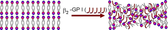
The mechanism of osteoarthritis (OA) is still not fully understood, but it has been established that this debilitating disease is often accompanied by a change in the synovial fluid composition, reduction in viscosity and deterioration of cartilage surface. Well-defined outermost bilayers were clearly visible on healthy cartilage surface, but OA may involve in the depletion of important joint molecules and SAPLs on the articular surface [2, 3]. Further, evidence for SAPL lining depletion was demonstrated by the cartilage wettability contact angle changing from 103 to 65 degrees. This insight led to the hypothesis that the SAPL is deactivated in the pathologic state of OA and remains present in synovial fluid but an inactive state (see Figure. 1). The pathological synovial fluid contains three times more phospholipids (PL), but the cartilage structure changes and its ability to lubricate, is remarkably poor. During normal functioning, the SAPL serves as a sacrificial perturbation bilayer, whereby it can improve by self-assembly mechanisms [10].
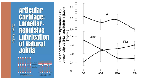
The concentration of components of synovial fluid (SF), such as hyaluronan (A-), lubricin, and surface-active phospholipids, from unaffected controls (or normal), eOA, lOA, and RA in the human synovial fluid are shown in Fig. 2 (B). During osteoarthritis (OA), and rheumatoid arthritis (RA), contain less hyaluronan (A-), and lubricin and 2 to 3 times more with phospholipids. Also, the MW distribution of (A-) shifted toward the lower range in OA and RA SF. These results indicate that activities in OA and RA SF are enhanced, leading to decreased levels of lubricin and high-MW hyaluronan (A).
Cartilage destruction in most rheumatic diseases and osteoarthritis has generally been accepted as a mechanism of deactivation of phospholipid bilayers. An acid-base interaction occurs between protonated amino acid group (-NH3+) of β2-Glycoprotein I and the phospholipid (–PO4-) group: (-NH3+) + (–PO4-) → (-NH3+ PO4--) that is strong enough to deactivate the PLs bilayer surface.
β2-Glycoprotein I (β2-GPI) is a protein that circulates in blood at variable levels (50–500 μg mL−1 with a molecular weight of 50 kDa. β2-Glycoprotein I (β2-GP I) can exist in (a) closed conformation and (b) the open hockey stick-like conformation when β2-GP I. β2-GP I in its hockey stick-like conformation is a strongly adhesive protein and binds to different receptors on cells. Binding of β2-GP I to anionic charged phospholipid (–PO4-) groups at pH ~ 7.4, results in a change in conformation and exposure of the epitope for the autoantibodies [11, 12, 13].
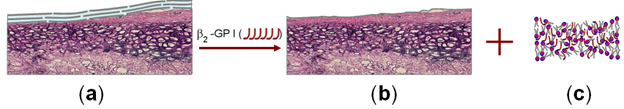
Softening of the cartilage is the first phase of cartilage deterioration [3]. Classic morphological changes of osteoarthritic articular cartilage begin with fibrillation and local surface disorganization involving splitting of the superficial layers of the cartilage, Figure 3. The early splitting is tangential with the cartilage surface, following the axes of' the predominant collagen bundles. Continued deterioration of articular cartilage leads to an exposure of the subchondral bone and more generalized synovial change. To understand the processes leading to cartilage failure is important to look at the cellular processes and biochemical structure of the normal cartilage.
A phospholipid involved in natural articular joints has a layered structure with weak inter-layer forces and low-strength shearing characterizes solid phospholipid lubricants. In this study, it was observed that osteoporosis significantly altered the phospholipid content so that the lipid profile was substantially changed from surface active to being deactivated (β2-GPI-NH3+) + (PLs–PO4-) → β2-GPI (-NH3+ PO4--PL) with a poor boundary-lubricating ability. Some have postulated or suggested that these lipids indicated “excessive tissue destruction”. Deactivated PL molecule is unable to form bilayers, lamellar phases, and liposomes.
We gratefully acknowledge Ms Joanna A. Pawlak for editing this manuscript and for the financial supports from the Tribochemistry Consulting (Grant no. 01. 2019) .
Dear Editorial Team, Clinical Medical Reviews and Reports. My experience with the journal was highly positive. The peer-review process was rigorous, constructive, and completed in a timely manner. The reviewers provided valuable comments that helped improve the quality and clarity of our manuscript. The editorial office was professional, responsive, and supportive throughout all stages of the publication process. Communication was clear and efficient, and any questions were addressed promptly. Overall, I found the journal to maintain high scientific standards and an excellent publication workflow. I would be pleased to consider submitting future work to this journal. Best wishes from, Elena Popa.

It was my pleasure to submit my testimonial concerning the Reviewer Board of our Scientific Journal “Brain and Neurological Disorders”. The Reviewers focused on some modifications and their contribution was helpful. The ladies of our Editorial Office were also supported my efforts. It was my honor to have such a co-operation and I am looking forward for more collaboration.

Dear Grace Pierce, Editorial Coordinator of Journal of Clinical Research and Reports, Thank you for the speedy and efficient peer review process. I appreciate the fact that your peer reviewers do not take months to respond like with some other journals. I would also like to thank the editorial office for responding quickly to my questions. It is an excellent journal. I plan to submit more manuscripts in the future. Best wishes from, Robert W. McGee

Dear Grace Pierce, Editorial Coordinator of Journal of Clinical Research and Reports, Working with you and your team on our recent publication in JCRR has been a truly wonderful and enjoyable experience. The responses were prompt, and the reviewers were patient, constructive, and highly professional. One reviewer in particular gave me the feeling that a professor was carefully reading and commenting on my coursework, which was deeply touching. The entire process was straightforward and hassle‑free, with no tedious online forms to complete. I highly recommend this journal. Best wishes from, DR Aibing Rao, Head of R&D

I Appreciate the Opportunity to Share my Experience with the Journal of Clinical Research and Reports. The peer review process was timely and constructive, and the feedback provided helped improve the quality of our manuscript. The editorial office was professional, responsive, and supportive throughout the process, ensuring smooth communication and efficient handling of the submission. Overall, it was a positive experience collaborating with your team.

Dear Mercy Grace, Editorial Coordinator of Obstetrics Gynecology and Reproductive Sciences, We would like to express our gratitude for your help at all stages of publishing and editing the article. The editors of the magazine answer all the necessary questions and help at every stage. We will definitely continue to cooperate and publish other works in the Obstetrics Gynecology and Reproductive Sciences! Best wishes from, Alla Konstantinovna Politova,
