
Review Article | DOI: https://doi.org/10.31579/2578-8965/218
1 Research and Clinical Center for Infertility, Yazd Reproductive Sciences Institute, Shahid Sadoughi University of Medical Sciences, Yazd, Iran.
2 Shahid Sadoughi University of Medical Sciences, Yazd, Iran.
3 Department of Microbial Pathogenesis and Immunology, Texas, A&M University Health Science Center, Bryan, TX 77807, USA.
*Corresponding Author: Fatemeh Rahimianfar, Research and Clinical Center for Infertility, Yazd Reproductive Sciences Institute, Shahid Sadoughi University of Medical Sciences, Yazd.Iran.
Citation: Fatemeh Rahimianfar, Mahtab Rahimianfar and Sahar Eshghjoo, (2024), The comparative analysis of Vascular Endothelial Growth Factor-A (VEGF-A) impacts in Ovarian Hyper Stimulation Syndrome and Obesity, J. Obstetrics Gynecology and Reproductive Sciences, 8(4) DOI:10.31579/2578-8965/218
Copyright: © 2024, Fatemeh Rahimianfar. This is an open-access article distributed under the terms of The Creative Commons Attribution License, which permits unrestricted use, distribution, and reproduction in any medium, provided the original author and source are credited.
Received: 03 May 2024 | Accepted: 17 May 2024 | Published: 03 June 2024
Keywords: OHSS; obesity; VEGF-A; ovarian hyper stimulation syndrome; vascular permeability; adipose tissue; angiogenesis; adipocytes; endothelial cell; IVF; infertility
Ovarian Hyper Stimulation Syndrome (OHSS) is an iatrogenic complication that arises during in-vitro fertilization (IVF) procedures for infertility treatment, representing a growing global concern. This disorder is characterized by several risk factors, clinical features and laboratory manifestations. The underlying pathophysiology involves proinflammatory mediators, with VEGF-A playing a pivotal role. VEGF-A significantly impacts vasculature and contributes to the resulting pathophysiology of OHSS and other related diseases. Interestingly, obesity, another example, demonstrates distinct VEGF-A effects on vasculature, differing from its role in OHSS. In this review article, our aim is to describe pathophysiology of OHSS and obesity as well as providing updated insights into these disorders, highlighting how VEGF-A modulates vasculature through unique intracellular signaling pathways in endothelial cells.
1.Introduction
Currently, a vast number of people suffer from infertility[1]. In the Unites States, around 12.1% of reproductive-age women have this problem [2]. It is defined as an individual’s inability to conceive after one year (or longer) through a natural process. Infertility is classified as primary (lack of history of pregnancy in the past) and secondary infertility (failure to repeat pregnancy after at least one conception) [3]. There are several risk factors for infertility including, genital infections, congenital uterine abnormalities, prolonged oral contraception, hormonal disorders, sociocultural factors, etc. Moreover, due to increasing life style factors attributed, in part, to smoking, sexual transmitted diseases (STDs), obesity, alcohol use, and lake of physical activity, the prevalence rate of infertility has grown [4]. Therefore, assisted reproductive technologies (ARTs) are utilized to help those infertile couples in treatment of this multifactorial disorder [5].
The uses of ARTs have increased during recent years. ARTs are defined as all procedures for commencing conception, which consist of numerous types such as in-vitro fertilization (IVF), intrauterine insemination (IUI), intracytoplasmic sperm injection (ICSI), gamete and embryo cryopreservation, preimplantation genetic screening, preimplantation diagnosis, and/or the use of fertility medication. Among these different kinds of ART, IVF is the most predominant type [6].
IVF procedure is categorized in 4 basic steps. To clarify, high- dose gonadotropins are used for controlled ovarian hyper stimulation (COH) in the first step. Additionally, patients are triggered by using such drugs as human Chorionic Gonadotropin (hCG) after reaching desired count /size of ovarian follicles so as to initiate the ovulatory cascade, yielding the final follicle maturation process. Afterwards, oocytes are retrieved from the ovaries in surgery rooms and are transferred to special laboratories to achieve fertilization. In step three, embryos are cultured in equipped mediums for a few days in order to prime the embryos for transfer (3 days to reach the eight-cell stage or 5 days to reach the developed embryo in blastocyst stage). In the final step, embryos are transferred into the uterus, which is known as Fresh-ET, and/ or frozen for the future transfer [6]. In spite of the fact that IVF is beneficial for most of infertile couples, ovarian hyper stimulation syndrome (OHSS) can occur as one of its complications [2, 6, 7]. In this study, our aim is to overview the pathophysiology of OHSS and its vasculature events and then dive into obesity which has both similarities and differences in some points about vasculature changes.
OHSS is an important iatrogenic condition during IVF procedure, which has the potential to be life-threatening in its severe forms [5-8]. In other words, OHSS often develops as the consequence of gonadotropins (mostly hCG) administration which facilitates oocytes maturation and is released during IVF treatment [1, 2]. There are several risk factors for OHSS (table 1). For better perception, researchers categorized OHSS risk factors into two different groups: primary risk factors and secondary risk factors. Primary risk factors include past history of OHSS, low body mass index (BMI), PCOS (which is also a risk factor for infertility), high antral follicle count (AFC) > 8, age < 30> 35 years old are at high risk of infertility), and high amounts of basal anti-mullerian hormone (AMH) > 3.36 ng/ml (high specifity and sensitivity (81.3%, 90.5%)). The secondary risk factors are associated with ovarian response to COH, and they consist of small follicle counts > 14 with the diameter of around 11mm (8-12 mm) in the triggering day, rapid elevation in serum estradiol concentration > 2500 pg/ml, and large retrieved oocytes count >20 [3, 4, 9-13]. Furthermore, hCG a complex heterodimeric glycoprotein hormone, is regarded as one of the main risk factors for OHSS development [14, 15]. In 2020, one study reported that patients with dyslipidemia are in the higher risk for development of OHSS [11]. Along with different types for categorization of these risk factors, numerous attempts have been carried out to classify OHSS.
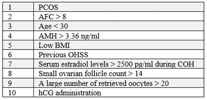
Table 1: The most common risk factors for OHSS.
Two sorts of classifications have been determined for this potentially lethal complication: Firstly, based on timing presentation in which two forms have been described: early and late. Early, OHSS is commonly trigger-related and it happens within 10 days of induced ovulation. The late OHSS is mainly pregnancy-related because of excessive amounts of endogenous hCG from a developing pregnancy, and it appears after ≥ 10 days following oocyte retrieval [8, 12, 14, 16]. Secondly, based on clinical, imaging plus laboratory features, in which OHSS is categorized into 4 stages: mild, moderate, severe, and critical. The milder form is the most prevalent among other stages of OHSS [2]. According to the second classification type, OHSS appears with various features related to severity.
There are an extensive range of clinical presentation for OHSS from mild to critical although these stages are not purely separated from each other. OHSS manifestations appear because of bilateral cystic enlargement of exceedingly luteinized ovaries [13]. Mild ovarian enlargement (ovarian diameter: 5-12 cm) makes the mild form of OHSS. Initial clinical presentations of mild OHSS (Figure 1) are commonly associated with abdominal distention, which include mild abdominal pain, poor appetite, nausea, vomiting, mild diarrhea, and bloating [1, 2, 8, 12, 13] even though The symptoms of this stage might be imperceptible.
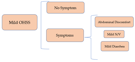
N/V; Nausea/ Vomiting. According to the figure, mild form of OHSS can be symptomatic or even without any symptoms in clinical presentation and laboratory findings. In these cases, mild OHSS will be distinguished by ovarian enlargement via ultra-sonography.
Figure 1: The most important clinical presentations and laboratory features of Mild OHSS.
In moderate OHSS (Figure 2), detectable ascites on ultrasound is added to the diagnostic criteria, and an apparent clinically ascites (grade 2) plus other more severe manifestations are revealed in the severe form. Indeed, clinical presentations in the severe forms (Figure 3) include severe abdominal discomfort, severe shortness of breath (SOB), rapid weight gain around > 1 kg in a day, severe nausea and vomiting, venous thrombosis, oliguria, pleural effusion (signs: tachypnea, crackle upon auscultation), intra-abdominal hypertension (IAH) which is defined as intra-abdominal pressure (IAP) > 12 mmHg, hemodynamic changes and so on. Also, the critical form manifestations (Figure 4) include arterial thrombosis, anuria, thromboembolism, pericardial effusion, acute respiratory distress syndrome (ARDS), massive hydrothorax, sepsis, shock, and maybe even death [8, 13, 14, 17]. Laboratory findings are different related to the severity of the disease. For instance, in the mild stage, there is no significant laboratory changes most of the times. In the moderate form of OHSS, laboratory findings are such as white blood cells (WBC) > 15000/ ml, hematocrit (HCT) > 41%, and hypoproteinemia (17). In the severe stage, the laboratory changes get more serious, including HCT> 55%, WBC > 25000/ ml, Na < 135> 5 meq/ml, serum creatinine (Cr) > 1.6 mg/dl, increasing liver enzymes, and clearance of Cr < 50>
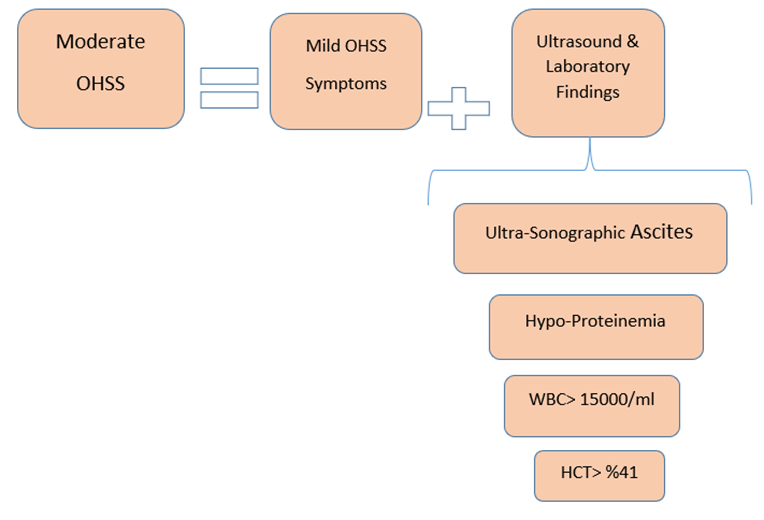
HCT; hematocrit, WBC; white blood cell. In moderate form of OHSS, in addition to mild clinical symptoms, there is ovarian ascites which will be diagnosed by Ultrasound. Moreover, in this stage, patients reveal laboratory changes.
Figure 2: The most important clinical presentations and laboratory features of Moderate OHSS.
In details, to explain more about the manifestations of OHSS, we should state that typically, as the severity increases, the signs and symptoms get more serious and even lethal. For instance, clinical ascites, intra-abdominal hypertension (IAH), hemoconcentration, infection, and severe hemodynamic changes which are highly dangerous for patients [2].
IAH, one of the severe manifestations, is defined as intra-abdominal pressure (IAP) > 12 mmHg. As a matter of fact, IAP is determined by the abdominal walls elasticity and the content of the abdominal cavity. IAP increase occurs due to accumulation of fluids or lesions which occupy the abdominal space. There are numerous methods for IAP measurement, in which trans-vesicular pressure measurement through a bladder catheter is one of the most prevalent and certain methods. Anyway, Normal IAP range is less than 5 mmHg. Also, in chronic obesity, IAP has been reported around 10-15 mmHg. Detrimental ranges of IAP start from more than 12 mmHg, which have been mentioned previously in IAH definition. IAH is separated into two different groups based on timing of IAP increasing: first of all, acute IAH which occurs mostly in patients who have undergone surgeries. In this type, IAP adds up over a few hours. Secondly, subacute IAH which happens along with medical disorders like pancreatitis. In this type, IAP rises over a few days. Additionally, four grades have been determined for IAH: 1) IAP 12-15 mmHg, 2) IAP 16-20 mmHg, 3) IAP 21-25 mmHg, and 4) IAP > 25 mmHg. In grade I, there are venous stasis and renal impairment. In grade II and III, physiologic impacts include decreased lung compliance and reduced splanchnic blood flow. Also, oliguria happens during grade II and III. Finally, in grade IV, anuria occurs with a more serious physiological effect known as decreased central perfusion pressure (CPP) (18). As a result, IAH induces oliguria (i.e. urinary output < 20> 5 meq/L, and metabolic acidosis), increased alanine & aspartate aminotransferase values, etc. [2, 8, 11]. According to some studies, there is another term known as abdominal compartment syndrome (ACS) which occurs by IAP elevation plus organ dysfunction.
Together, ACS might occur in severe OHSS and there are four grades for ACS a little similar to IAH groups: 1) IAP 10-15 mmHg, 2) IAP 16-25 mmHg, 3) IAP 25-35 mmHg, and 4) IAP > 35 mmHg. In other words, ACS with IAP > 20 mmHg is accompanied by organs dysfunction. Moreover, there is another classification for ACS as primary or secondary. Primary ACS is due to operation or trauma. However, secondary ACS is because of capillary leakage. Indeed, OHSS is regarded as a form of secondary ACS. In addition, a triad for ACS has been determined: 1) Respiratory effects: when IAP increases, the diaphragm comes up so that patients suffer from dyspnea and sometimes pleural effusion due to negative impacts on lungs. 2) Venous effects: when IAP raises, decreased venous return happens due to inferior vena cave (IVP) compression. 3) Intestinal effects: when IAP increments, it causes visceral compression which is the result of intestinal obstruction and in this part, patients complain about loss of appetite as well as nausea and vomiting which occur gradually. Overall, parts 1 and 3 (respiratory and intestinal effects) are observed in OHSS [18].
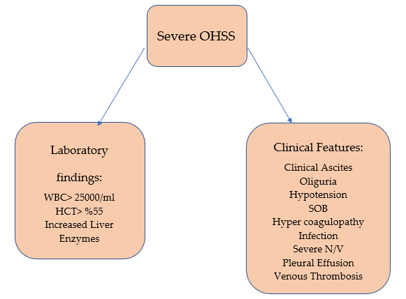
WBC; white blood cells, HCT; hematocrit, SOB; shortness of breath, IAH; intra-abdominal hypertension, ACS; abdominal compartment syndrome, N/V; Nausea/Vomiting. According to the figure, the severe form of OHSS is accompanied by obvious clinical ascites as well as other dangerous clinical and laboratory findings.
Figure 3: The most important clinical presentations and laboratory features of Severe OHSS.
In addition, another severe sign of OHSS, hemoconcentration, is described as increased hematocrit (HCT), thrombocytosis, and leukocytosis. Hemoconcentration is a predisposing factor for thrombosis and hyper coagulopathy. Thrombosis mainly occurs in the venous system in which the jugular and subclavian veins are the most frequent venous locations [2, 19].
Moreover, OHSS patients are at higher risk for different kinds of infections in different sites, including urinary tract, lungs, and upper respiratory tract, etc. Furthermore, these patients might experience some other infections consisting of intravenous line phlebitis, gluteal abscess due to progesterone injection, and post-operative wound infections [2, 20]. Causative organisms for these such infections in severe OHSS include Klebsiella pneumonia, Escherichia coli, Pseudomonas aeruginosa, Morganella morganii, Proteus mirabilis, and Proteus vulgaris. [2, 21]
OHSS also might be accompanied by hemodynamic changes from a simple hypotension until different types of shocks such as hypovolemic, obstructive, distributive, and septic shock related to severity of the disease. Hypovolemic shock happens because of third space or gastro intestinal loses, obstructive shock due to pulmonary embolism or cardiac tamponade, distributive shock as the result of inclement inflammation, and septic shock because of infections [2, 11].
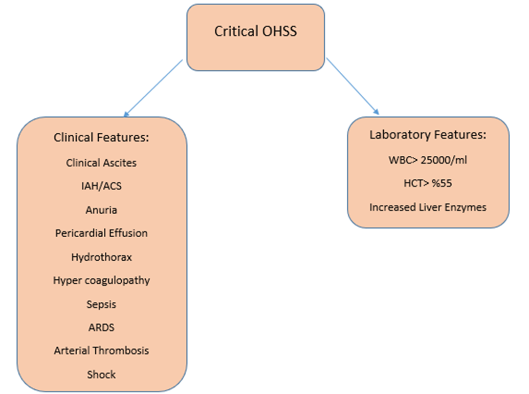
HCT; hematocrit, WBC; white blood cell, ARDS; acute respiratory distress syndrome, IAH; intra-abdominal hypertension, ACS; abdominal compartment syndrome. As you notice in the figure, the critical stage of OHSS possesses much more serious manifestations than other stages which is considered to be further life-threatening.
Figure 4: The most important clinical presentations and laboratory features of Critical OHSS.
To summarize, a number of most important clinical and laboratory findings in OHSS have been shown in figure 1. These manifestations of OHSS specially in the severe forms, predominantly are caused by fluid shifting into the third space of the body (mostly in peritoneal compartment, less frequently in pleural and pericardial space) [2, 19]
1.1.1 Third spacing phenomenon
Third spacing in OHSS is the result of increased vascular permeability. There are two main proposed models for increased vascular permeability: first, formation of vesiculo-vacuolar organelles (VVOs) which are a kind of trans-endothelial channels generated from vesicles or vacuoles. The next model which is deserved to be taken into account is transient dissolving of endothelial junctions [22]. For more explanation, endothelial cells are the bulk of the innermost layer of vessels; they are actually simple squamous cells. Three sorts of endothelial cell junctions have been discovered: gap, tight, and adherence junctions. Each of these different cell junctions possess their own constituents. For instance, in gap junctions, connexins are participated as the junction generators. Nevertheless, in tight junctions, some other molecules are involved such as claudins, nectin, occluding, and so on.
The constituents of adherence junctions are such as nectin, VE-cadherin, etc. Furthermore, inner side of endothelial cells are covered by glycocalyx which is made up of two layers of fiber matrix and full of proteoglycans. The glycocalyx layer is bound to the membrane of endothelial cells and contributes to the modulating of vascular permeability so that damaged glycocalyx may be also associated with increased permeability of vessels [23]. Plasma possesses three principle macromolecules, including globulins, fibrinogen, and albumin. They are in charge of different tasks like maintaining the pressure balance between blood and interstitial space. In addition, inflammatory cells adhere to endothelial cells so as to pass through endothelial junctions or via the thin endothelial cells directly [22]. Somehow the same probably occurs in OHSS.
In OHSS, the increased capillary permeability is fundamentally the main pathophysiologic feature of massive luteinization of enlarged-ovarian granulosa cells due to high dose hCG (exogenous or endogenous). In addition, some proinflammatory and vasoactive factors have directly or indirectly been involved in this process, resulting in shifting of intravascular fluid into extravascular compartment which is known as third space. The vasoactive factors include interleukin (IL)-1β, transforming growth factor (TGF)-α, TGF-β, endothelin-1, insulin-like growth factor 1 (IGF-1), epidermal growth factor (EGF), von Willebrand factor (VWF), prostaglandin, basic problast growth factor (BPGF), inhibin, prolactin, estrogen, histamine, serotonin, renin-angiotensin-aldosterone system, and most importantly, vascular endothelial growth factor (VEGF) [1, 13, 24-28]. Among the factors mentioned above, a few of them are not strongly evidenced to approved as key mediators for increasing vascular permeability in OHSS, despite of being supposed as possible factors for enhancing vascular permeability in the past [17]. Therefore, we explain more about common recent proved mediators affecting vascular permeability, of which VEGF is the most principle.
1.1.1.1 VEGF
VEGFs also called vascular permeability factors (VPFs), are signal proteins accompanied by angiogenesis as well as vasculogenesis properties. Indeed, they are the member of the platelet-derived growth factor family of cysteine-knot growth factors. There are 7 types of VEGF, including VEGF-A (i.e. VEGF), VEGF-B, VEGF-C, VEGF-D, the placental growth factor (PlGF), the viral genome-derived VEGF-E, and VEGF-F which is encoded in the venom of some snakes [29-33]. Moreover, there are 3 types of VEGF receptor (VEGFR). To clarify, VEGFR-1 is the receptor for VEGF- A, B, PlGF. VEGFR-2 is the receptor for types A, E, C, and D and VEGFR-3 is the receptor for VEGF-C and D. In humans, VEGFRs are encoded by the FLT genes such as FLT-1 which encodes VEGFR-1, and FLT-4 which encodes VEGFR-3. VEGF- C/D are essential for the regulation of lymph-angiogenesis plus angiogenesis at an early embryogenesis less importantly [32-35]. VEGF-B is involved in embryonic angiogenesis (specifically, myocardial tissue) [36]. In addition, PlGF plays a main role in pathological angiogenesis (like retinopathy especially those with diabetes retinopathy) and inflammation [37]. Among different kinds of VEGF, type A was discovered initially and is the most responsible type of VEGF playing an important role in this disorder.
1.1.1.1.1. VEGF-A; increasing vascular permeability
VEGF-A actually gets involved in capillary permeability and pro-angiogenic activity. Indeed, it is made by those cells underwent stress conditions, such as endothelial cells. It is expressed during female reproductive cycle remodeling as well as atherosclerosis and cancer [29, 34]. During OHSS, VEGF-A after getting released into the blood circulation, binds to its specific receptor on the cell surface to exert its effects on the body (figure 5). For more specific explanation, endothelial cells connect to each other tightly by some protein complexes which are known as adherens junctional proteins (AJ proteins). There are several kinds of AJ proteins like β-catenin, VE-cadherin, α-catenin, and p 120-catenin. These proteins maintain vascular integrity so as to make a vascular barrier. The barrier is able to breakdown due to some reasons such as endocytosis, phosphorylation, S-nitrosylation, and/or cleavage of these proteins. The vascular barrier integrity is dynamically modulated by VEGFR signaling. As a matter of fact, VEGF enhances VE-cadherin and β-catenin tyrosine (Y)-phosphorylation, followed by capillary leak. Also, it is recently evident that a kind of kinase known as Src-family protein-tyrosine kinase (PTK), is activated by VEGF and plays an important role in promoting AJ phosphorylation. Src-family PTK is also able to get actuated by integrin receptors which cross-talk with VEGF in vascular permeability controlling. To clarify, VE-cadherin phosphorylation at Y658, Y731 or β-catenin phosphorylation at Y654, both by Src mediation, induce complicated AJ stability regulation. Moreover, β-catenin phosphorylation at Y142 disrupts α-catenin binding. In spite of the fact that in-vitro phosphorylation of β-catenin at Y142 by PTK6 has been demonstrated, the modulating of this kind of AJ protein phosphorylation remains unknown in-vivo. There is another type of PTK, called focal adhesion kinase (FAK) which is actuated by both integrins and VEGF. When FAK binds to paxilin, followed by its localization to cell adhesions, and a FAK/αvβ5 integrin signaling structure formation, actin-myosin tension is generated and cell motility increases [32, 38-40]. Consequently, VEGF-A binds to VEGFR and activates the receptor so that cell signaling induces phosphorylation of AJ proteins resulting in vascular barrier breakdown and increased vascular permeability.
As mentioned above, VEGF-A holds two important impacts on vasculature. First, the increase in vascular permeability, which was explained in the previous paragraphs by the description of OHSS as an example. The next role that should be taken into account is that VEGF-A also plays a principal role in physiological and pathological angiogenesis which occurs in obesity as an instance [32, 33, 41].
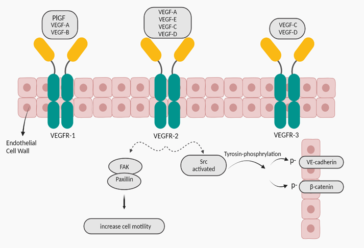
PlGF; placental growth factor, FAK; focal adhesion kinase
Figure 5: VEGFRs and cell signaling; after binding VEGF-A to VEGFR-2, a special cell signaling sets out to degrade AJ proteins between endothelial cells so that yields increased vascular permeability (32, 38-40, 42).
1.2. Obesity
Obesity incidence is aggregating over the last few decades, leading to be one of the main health problems worldwide [43, 44]. According to WHO, approximately 20% of adult population around the world, will suffer from obesity by 2025 [44, 45]. To clarify the concept of obesity, we should state that the imbalance between energy intake and energy consumption underlies obesity and overweight. [46-50]. The next thing which is deserved to be pay attention is that changes in diet quality might also cause obesity [51]. Obesity is in fact a complex chronic disorder, which is specified as three sorts; generalized obesity (BMI ≥ 30 kg/m2), extreme obesity (BMI > 40 kg/m2), and central obesity in which the abdomen fat gets accumulated excessively [48, 52, 53]. There are a number of factors such as epigenetic changes, increasing the risk of obesity. Epigenetics is in fact a phenomenon in which some variables like nutrition and lifestyle lead to make some alterations in gene expression in terms of histone modification, non-coding RNA, and DNA methylation, without any changes in DNA sequence [54, 55]. It should be described into more details that the anti-aging gene Sirtuin 1 has been shown to be involved in ovarian function and OHSS. Then, this is critical to epigenetic alterations in obesity, and under hypoxia Sirtuin 1 regulates VEGF via HIF alpha. The use of Sirtuin 1 activators versus inhibitors may be critical to VEGF regulation and vasculature in OHSS and related diseases such as obesity. Diet, environment, stress and lifestyle factors literally alter Sirtuin 1 which affects the pathophysiology of OHSS and obesity [56-64]. Other predisposing factors include genetic factors, inadequate physical activity, socioeconomic status, excess caloric consumption, sleep deprivation, medical conditions like hypothyroidism and insolinoma, psychosocial stress and mental disorders [47, 48, 65, 66]. Furthermore, it would be such an interesting point that researchers categorized the genetic type into three groups: first, monogenic obesity which is caused by a mutation of a single gene that is involved in the leptin-melanocortin pathway that regulates energy homeostasis. In this type, the major characteristically feature is hyperplasia: secondly, polygenic obesity which results from the occurrence of multiple gene variants concurrently; third, syndromic obesity which is accompanying other signs of developmental diseases such as, Bardet-Biedel, MOMO, Cohen, and Prader will syndrome [53, 65, 67-69]. For management of genetic obesity, a combination treatment is required, including administering medicine, rectifying patients’ lifestyles, plus bariatric surgeries [53]. Scholars demonstrated that obesity particularly causes several adverse health consequences for human body. As a matter of fact, obesity promotes some undesirable outcomes attributed, in part, to type two diabetes mellitus, hypertension, sleep apnea, endometriosis, liver cirrhosis, dyslipidemia, PCOS, infertility, metabolic syndrome, arthritis, cardiovascular diseases, etc. [65-67, 70]. Evidence demonstrates that obesity is able to elevate the risk of some pregnancy-related complications, such as pregnancy loss, gestational hypertension, gestational diabetes, miscarriage, maternal death, and so on [71, 72]. Moreover, obesity, as a systemic inflammatory condition, adversely increases the risk of some types of cancers which are related to vascular dysfunctioning, including liver and gastrointestinal cancers [43, 48, 49, 55, 67]. As get informed, Obesity has been revealed as a metabolic state in which excess in storage of triglycerides in adipose tissues occurs gradually [50, 72].
1.2.1. Adipose tissue
Adipose tissue is distinguished as an endocrine organ [46, 49, 67]. It comprises of mixtures various adipocytes which are covered by stromal vascular cells, as well as fibroblasts and macrophages. These vasculature yields a condensed network of blood capillaries around the adipocytes [46, 67, 73]. Additionally, scholars recognized that there are different kinds of adipocytes in their functions despite of similar morphology. For instance, thermogenic adipocytes which conserve core body temperature. This kind of adipocyte exists around central organs. Some other examples include perivascular, mesenteric, and subcutaneous adipocytes. the latter instance is regarded as the largest depot and is capable of getting expanded as a reaction to excess food intake [73]. If we take a more totally look at adipose tissues, we realize that they comprise two types in human body; white adipose tissue (WAT), and brown adipose tissue (BAT). Researchers found out some different features between these two kinds. WAT is assigned for energy storage in the form of triglycerides. Moreover, WAT works as an endocrine organ, secreting some hormones such as gerlin and leptin, which are called adipokines. Nevertheless, BAT is specialized for thermogenesis in addition to energy expenditure due to mitochondria function which exist in this sort of adipocytes at high concentrated levels plus high density vasculature [41, 67, 74-76]. Overall, adipose tissue has the capability to get expanded by some triggering situations which include fasting and excess food intake. During the former condition, the body attains benefits from the adipose tissue expansion due to providing the ability to store fuels for using. The latter situation actually brings some drawbacks for the body so that causes obesity plus in turn its comorbidities [73]. The adipose tissue expansion can occur through two distinct mechanisms which consist of hypertrophic expansion (increase in size of adipocytes) and hyperplastic expansion (formation of new adipose cells) [67, 72, 75, 77]. Both of these mechanisms are able to happen in adults, specifically hypertrophy which sets out to get generated by excess caloric intake [73, 75]. During hypertrophy in adipose tissues, angiogenesis occurs because of overexpression of VEGF-A [67, 73].
1.2.1.1. VEGF-A Leads to Angiogenesis
As described above, overfeeding leads to hypertrophy of adipocytes eventually. The increase in size of adipocytes yields several negative impacts on the adipose tissues which include tissue hypoxia (55, 67, 75, 76). Indeed, adipose tissue dysfunctions contribute to adiposopathy for which there are two parts (anatomic & functional). For more explanation, the anatomic changes of adiposopathy are such as adipose tissue expansion and augmented levels of fat deposition in organs of the body. Nevertheless, functional changes include adipose tissue dysfunctions through endocrine and immunology responses, increased amounts of reactive oxygen species, tissue hypoxia, etc. [46, 77]. actually, some researchers state that these results especially adipocyte hypoxia might be because of either the compression effect of large adipocytes on vasculature by creating a niche or impairing capillary function through depressing capability of vessels to eliminate extracellular fatty acids. Furthermore, some other researchers hypothesized that the event of hypoxia in adults’ adipose tissues following adipocytes hypertrophy occurs due to the impacts of excess calories on both vasculature function and adipocytes [46, 73, 78]. Anyway, after generating tissue hypoxia in which the tissue receives inadequate blood flow, in turn the adipose tissue function gets disrupted and here is an alarm for the body, since hypoxia finally can lead to endothelial cells death. Therefore, the endothelial cells begin to overexpression of cytokines and angiogenic factors such as VEGF-A [32, 73, 79]. It should be emphasized that the VEGF-A is able to yield both angiogenesis and anti-angiogenesis on the capillaries [41, 73]. This against effect on endothelial cells comes from conversed effects of receptors on the cells. As a matter of fact, when VEGF-A locates on VEGFR 1, the anti-angiogenic property starts to happen. Nevertheless, when VEGFR 2 gets activated by VEGF-A, it shows angiogenic features [33, 34, 41]. As hypoxia is one of the stimulators of endothelial cells due to capillary rarefaction, the aim of releasing high amounts of VEGF-A would consequences angiogenesis in order to take the adequate blood flow to the tissue [32, 46, 76, 78]. For more explanation, VEGF-A sits on VEGFR 2 on endothelial cell wall. The mentioned receptor gets phosphorylated and is activated to generate intra cellular signaling such as PLC (phosphoinositide phospholipase C)- dependent pathway. This pathway consists of two mechanisms, including NOS-dependent signaling and mitogen-activated protein kinase (MAPK) cascade that both of them Finally, induce gene expression, mitogenesis, cell proliferation, and cell survival, thereby angiogenesis [32, 34, 80]. To clarify, VEGFR-2 activated leads tyrosine phosphorylation of PLCγ1 for which the activity of Src- family kinases are the requirements. phosphorylated PLCγ1 aggregates the level of inositol triphosphate (IP3) plus Ca2+ inside the endothelial cells which in turn leads to increased production of NO. Additionally, phosphorylated PLCγ1 activates MAPK cascades. Both NOS and MAPK cascade result in endothelial cells proliferation (32, 34). Then the basement membrane of the capillaries gets cleaved, allowing proliferated endothelial cells to incur into the adipose tissue stroma and make a lumen by endothelial cells migration. The following results include arranging extended blood capillaries which makes a dense network of vasculature in the adipose tissue [33, 67, 78].
We reviewed the two varied impacts of VEGF-A on blood vessels during two distinct situations; OHSS is one of the important IVF complications; leads to secretion of some mediators like VEGF, cytokines, renin-angiotensin system substances, etc. Among these vasoactive mediators, VEGF is considered as the main factor resulting in vascular permeability increase. In order to achieve this goal, VEGF-A binds to its receptor. Then, VEGFR2 activation generates a cell signaling which includes AJ protein phosphorylation. Since AJ stability underlies tight endothelial cell junction, the vascular barrier breakdown occurs due to phosphorylation of AJ proteins. Therefore, vascular permeability increases and intravascular fluid simply shifts into third space. On the other hand, during obesity something different occurs by VEGF-A; obesity prevalence has been increased during recent years due to environmental, genetic factors and so on. Due to adipose tissue expansion, tissue hypoxia occurs thereby cytokines and angiogenic factors such as VEGF-A release in order to repair capillary rarefaction. During this process, VEGF-A locate on VEGFR2, thereby activates NOS-dependent signaling and MAPK cascade by phosphorylation of PLCγ1. This event results in endothelial proliferation which then migrate and create denser network of capillaries in adipose tissue.
This research received no external funding.
The authors declare no conflict of interest.
Dear Editorial Team, Clinical Medical Reviews and Reports. My experience with the journal was highly positive. The peer-review process was rigorous, constructive, and completed in a timely manner. The reviewers provided valuable comments that helped improve the quality and clarity of our manuscript. The editorial office was professional, responsive, and supportive throughout all stages of the publication process. Communication was clear and efficient, and any questions were addressed promptly. Overall, I found the journal to maintain high scientific standards and an excellent publication workflow. I would be pleased to consider submitting future work to this journal. Best wishes from, Elena Popa.

It was my pleasure to submit my testimonial concerning the Reviewer Board of our Scientific Journal “Brain and Neurological Disorders”. The Reviewers focused on some modifications and their contribution was helpful. The ladies of our Editorial Office were also supported my efforts. It was my honor to have such a co-operation and I am looking forward for more collaboration.

Dear Grace Pierce, Editorial Coordinator of Journal of Clinical Research and Reports, Thank you for the speedy and efficient peer review process. I appreciate the fact that your peer reviewers do not take months to respond like with some other journals. I would also like to thank the editorial office for responding quickly to my questions. It is an excellent journal. I plan to submit more manuscripts in the future. Best wishes from, Robert W. McGee

Dear Grace Pierce, Editorial Coordinator of Journal of Clinical Research and Reports, Working with you and your team on our recent publication in JCRR has been a truly wonderful and enjoyable experience. The responses were prompt, and the reviewers were patient, constructive, and highly professional. One reviewer in particular gave me the feeling that a professor was carefully reading and commenting on my coursework, which was deeply touching. The entire process was straightforward and hassle‑free, with no tedious online forms to complete. I highly recommend this journal. Best wishes from, DR Aibing Rao, Head of R&D

I Appreciate the Opportunity to Share my Experience with the Journal of Clinical Research and Reports. The peer review process was timely and constructive, and the feedback provided helped improve the quality of our manuscript. The editorial office was professional, responsive, and supportive throughout the process, ensuring smooth communication and efficient handling of the submission. Overall, it was a positive experience collaborating with your team.

Dear Mercy Grace, Editorial Coordinator of Obstetrics Gynecology and Reproductive Sciences, We would like to express our gratitude for your help at all stages of publishing and editing the article. The editors of the magazine answer all the necessary questions and help at every stage. We will definitely continue to cooperate and publish other works in the Obstetrics Gynecology and Reproductive Sciences! Best wishes from, Alla Konstantinovna Politova,
