
Review | DOI: https://doi.org/ 10.31579/2768-0487/004
*Corresponding Author: Janos Vincze, Health Human International Environment Foundation, Budapest, Hungary.
Citation: J Vincze, Gabriella V Tiszay. (2021) The Biophysical Modeling of the Sleep Regulation in the Human Organism. Journal of Clinical and Laboratory Research. 2(4) DOI: 10.31579/2768-0487/004
Copyright: ©2021 Janos Vincze. This is an open-access article distributed under the terms of the Creative Commons Attribution License, which permits unrestricted use, distribution, and reproduction in any medium, provided the original author and source are credited.
Received: 26 January 2021 | Accepted: 06 March 2021 | Published: 30 April 2021
Keywords: sleep; biophysical modeling; histogram of the sleep; EEG
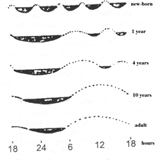
A seventy-year-old human spends approx. twenty years, or one-third of their life, asleep. During sleep, their connection with the outside world is more or less lost, but in the meantime they experience exciting dreams that are difficult for rational thinking to grasp. Analysis of EEG recordings revealed that sleep can be divided into two sharply separated states: one is the so-called slow wave sleep; the other is the so-called paradoxical sleep (REM), e.g. characterized by strong eyeball movement. The slow sleep period can be divided into 4 periods based on the EEGs, and these follow each other in a well-defined order, then comes the paradoxical sleep and the whole thing is repeated; during a full sleep of approx. 4–6 times. The duration of paradox sleep depends on the individual, but it lasts for approx. 6–15 minutes. Compared to the literature, one of the significances of this article is also that we wrote a mathematical model of the sleep. This model also allows us to characterize different forms of particular sleep.
The significant time spent sleeping also indicates that it is a matter of meeting some important biological need, as evidenced by the fact that if we prevent sleep for days, the body then makes up for the omission, and even completes paradoxes with particular precision [1]. More recently, the notion that classifies sleep as an instinctive action has become widespread. Instinctive actions are species-specific, genetically coded, rather rigidly precise behaviors that satisfy an important biological need, and which are under the control of a certain internal driving force – the so-called motivation [2].
During deep sleep muscular tone is sharply reduced. Relaxation of the muscles and the lowering of their tone, howeever, are not constant and necessary components of sleep [3]. All the senses, i.e. vision, hearing, taste, smell, and the tactile sense are greatly reduced, and a much stronger stimulus is required to induce a reaction in a sleeping person than when he is awake. In a deep and calm sleep respiratory periods are much less frequent and more regular; gas exchange and basal metabolism are somewhat reduced; the pulse is slower: arterial pressure falls; secretion of urine by the kidneys is reduced [4]. During night sleep the body temperature of man is lowered
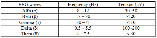
The possibility of objective experimental studies regarding sleep was provided only by the development of technology in the 1950s. This was mainly due to the widespread use of electroencephalographic examinations [5]. An electroencephalogram (EEG) is a graphical representation of the electrical activity of brain neurons; a measurement signal on the order of microvolts that shows the change in activity of thousands of neurons over time. The EEG is registered with low-resistance electrodes placed on the scalp [6]. The EEG curve is a measure of the event-dependent potential measured on the scalp. The formed synchronized currents (which are synchronized by the afferents of the thalamus) can be measured by adding them up. As a result of synchronization, measurable EEG waves are created. EEG waves are classically grouped according to frequency and amplitude.
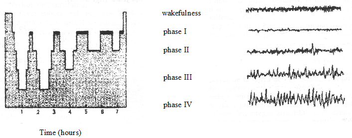
Slow-wave sleep, as its name implies, is caused by slow waves occurring in increasing numbers and at lower and lower frequencies in the EEG, as well as periods of 2–3 seconds consisting of more frequent waves, being characterized by the so-called “sleep spindles”. Based on the electrical activity of the brain, several stages of slow-wave sleep can be distinguished, e.g. four sleep states are usually defined from EEG curves, denoted by numbers 1–4 from superficial sleep to deep sleep. Although the different origins of each stage arise from time to time, slow-wave sleep is mostly considered qualitatively one, with each stage denoting different sleep depths [7, 8].
Characteristics of each phase:
Under normal conditions, electrical activity in the brain follows the development of sleep well. Sleep progresses from stage 1, i.e., from the most superficial stage, to stage 4, then its depth decreases again, and – before another stage 1 occurs – paradoxical sleep appears. Sleep is deepest in the first half of the night, especially in the first sleep cycle. In later cycles, the amount of stage 4 is less and less, towards the morning the slow-wave sleep does not even go beyond stage 2.
Paradoxical sleep is a qualitatively different state from slow-wave sleep. Cerebral electrical activity is characterized by vigorous, rapid activity, which can sometimes exceed wakefulness activity. The muscles relax completely.
During deep sleep muscular tone is sharply reduced. Most muscles in a sleeping person are completely relaxed, which is why objects fall from the hands of a person sleeping in a sitting position and why his head sinks onto his chest, and his trunk sags.
Relaxation of the muscles and the lowering of their tone, howeever, are not constant and necessary components of sleep [9]. Even during normal sleep various movements are possible, and a definite pose can be maintained for a long time, as when sleeping in a sitting posture. In certain forms of so-called hypnotic sleep, e.g. in cataleptic sleep, a marked increase of muscular tone is even observed [10]. All the senses, i.e. vision, hearing, taste, smell, and the tactile sense are greatly reduced, and a much stronger stimulus is required to induce a reaction in a sleeping person than when he is awake.
The reflex function is sharply reduced during sleep. Conditioned reflexes are inhibited, unconditioned ones are considerably weakened. Their thresholds of stimulation are considerably raised, and the latent period lengthened. Sensomotor changes dominate the picture of sleep as compared with changes in vegetative functions [11].
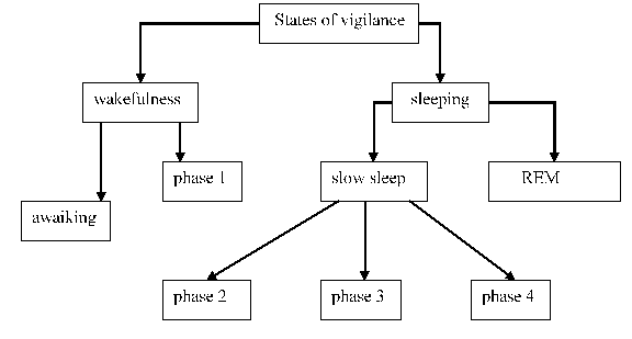
The vigilance
The biophysical modeling
Sleep is one of the fundamental functions of brain; we will present a modelling of this phenomenon. Sleep is a natural phenomenon in which the lowering until the disappearance of the superior adaptation reaction takes place, the sensitive-motor relations with the environment and the decrease of most of the bodily functions. Sleep manifests like an imperious need, like a habit, maintained with the price and the rigours of a true biorhythm; it differs from the pathological states of loosing consciousness by the fact that while sleeping the subject can be woken up through sensorial stimulations [12].
The modelling of the wakeful – sleep state is made through the following functions:
y = a(a) + b(b).sin [d(d)t + w(f)]
Where: t e [0, 2p] ® [0h, 24h] ; a(a) » ½.
So, two thirds of the function’s value domain for the definition domain of the t parameter is found in the positive region and only a third is found in the negative region, which corresponds to the report for the wake state and sleep in the case of normal person. This report can be modified through a(a). We can make a translation of this modelling function through the modification of w(f), so this way the beginning of the sleep state can be determined by the observer. If the value d(d) = 2, it means that in 24 hours that person had the sleeping state for 2 hours.
This model also allows us to characterize different forms of particular sleep. Experiments in humans have shown that memories first enter the hippocampus and then, after replay and filtering, they end up in the external cerebral cortex, i.e. the gray matter. Dream is a psychic activity that occurs in the state of sleep and is related to the emotional state, experienced events, traumas, stress that are recorded in memory.
Dear Editorial Team, Clinical Medical Reviews and Reports. My experience with the journal was highly positive. The peer-review process was rigorous, constructive, and completed in a timely manner. The reviewers provided valuable comments that helped improve the quality and clarity of our manuscript. The editorial office was professional, responsive, and supportive throughout all stages of the publication process. Communication was clear and efficient, and any questions were addressed promptly. Overall, I found the journal to maintain high scientific standards and an excellent publication workflow. I would be pleased to consider submitting future work to this journal. Best wishes from, Elena Popa.

It was my pleasure to submit my testimonial concerning the Reviewer Board of our Scientific Journal “Brain and Neurological Disorders”. The Reviewers focused on some modifications and their contribution was helpful. The ladies of our Editorial Office were also supported my efforts. It was my honor to have such a co-operation and I am looking forward for more collaboration.

Dear Grace Pierce, Editorial Coordinator of Journal of Clinical Research and Reports, Thank you for the speedy and efficient peer review process. I appreciate the fact that your peer reviewers do not take months to respond like with some other journals. I would also like to thank the editorial office for responding quickly to my questions. It is an excellent journal. I plan to submit more manuscripts in the future. Best wishes from, Robert W. McGee

Dear Grace Pierce, Editorial Coordinator of Journal of Clinical Research and Reports, Working with you and your team on our recent publication in JCRR has been a truly wonderful and enjoyable experience. The responses were prompt, and the reviewers were patient, constructive, and highly professional. One reviewer in particular gave me the feeling that a professor was carefully reading and commenting on my coursework, which was deeply touching. The entire process was straightforward and hassle‑free, with no tedious online forms to complete. I highly recommend this journal. Best wishes from, DR Aibing Rao, Head of R&D

I Appreciate the Opportunity to Share my Experience with the Journal of Clinical Research and Reports. The peer review process was timely and constructive, and the feedback provided helped improve the quality of our manuscript. The editorial office was professional, responsive, and supportive throughout the process, ensuring smooth communication and efficient handling of the submission. Overall, it was a positive experience collaborating with your team.

Dear Mercy Grace, Editorial Coordinator of Obstetrics Gynecology and Reproductive Sciences, We would like to express our gratitude for your help at all stages of publishing and editing the article. The editors of the magazine answer all the necessary questions and help at every stage. We will definitely continue to cooperate and publish other works in the Obstetrics Gynecology and Reproductive Sciences! Best wishes from, Alla Konstantinovna Politova,
