
Research Article | DOI: https://doi.org/10.31579/2766-2314/079
1Department of Histology, Faculty of Medicine, Sabratha University, Libya.
2Department of Histology, Faculty of Medicine, Zawia University, Libya.
3Department of Physiology, Faculty of Medicine, Sabratha University, Libya.
4Department of Histology, Faculty of Medicine, Ain Shams University, Egypt.
*Corresponding Author: Azab Elsayed Azab, 3Department of Physiology, Faculty of Medicine, Sabratha University, Libya.
Citation: Marwan T M Abofila, Asma N Ali, Azab E Azab, Adel Salah El Din Zohdy, Ghada F Mohammed, A A M Attia. (2022). The Ameliorating Effect of Dexamethasone against Pancreatic Histopathological changes Induced by L-Arginine in Adult Male Albino Rats. J, Biotechnology and Bioprocessing 3(3); DOI:10.31579/2766-2314/079
Copyright: © 2022 Azab Elsayed Azab. This is an open access article distributed under the Creative Commons Attribution License, which permits unrestricted use, distribution, and reproduction in any medium, provided the original work is properly cited.
Received: 10 May 2022 | Accepted: 03 June 2022 | Published: 08 June 2022
Keywords: histological study; acute pancreatitis; pancreas; L-Arginine; dexamethasone; adult male albino rats
Background: Acute pancreatitis (AP) is an inflammatory process of the pancreas with possible peri-pancreatic tissue and multiple-organ involvement inducing multi-organ dysfunction syndrome with an increased mortality rate. Acute pancreatitis progresses with a local production of inflammatory mediators.
Objectives: The aim of this study was to investigate the time courses of the effects of dexamethasone on microscopical changes occurring in the pancreas of rats used as models of AP induced by L-arginin.
Materials and Methods: 60 adult male albino rats weighing 150-200 gm were used. They were divided into 3 groups: Control group: Which is also divided into 2 subgroups (a & b) each of animals of the first were IM injected with 0.5ml/100gm B.W saline and those of second were injected by 0.5mg/100gm B.W dexamethasone. L-Arginine group: which received L-Arginine to induce AP. The animals of this group were divided into 3 subgroups a, b and c the animals of which were sacrificed 3 days, 2 weeks and 1 month after L-Arginine injection respectively. Dexamethasone and L-Arginine group: in which the animals were injected with both L-Arginine and dexamethasone. They were also divided into 3 subgroups a, b and c, the animals of which were sacrificed 3 days. 2 weeks, one month after the injection of the drugs. The pancreas of the scarified animals were dissected out and prepared for microscopical examination.
Results: Three days after the injection of L-Arginine, the dispersed cells of the exocrinal part of the gland had showed lost of their acinar arrangement and signs of cell death and some small aggregates of fat cells and increase in collagenous fibres in the stroma. Two weeks after L-Arginine injection, the gland was composed mainly of stroma of loose C.T. in which widely dispersed groups of ducts were present with more intense mononuclear cellular infiltration. Large aggregates of fat cells were also seen. No other parenchymal cells or acini could be detected. One month after injection of L-arginin, the gland appeared to be composed mainly of stroma of white adipose C.T. among which islands of glandular tissue were seen forming small irregular separate lobules. Most of these lobules were composed of an islet of Langerhans of normal structure with related group of normal pancreatic acini and ducts. The collagenous fibres density increased progressively in the stroma of the gland along the experiment. Injection of dexamethasone in AP model animals greatly ameliorated the degenerative changes that occurred in the gland. It also fastened the process of glandular regeneration. Three days after the injection of L-Arginine and dexamethasone, the exocrinal secretory cells of the pancreas were distorted. Most of them had lost their acinar arrangement and showed variable signs of cell death. On the other hand, some of the cells kept their acinar arrangement specially in areas close to islets of Langerhans. These exocrinal cells were present among stroma of C.T infiltration with mononuclear cells, collagenous fibres and few fat cells. Two weeks after the injection of the L-Arginine and dexamethasone, the gland had partially regained its normal structure, a process which was completed in the animals one month after the injection of drugs.
Conclusion: It can be concluded that treatment of AP with dexamethasone is beneficial in ameliorating the pancreatic affection and helps in its rapid regeneration.
Pancreatitis means inflammation of pancreas, necro-inflammatory disease of pancreas that can manifest as either an acute or chronic disease [37]. Chronic pancreatitis is progressive and potential fatal disease caused by persistent unresolved inflammation and pancreatic fibrosis typically accompanied by intractable abdominal pain, particularly during the early phase [11, 27]. Acute pancreatitis (AP) is sudden acute inflammatory processes of pancreas [24]. It is defined as inflammatory process of the pancreas with possible peripancreatic tissue and multiple-organ involvement inducing multi-organ dysfunction syndrome (MODS) with an increased mortality rate [10].
Acute pancreatitis present as patient with an acute abdominal pain, sudden and severe, continuous poorly localized and radiating to back, vomiting may be early feature. In mild attack, the acute abdominal pains are minimal with rapidly resolving abdominal signs like abdominal distension, some abdominal tenderness, and guarding, absent bowel sounds, minimal systemic illness, moderate tachycardia. The severe attack is characterized by severe acute abdominal pain, severe toxaemia and shock, generalised peritonitis, diffuse abdominal tenderness, guarding, rigidify, absent bowel sounds, acute respiratory distress syndrome [31].
Alcohol and gall stones are the etiology of acute pancreatitis in many adults and although some differences exist based on sex and ethnicity [6, 39].
The etiology in children is often drugs, infectious, trauma, and anatomic anomalies such as choledochal cyst and abnormal union of pancreatobiliary junction [13, 9, 38, 28].
The majority of acute pancreatitis patients present with mild disease, however, approximately 20% run a severe course and require appropriate management in an intensive care unit [2]. The current ideal drug for the treatment of severe acute pancreatitis (SAP) is still in need and standardized treatment of consensus is only confined to fluid therapy, nutritional support, treatment of necrosis and infection, and endoscopic procedure of biliary stones [41].
In the past years, a number of researches about the initiation and propagation of AP have been done, but to date, no fully effective drugs or treatment is available [47].
AP is a multi-system disease with alterations not only in the pancreas, but also in liver, lungs and kidneys, which may lead to distant organ dysfunction and death. Although its exact nature is still unknown, acute panreatitis progresses with a local production of inflammatory mediators, eventually leading to systemic inflammatory response syndrome.
The effects of glucocorticoids on acute pancreatitis have remained contradictory. So, the study aimed to investigate the time courses of the effects of the exogenous glucocorticoids agonist dexamethasone on microscopical changes occurring in the pancreas of rats used as a model of AP induced by L-Arginine.
Animals
Sixty adult male albino rats weighing 150-200 gm were used in this study. The animals were maintained in medical research center in Ain Shams University.
They were housed in plastic cages with mesh wire covers and allowed water and standard rat chow adlibitum for the period of experiment. The practical work was performed in accordance to guide for care and use of laboratory animals and approved by the animal ethical committee of Ain Shams University.
Induction of pancreatitis
Pancreatitis was induced by L-arginine hydrochloride powder of highest purity. It was supplied by med pharma. 20% L-Arginine hydrochloride solution was prepared by dissolving 2gm L-arginine hydrochloride in 8ml saline. PH was adjusted to 7 and then the volume was increased to 10 ml solution.
Animals were weighted accurately and injected by L-arginine using a dose of 500 mg / 100 gm body weight (2.5ml/100gm). The calculated dose for each animal was divided into two halves (1.25 ml/100gm), each one of them was injected followed by the second dose after an hour interval. The same procedure was repeated in following day [12].
Animals groups
The animals were divided into three groups:
Group I (control group): It included 10 male albino rats. This group was subdivided into 2 subgroups, 5 rats each. Sub group IA: It included 5 male albino rats received IM injection of normal saline 0.5 ml/100gm body weight. Sub group IB: 5 male albino rats received IM injection of dexamethasone 0.5mg/100gm of body weight in single dose [43].
Group II (L-argnine group): Twenty five rats received 2 intraperitoneal injections of L-Arginine (250 mg/100gm BW) as one hour apart. The same dose was repeated in following day. They were subdivided into three subgroups: Subgroup IIA: Five animals were scarified after three days from last injection of L-Arginine. Subgroup IIB: Five animals were scarified after two weeks after last injection of L-Arginine. Subgroup IIC: Six animals were scarified one month after last injection of L-Arginine.
Group III (dexamethasone and L-Arginine group): It's included 25 male albino rats that received IM injection of dexamethasone (0.5mg/100gm BW) one hour after receiving 2 intra-peritoneal injection of L-Arginine (250mg/100gm BW) one hour apart. The same dose was repeated in following day [43]. They were subdivided into 3 subgroups: Sub group IIIA: 5 animals were scarified after 3 days from last injection of dexamethasone. Sub group IIIB: 5 animals were scarified after 2 weeks from last injection of dexamethasone. Sub group IIIC: 14 animals were scarified after 1 month after last injection of dexamethasone.
Methods
At the end of the experimental of each group, the animals were sacrificed by decapitation. Pancreas of animals were dissected out and were put immediately in fixation. Pancreas were fixed in Bouin's solution for 4-5 hours. The paraffin sections of 5Mm thick were prepared and stained by the following stains (H&E, Periodic acid schiff reaction (PAS) [14], Maisson's trichrome stain [4], Immuno-histochemical study, Proliferating cell nuclear antigen (PCNA) [5], and Capsae-3 Technique [3].
The survival rate:
In group I, no animals died throughout the study, with a survival rate 100%. In group II, nine animals died and sixteen animals survived out of 25 animals with a survival rate 64%. In group III one animal died and 24 animals survived out of 25 with a survival rate 96%.
Statistical analysis of serum amylase levels:
Statistical analysis of serum amylase levels in the different groups 1h, 3hs, 6hs and 24 hours after the injection of the drugs came to the conclusion which is summarized in histogram (I).
sFrom this histogram it is apparent that serum amylase level was in normal level in the control subgroups (a-injected with saline and (b) injected with dexamethasone) in the four times studied. On the other hand, the enzyme level slightly and insignificantly increased after 1-3-6 hours after injection of L-Arginine where as it is severely and significantly elevated 24 hours after injection of the drug. Dexamethasone injection with L-Arginine caused mild non significantly increase of serum amylase level in the 1st and 3rd hours, but moderate significant increase in the 6th and 24th hours. It is worlly to note that the severe enzyme level on the 24th hour was much and significantly lower in the group of animals injected with dexamethasone and L-Arginine compared to that of the animals injected with L-Arginine alone.
Control group I (A+B) (a; animals injected with normal saline, b; animals injected with Dexamethasone).
Sections of the pancreas of the animals of both these groups were almost
similar and showing the characters of normal pancreas. The pancreas appeared surrounded by a thin capsule from which complete and irregular septa arise dividing the gland into lobules. The capsule and septa are composed of loose connective tissue. In the sections the lobules appeared widely separated from each other indicating the loose attachment of the capsule and septa to the lobules.
The lobules are heavily crowded with the elements of the parenchyma including the exocrinal and endocrinal parts. The exocrinal part of the pancreas appeared as a pure serous gland formed of heavily crowded serous pancreatic acini. Intralobular ducts could hardly be seen. The interlobular ducts occupied the interlobular septa and are lined by one row of cubical cells.
Each of the acini is small triangler or polyhydral in shape. It is lined by numerous pyramidal cells. The lumen is narrow and lined by one row of low cubical centroacinar cells whose nuclei appeared oval or rounded in shape. Each acinar cell contains a single, rounded, vesicular nucleus with prominent nucleolus. The nucleus is shifted to the base of the cell. The cytoplasm shows deep basal basophilia and apical acidophilic zymogen granules.
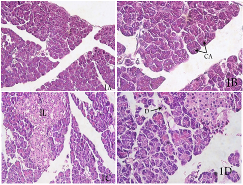
The endocrinal part of the gland (Islets of Langerhans) appears as pale-stained masses of cells present inside the lobules among the pancreatic acini. These masses are formed of branching and anastmosing cords of cells with blood capillaries in between. The cells are rounded or polygonal in shape and variable in size and stainability (figure 1).
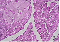
Masson trichome stain showed the minimal content of collagenous stroma in-between the acini (figure 2) PAS stain demonstrated the thin continuous PAS +ve basement membrane surrounding the acini. Also thin PAS +ve capsule of reticular fibers are seen surrounding the islets of Langerhans and separating them from the surrounding exocrinal part (figure 3).

Group II (acute pancreatitis) by L-Arginine
Sub group II (A) 3 days after injection of L-Arginine:-
In sections of pancreas of this sub group, the gland appeared keeping its lobular architecture. The lobules were inter-communicating and separated by thin, incomplete and irregular interlobular septa of loose C.T.
The parenchyma of the exocrinal part of the gland was greatly distorted. It was formed of dispersed cells which had lost their acinar arrangement. These cells were of a heterogenous appearance of variable shape and size.
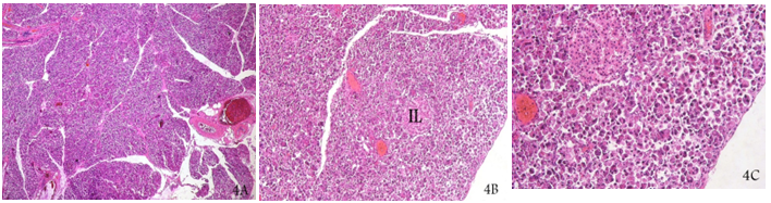
Their cytoplasm was of variable degree of acidophilia. Each cell had a peripheral deeply stained pyknotic nucleus. Some of the cells were bincleated. The blood vessels appeared congested with blood. The endocrinal part of the gland was unaffected and kept its normal appearance (figure 4).
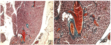
The collagenus fibers stroma was increased specially in the connective tissue surrounding the blood vessels and the interlobular ducts. Also the collagen fibers content was relatively increased intralobulerly in between the parenchymal cells. Some small aggregates of unilocular fat cells were seen in stroma (figure 5).
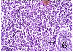
The PAS staining showed the loss of the basement membrane material in the parenchyma inspite of the intact PAS +ve thin capsule of reticular fibers surrounding the islets of Langerhans (figure 6).
Subgroup II(B): 2 weeks after injection of L-arginine:
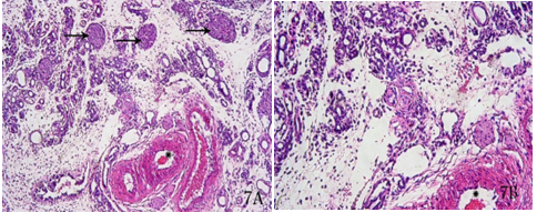
Sections of pancreas of this subgroup showed that the gland was composed mainly of stroma of loose C.T in which widely dispersed groups of ducts are present. These ducts were lined by single row of cubical cells and surrounded with mononuclear cellular infiltration. No other parenchymatous cells or acini could be detected. The blood vessels were intact and congested with blood. Also the endocrinal part of the gland was intact in the form of widely separated islets of Langerhans of normal structure among the stroma of C.T (figure. 7). Aggregations of unilocular fat cells with their signet ring appearance were also seen with more presentation than that in subgroup IIA. Masson trichome stain showed that the collagenous stroma was relatively dense surrounding the blood vessels and the ducts (figure. 8).
PAS stain showed the positively stained basement membrane material as a very delicate interrupted membrane surrounding some fat cells (figure. 9).


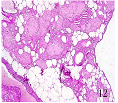
Subgroup IIC:- 1 month after injection of L-Arginine:-
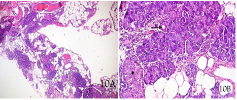
Sections of the pancreas of the animals of this sub-group showed that it was composed mainly of stroma of white adipose C.T. It was formed of crowded aggregates of unilocular fat cells in stroma of loose C.T. Among this fatty stroma, islands of glandular tissue were seen forming small irregular, separate lobules. Most of these lobules were composed of an islet of Langerhans of normal structure with related group of pancreatic acini and interlobular ducts. The acini showed their normal appearance. Each was small triangular or polygonal in shape. It was lined by numerous pyramidal cells. The lumen was narrow and lined by one row of low cubical centroacinar cells whose nuclei appeared oval or rounded in shape. Each acinar cell contained a single, rounded, vesicular nucleus with prominent nucleolus. The nucleus was shifted to the base of the cell. The cytoplasm showed deep basal bosophilia and apical acidophilic zymogen granules. The blood vessels were congested with blood (figure. 10).
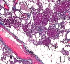
Masson trichome stain showed stroma more densely crowded with collagen fibers compared to the subgroup IIB (figure. 11).
No PAS +ve basement membrane could be detected surrounded the acini, but it was clear and well developed around most of the fat cells. Also the reticular C.T. PAS +ve capsules surrounding the islets of Langerhans were apparent (figure. 12).
Group III (acute pancreatitis); induced by L-Arginine and treated by Dexamethasone:-
Subgroup IIIA 3 days after injection of L-Arginine
The lobular architecture of pancreas was kept where it appeared formed of lobules separated by thin, incomplete and irregular septa of loose C.T.
The parenchyma in the lobules was formed of islets of Langerhans of normal structure present among distorted parenchyuma of the exocrinal part. This exocrinal part appeared as numerous ducts lined by simple cubical epithelium. The exocrinal secretory cells were greatly distorted. Most of them had lost their acinar arrangement and appeared small in size, polygonal or round in shape. Each had a deeply stained pyknotic nucleus peripherally situated. The cytoplasm showed variable degree of acidophlia. On the other hand, some of the cells kept their acinar arrangement especially in areas close to the islets of Langerhans. These exocrinal cells were present among stroma of C.T infiltrated with mononuclear cells, collagen fibers and few fat cells (figure. 13).
Delicate PAS +ve basement membrane could be seen surrounding the ducts and the acini, as well as the fat cells (figure. 14).
Subgroup IIIB (2 weeks after injection of L-arginin:-
Sections of the pancreas of the animals of this sub-group showed that the gland had regained its normal structure with the exception, that the interlobular septa contained aggregates of unilocular fat cells specially close to the blood vessels (figures. 15).


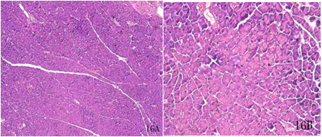
Subgroup IIIB (1 month after injection of L-arginine:-
Sections of the pancreas of animals of this subgroup showed that the gland had completely regained it is normal structure (figures. 16).
Immunehistochemical results:
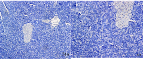
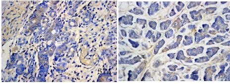
Examination of the sections of the pancreas of the animals of subgroup IIA showed that they contained no immune-positive nuclei by PCNA technique (figure. 17).

Whereas the regenerating lobules of the gland in animals of subgroup IIC showed a moderately crowded immune-positive cells in the developing acini (figure. 18).

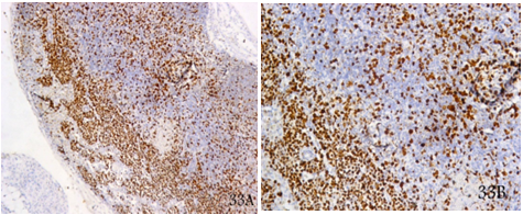
On the other hand, examination of the sections of the pancreas of animals of subgroup IIIA showed that the gland is populated by a markedly, heavily crowded PCNA immune-positive cells (fig. 19), where as in subgroup IIIC the gland was completely devoid of any immune-positive cells (figure. 20).
Concerning casapse-3 immuno-reactivity, it was found to be, positive in all degeneratively cells of the pancreas of subgroup IIA (figure. 21) and intensely positive in the cells of the newly formed acini in subgroup IIC (figure. 22).


On the contrary, in the pancreas of animals of subgroups IIIA and IIIB the reaction was only confined to the cells of regenerating acini in (figure. 23, 24) where as it was completely negative in fully regeneration globe of subgroup IIIC.

Pancreatitis is an inflammatory metabolic disorder, which is a major cause of physical and socioeconomic loss worldwide. Generally, pancreatitis is categorized into two different entities of acute and chronic [15].
Acute pancreatitis (AP), a pancreatic inflammatory disease, is one of the most catastrophic upper abdominal disorders. The incident rate of this inflammatory disease is about 150 to 420 and 700-800 per million/year in United Kingdom and United States, respectively, whereas it ranges from 106 to 205 per million/year in Japan. Severe acute pancreatitis (SAP) with mortality approaching 30% occurred in approximately 20% of the patient with AP because of multiorgan dysfunction and local complication [47]. For many years, treating AP (specially its severe form) has been difficult. The effects of glucocorticoids on acute pancreatitis (AP) have remained contradictory [29].
L-Arginine-induced pancreatitis is an experimental model of severe necrotizing acute pancreatitis. It is a slowly – developing experimental model in which characteristic laboratory changes are observed 24-hrs after induction of the disease.
It was found that the enzyme level slightly and insignificantly increased after 1-3-6 hours after injection of L-Arginine where as it is severely and significantly elevated 24 hours after injection of the drug. Dexamethasone injection with L-Arginine caused mild non significantly increase of serum amylase level in the 1st and 3rd hours, but moderate significant increase in the 6th and 24th hours. It is worlly to note that the severe enzyme level on the 24th hour was much and significantly lower in the group of animals injected with dexamethasone and L-Arginine compared to that of the animals injected with L-Arginine alone. This is in accord with the opinion of [25, 35] who mentioned that serum amylase is one of inflammatory markers which increased early (after 24hs) in the course of both mild and severe acute pancreatitis and were significantly lowered by dexamethasone.
The survival rate of animals in each group was also estimated. In group II, 7 animals died in the first day of the experiment and 2 animals died on the second day, with survival rate of 64%. While in group II only one animal died on 3rd day of the experiment with survival rate of 96%. This is in accord with the findings of [43]. Also, [2] mentioned that approximately 20% of patients with acute pancreatitis develop a severe disease with complications and high risk of mortality. On the other hand, [29] found that in the Dexamethasone treated group, the survival rate increased, compared to the untreated acute pancreatitis group.
Microscopically, sections of the pancreas of the animals of subgroups IA & IB were almost similar and showing the characters of the normal pancreas. On the other hand, sections of the pancreas of the animals of subgroup II(A) (3 days after injection of L-arginine), showed the characters of acinar necrosis. The pancreas had kept its lobular pattern, but the lobules were intercommunicating and separated by thin incomplete irregular septa of loose C.T. the parenchyma was greatly distorted and formed of widely dispersed acinar cells indicating the increase of the intercellular edema.
The cells had lost their acinar arrangement and appeared heterogeneous of variable shape and size and their cytoplasm showed variable degrees of acidophilia. The nuclei of the cells were pyknotic and peripheral. None of these nuclei showed PCNA +ve immune-reactivity indicating no regeneration activity. The blood vessels were congested with blood. The ducts and the islets of Langerhans, were not affected. This was accompanied with signs of early collagen fibrosis of the gland and distortion of the acinar basement membrane. Small aggregates of fat cells was also seen in the stroma. Two weeks after last injection of L-arginine, the gland was more severely affected. It was composed mainly of stroma of loose C.T. with widely dispersed groups of ducts and islets of Langerhans, no other parenchymal cells or acini could be detected. The C.T stroma was heavily populated with mononuclear cellular infiltration. The fat cells aggregation and fibrosis were more prominent. This is in agreement with the picture described by [45,47] who mentioned that AP is associated with parenchymal edema, tissue necrosis, haemorrhage and inflammatory cells infiltration [2, 24]. suggested that inflammatory cytokines and neutrophil-mediated oxidative stress have a central role in the pathogenesis of acute pancreatitis induced by L-arginine. The later added that this inflammatory process is associated with acinar cell damage and an inadequate immune response mediated mainly by monocyte, macrophage, polymorph nuclear cells (PMNs), lympohcytes, mast cells and endothelial cells [44]. mentioned that a lot of factors are involved in the local and systemic complications of SAP, such as systemic inflammatory response syndrome which can lead to multiple organ failure and pancreatic necrosis. Among them, an intensive systemic inflammatory response mediated by overproduction of pro-inflammatory cytokines and pancreatic autodigestion by activated trypsin plays a very important role. In addition, oxidative stress, secondary infection, endotoxemia are also risk factors [30, 17]. reported that, activation of the exocrine enzyme, especially trypsin, induces various activation of protease within the pancreas which would degrade lots of cellular protein and eventually result in pancreatic lesion. In addition, the autoantigens in injured cells trigger immune system, leading to aseptic inflammation of the pancreas, and eventually tissue damage and necrosis, 18 observed that ER stress is early paralleling trypsinogen activation in caerluein model as well as L-Arginine model of AP leading to pathological sustained global rise of cytosolic Ca++. The process of fibrogenesis in the pancreas insulted by AP had been discussed by [36]. They reported the presence of stellate cells in the pancreas which exhibit morphological and functional features similar to cultured hepatic stellate cells including positive desmin and smooth muscle actin (SMA) staining after a period of time in culture. In the normal rat pancreas, these stellate cells stain positively for desmin but do not stain for SMA, indicating a quiescent non activated state. They also noted that the pancreatic stellate cells are involved in collagen production during acute pancreatitis. It is possible that the cytokines TGFb released by acinar cells secondary to cell injury may be one of the predominant factors promoting a fibrotic response in pancreatic stellate cells. Also these cells can become active, during AP, through the action of angiotensin II produced by renin-angiotensin.
Sections of the pancreas of the animals of subgroup II(C) (1 month after L-Arginine injection) showed that the gland is composed mainly of stroma of white adipose C.T. Among this fatty stroma, islands of glandular tissue were seen forming small irregular separate lobules. Most of these lobules were composed of islets of Langerhans with their normal structure and a related group of heavily crowded pancreatic acini and ducts. The acini were of normal appearance and collagen fibrosis was more marked. The finding that the stroma of the pancreas was formed of white adipose connective tissue lately after induction of AP, is in agreement with [1] who considered this phenomena as a sign of atrophy.
The re-appearance of new lobules of normal pancreatic acini with the pre-existing ducts, which kept their normal structure, indicates the early spontaneous regeneration of the gland. This was confirmed by the finding that these regenerating lobules showed a moderately crowded PCNA +ve nuclei. It was observed that these newly formed lobules are born close to previously existing unaffected islets of Langerhans. This finding must raise the question of the role of islets of Langerhans and the ducts in process of regeneration of the pancreatic acini, and lobules.
In this study it was observed that during AP, the structure of the various pancreatic ducts was not affected. On the other hand, [33] mentioned that the injurious stimulus in AP alters the normal physiological functions of ducts cells as much as it does for the acinar cells. [16] had put forth an interesting hypothesis that enhanced ductal fluid secretion during early stages of pancreatitis may defined against damage by washing out toxic agents and digestive enzymes, while overwhelming of this ductal defense mechanism leads to pancreatic injury [8, 32, 16]. mentioned that; inhibited bicarbonate secretion by the ductal cells may lead to reduction in luminal pH which has been shown to contribute intra-acinar zymogen activation.
Examination of the sections of the pancreas of animals of subgroup III(A) showed that the gland kept its lobular architecture being formed of lobules separated by thin, incomplete and irregular septa of loose CT. The parenchyma of the lobules was formed of islets of Langerhans of normal structure; present among distorted parenchyma of the exocrinal part. This exocrinal parenchyma appeared as numerous ducts lined by simple cubical epithelium. The exocrinal secreatory cells were greatly distorted and showing the characters of necrosis. On the other hands some of these cells had kept their acinar arrangement especially in areas close to islets of Langerhans the gland also showed heavy crowded population with PCNA +ve nuclei indicating the high and early regenerative process. The stroma of the gland was formed of C.T infiltrated with mononuclear cells, collagen fibres and few fat cells, delicate PAS +ve basement membrane could be seen surrounding the ducts, the acini and the fat cells. However, the sections of the pancreas of the animals of subgroup III(B) showed that the gland had regained most of its normal structure. This normal structure of the gland was almost completely regained in the animals of sub group III(C), with no regenerative process as indicating by the absence of any PCNA in nuclei. This picture indicates that the gland in group III showed signs of AP milder than those occurring in group II and also shown signs of more rapid regeneration.
[24] came to conclusion that treatment of AP with triterpene a, b-amyrin and glucocorticoid methyl prednisolone resulted in a significant decrease of serum amylase and lipase, pancreatic edema and serum TNF-a and IL-6 levels as well as the TNF-a and iNOS expression induced by L-Arginine. The drugs effectively improved the pancreatic morphology, and the results clearly point out their anti-inflammatory potential in obliterating the pancreatic inflammation and the associated tissue injury.
[45] reported that in rats suffering from severe acute pancreatitis, and treated with Dexamethasone, the degree of pathological changes (including inflammation, haemorrhage and necrosis, was milder than that in the untreated groups, at the same time interval. They also mentioned that Dexamethasone is a kind of long-acting glucocorticoid. In 1952, Stephen et al., reported, for the first time, a case in which cortisone treatment for acute haemorrhagic necrosis pancreatitis had a certain treatment effect and established the milestone of glucocorticoid treatment in acute pancreatitis.
It's mechanism in treating AP was postulated by many authors. This mechanism includes inhibiting the inflammatory mediators [7], resisting endotoxins [34] improving micro-circulation (42), removing oxygen free radicals [22], inhibiting NO [26] inhibiting NF-Kappa B [20, 23] inducing apoptosis of acinar cells [22, 21, 40]. Also, [46] came to the conclusion that the alleviation of pancreatic tissue injury by dexamethasone during SAP might be closely related to its role in inhibiting NF-Kapa b expression and regulating cytokines [43]. Also mentioned that dexamethasone can improve the pancreatic injury of SAP rats by directly inducing pancreatic cell apoptosis, or indirectly inducing apoptosis through inhibition of excessive rise of TNF-a, IL-6, PLA2 ….. etc. this is in accord with our fndings that the newly formed acinar cells in all the groups were +ve for caspase reaction indicating the apoptotic process. This character in completely about is full mature acini [43, 33]. mentioned that; both necrosis and apoptosis are death mode of injured cells, however, substantially different form necrosis, apoptosis will not release the harmful substance in lysosomes or cause interse inflammatory reaction. Apoptosis is known to eliminate damaged cells without eliciting much inflammation, while overwhelming cell injury shifts the cell death to necrosis which leads to wide spread inflammation. Necrosis prevails in SAP. The illness can be alleviated by apoptosis induction and aggravated by apoptosis inhibition, therefore, the apoptosis of pancreatic acinar cell might be a reaction beneficial to the body after the occurrence of pancreatitis. They also noted that severity of pancreatitis is considerably less when apoptosis is the predominating mechanism compared to when necrosis is predominant [43], in their study on rats with experimentally induced AP, they found that the apoptotic index was higher in animals treated with dexamethasone than in non-treated animals. They came to the conclusion that dexamethasone can promote the apoptosis of pancreatic cells and protect the pancreatic tissue.
It can be concluded that treatment of AP with dexamethasone,one is beneficial in ameliorating the pancreatic affection and helps in its rapid regeneration.
Dear Editorial Team, Clinical Medical Reviews and Reports. My experience with the journal was highly positive. The peer-review process was rigorous, constructive, and completed in a timely manner. The reviewers provided valuable comments that helped improve the quality and clarity of our manuscript. The editorial office was professional, responsive, and supportive throughout all stages of the publication process. Communication was clear and efficient, and any questions were addressed promptly. Overall, I found the journal to maintain high scientific standards and an excellent publication workflow. I would be pleased to consider submitting future work to this journal. Best wishes from, Elena Popa.

It was my pleasure to submit my testimonial concerning the Reviewer Board of our Scientific Journal “Brain and Neurological Disorders”. The Reviewers focused on some modifications and their contribution was helpful. The ladies of our Editorial Office were also supported my efforts. It was my honor to have such a co-operation and I am looking forward for more collaboration.

Dear Grace Pierce, Editorial Coordinator of Journal of Clinical Research and Reports, Thank you for the speedy and efficient peer review process. I appreciate the fact that your peer reviewers do not take months to respond like with some other journals. I would also like to thank the editorial office for responding quickly to my questions. It is an excellent journal. I plan to submit more manuscripts in the future. Best wishes from, Robert W. McGee

Dear Grace Pierce, Editorial Coordinator of Journal of Clinical Research and Reports, Working with you and your team on our recent publication in JCRR has been a truly wonderful and enjoyable experience. The responses were prompt, and the reviewers were patient, constructive, and highly professional. One reviewer in particular gave me the feeling that a professor was carefully reading and commenting on my coursework, which was deeply touching. The entire process was straightforward and hassle‑free, with no tedious online forms to complete. I highly recommend this journal. Best wishes from, DR Aibing Rao, Head of R&D

I Appreciate the Opportunity to Share my Experience with the Journal of Clinical Research and Reports. The peer review process was timely and constructive, and the feedback provided helped improve the quality of our manuscript. The editorial office was professional, responsive, and supportive throughout the process, ensuring smooth communication and efficient handling of the submission. Overall, it was a positive experience collaborating with your team.

Dear Mercy Grace, Editorial Coordinator of Obstetrics Gynecology and Reproductive Sciences, We would like to express our gratitude for your help at all stages of publishing and editing the article. The editors of the magazine answer all the necessary questions and help at every stage. We will definitely continue to cooperate and publish other works in the Obstetrics Gynecology and Reproductive Sciences! Best wishes from, Alla Konstantinovna Politova,
