
Review Article | DOI: https://doi.org/10.31579/2688-7517/047
1Department of Pharmacy, Mathematics and Natural Science of Faculty, Tadulako University, Palu, Central Sulawesi, Indonesia.
*Corresponding Author: Jamaluddin Department of Psychology, Mathematics and Natural Science of Faculty, Tadulako University, Palu, Central Sulawesi, Indonesia.
Citation: Yusriadi, Nurzamza, Rifal Setiawan, Agustinus Widodo, Jamaluddin (2022) The Activity of The Water Phase Extract of Anguilla marmorata Q. Gaimard from Lake Poso and Anguilla bicolor from Palu River in The Healing Process of Rattus norvegicus Cuts. Addiction Research and Adolescent Behaviour. 5(4); DOI: 10.31579/2688-7517/047
Copyright: © 2022 Jamaluddin, This is an open access article distributed under the Creative Commons Attribution License, which permits unrestricted use, distribution, and reproduction in any medium, provided the original work is properly cited.
Received: 15 April 2022 | Accepted: 22 April 2022 | Published: 06 May 2022
Keywords: extract of water-phase eel fish; anguilla marmorata, anguilla bicolor; palu River; lake Poso; incised wound; white rats
Eel fish are very widely distributed in Indonesia, especially in the region of Central Sulawesi. Eel fish are widely cultivated since they are rich in high nutritional content such as vitamins and albumin. This research aimed to examine activities of the extract of water-phase eel fish (Anguilla marmorata and Anguilla bicolor) on the incised wound healing in white rats. The eel fish extract is used as a base for ointments, where the ointment base used was Adeps lanae. The treatments were divided into five test groups, namely the concentration of 5%, 10%, 15%, positive control, and negative control. The test animal used in this study was white rats, then on the back of the rat a 2 cm long incision was made, and ointment was applied twice a day for 21 days. The experimental animals used in this research were white male rats in which their backs were injured 2-cm long, and the ointment was applied twice a day for 21 days. The concentration that had the largest healing percentage was 15%. The measurement data were analyzed using One-Way ANOVA and continued with the Post Hoc Test (Duncan). The results showed that the eel fish Anguilla marmorata had the incised wound healing activity in the white rats where the highest healing concentration on the 21st day was seen in the 15% group with the percentage of 85.3%, and the lowest concentration was found in 5% group, with the percentage of 72.1%. Whereas for the eel fish Anguilla bicolor on the 21st day, the maximum healing obtained was with the 15% group (83.5%), and the minimum healing was found in the 5% group (66.5%). From the results of these percentages, it can be concluded that the extract of water-phase eel fish Anguilla marmorata and Anguilla bicolor has the incised wound healing activity in white rats, and the most effective concentration for the incised wound healing process of each fish for 21 days is the concentration of 15%, with the percentage of healing for the eel fish Anguilla marmorata is 85.3%, and the eel fish Anguilla bicolor is 83.5%.
Eel fish (Anguilla sp.) are very well sold in international markets such as in Japan, Hong Kong, South Korea, China, Taiwan, and several other countries. There are 350 types of eel fish in the world, 12 species of which are found in the tropics, 6 species found in Indonesian, namely Anguilla marmorata, Anguilla bicolor, Anguilla celebensis, Anguilla borneoensis, Anguilla ancertalis and Anguilla Mauritania. However, in Indonesia, there are only 2 types often cultivated, Anguilla marmorata (Q.) Gaimard and Anguilla bicolor (Antonies, 2013)[1]. Eel is a consumption fish that is in great demand in international markets because the meat of eel contains the nutrients the body needs with a protein content of 16.4% and a high vitamin A content of 4,700 IU/100 grams. Even the DHA (Docosahexaenoic acid) content of eel is 1,337 mg/100 grams, beating salmon, which is only 820 mg/100 grams and EPA (Eicosapentaeonic acid) levels of eel reach 742 mg/100 grams, far above salmon which is only 492 mg. / 100 grams and mackerel fish which is only 492 mg / 100 grams (Antonies, 2013)(1).
Eel fish (Anguilla sp.) contain albumin protein which functions to regulate the osmotic pressure in the blood and also as a means of transportation in blood. According to Iriana (2016)[2], albumin is useful in helping the formation of new body cell tissues during development and can accelerate the wound healing process. Apart from albumin, eel fish meat also contains vitamin c, which plays an essential role in wound healing in the proliferative phase. Without vitamin c, collagen synthesis will stop, and the new blood capillaries will be re-damaged and will experience bleeding, so that wound healing will take a long time (Morison, 2004)[3].
A wound is a form of tissue damage to the skin caused by contact with heat sources (such as chemicals, hot water, fire, radiation, and electricity), the result of medical action, exposure to blunt objects, or changes in physiological conditions (Sjamsuhidajat, 2005)[4]. When there is a wound to skin tissue, the skin will show signs of inflammation where foreign objects from outside the body can enter through an open wound such as an incision. The presence of foreign bodies triggers a hydrostatic pressure disorder, namely that extracellular fluid will enter the cell due to a difference or imbalance in the concentration inside and outside the cell through the osmotic route, causing the cell to experience edema or swelling. This will cause the cell to experience edema or swelling. This condition requires albumin nutrition which can maintain the osmotic pressure inside and outside the cell so that the edema does not get worse (Rehatta, 2019)[5].
The wound healing process generally consists of several phases, each of which is interrelated, namely the inflammatory, destructive, proliferation, and maturation phases. The first is the inflammatory response stage; this stage begins at the time of the injury. At this stage, there is hemostasis that frightens with the release of histamine and other mediators of the damaged cell, processes and processes white blood cell to the damaged area. Usually, this stage of the inflammatory response occurs 3 days after the injury. After this, the destructive phase occurs when the death of the tissues occurs by polymorphonuclear leukocytes and macrophages. Usually, this destructive phase occurs after 3 days of injury to the 14th day after the injury. Then the proliferation phase takes place, where new blood comes out of the connective tissue and infiltrates the wound. Usually, this proliferation phase occurs after 14 days of injury to the 21st day after the injury. In the last stage, namely the maturase phase, in this phase there is a process of forming new epithelium, contracting wounds and is the stage of completing connective tissue and new cells until their function returns to normal. This stage occurs on the 21st day to more than one year (Hidayat, 2008)[6].
Based on the information above, the researchers conducted research related to the healing process of cuts in white rats using betadine ointment as a positive control and adeps lanae as a negative control by using variations in the concentration of eel extracts 5%, 10% and 15%.
Materials
The main objects used in this research were the silver-eel-phase Anguilla marmorata obtained from Lake Poso and the silver-eel-phase Anguilla bicolor obtained from Palu River. The ingredients for the open wound healing activity test were ketamine, 70% alcohol, Betadine ointment, and Adeps lanae, where the bases used were hydrocarbon ointment bases, absorption ointment bases, and ointment bases in water (Ministry of Health of the Republic of Indonesia, 1995)[7]. The experimental animals used in this research were 25 white rats aged ±3 months and weighing 275-300 grams.
Methods
Sample Preparation and Collecting Technique
The sampling technique procedure was carried out using the purposive sampling method, i.e., a sampling technique by selecting samples from among the population according to what researchers wanted, namely based on the type, weight, size, and location of the collection. The eel fish was cleaned (removed scales, gills, and stomach contents), washed until there was no more blood and mucus, deboned, and cut into small pieces. Then they were mashed using a blender, and a solvent was added with a ratio of 1: 1 weight per volume (100 grams of fish: 100 ml of water solvent) for crude extract (Putri, 2016; Jamaluddin, 2019; Jamaluddin, 2021)[8][9].
Extraction Stage
The prepared crude extract was then extracted by steaming it at 40°C for 10 minutes. Next, it was filtered to separate the filtrate and residue. The extract obtained was then centrifuged at 3000 rpm for 10 minutes to separate the water phase and the oil phase. The water phase of the eel was then taken and ready to be tested (Nugroho, 2012)(10).
Compound Identification Stage
The water phase extract of the eel was further identified water-soluble compounds. The water-soluble compounds identified in the water extract of eel include vitamin C and albumin, which were identified using (biuret test, xanthoprotein test and visual test).
1. Vitamin C
The vitamin C test was performed by taking 2-ml of the eel fish extract filtrate, then adding 4 drops of methylene blue Lp, then heating it to a temperature of 40°C, so that within 3 minutes would form a dark blue color, and then became light blue or disappeared (Ministry of Health of the Republic of Indonesia, 1995)(7).
2. Albumin
The eel fish extract was tested qualitatively by the biuret, xanthoprotein, and visual tests for albumin protein presence.
Making of Eel Fish Extract Base
The water phase eel extract is made with concentrates of 5%, 10%, and 15%, added with a base of Adeps lanae ointment up to 100 grams. Starting with weighing the materials needed, namely adeps lanae and water phase extract of eel. The 5% concentrate consisted of 5-gram eel fish extract and 95-gram Adeps Lanae, the 10% concentrate consisted of 10-gram eel fish extract and 90-gram Adeps Lanae, and the 15% concentrate consisted of 15-gram eel fish extract and 85-gram Adeps Lanae. Next, Adeps lanae was added to the mortar, then the extract of the water-phase eel fish was added and crushed until it was homogeneous. Eel fish extract ointment with a concentration of 5%, 10%, and 15% were put in ointment pots (Karuniawan, 2016)(11).
Wound Induction on White Rats
For the wound induction, each rat was treated the same way as follows: first, the back hair of the rats was shaved using a 4-cm-diameter razor for simplifying the treatment; next, the mice were anesthetized using ketamine; then, an incision was made using a scalpel, with 2-cm long and 2-mm deep into the back of the rats.
Administration of Extract of Water-Phase Eel Fish
The administration of the preparations was done by applying twice a day, morning and evening, with a dose of 0.2 gram per application, from the 1st to the 21st day (Andrie, 2017)(12). In each treatment group, there were 5 white rats. The treatment of administering the extract of the water-phase eel fish in the white rats are as follows:
Group I : Betadine ointment was applied as the positive control with 5 rats.
Group II : Adeps lanae ointment base was applied as the negative control with 5 rats.
Group III : 5% concentrate of extract of water-phase eel fish was administered control with 5 rats.
Group IV : 10% concentrate of extract of water-phase eel fish was administered control with 5 rats.
Group V : 15% concentrate of extract of water-phase eel fish was administered control with 5 rats.
Measurement of Incised Wound Healing on White Rats’ Back
The wound healing was assessed by measuring the average length of the wound each day, starting from the first day of making the wound to the 21st day, and by calculating the percentage of wound healing.
The formula that calculated the percentage of open wound healing on the backs of white rats:
Information:
P% (for percentage of wound healing)
do (for the length of the initial wound)
dx (for the length of the wound on the last day)
Data Analysis
The data obtained from the incised wound healing activity test were then processed by the statistical test using One-Way ANOVA, continued by Post Hoc Duncan on 95% trust level for determining the relationship between one variable and another.
Qualitative Test Results of Eel Fish Water Phase (Anguilla marmorata and Anguilla bicolor)
The extract of water-phase eel fish Anguilla marmorata and Anguilla bicolor contained bioactive compounds that played a role in providing biological effects. The test results are presented in Table 1 below.
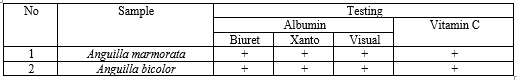
Information:
(+) = Positively contains compounds
(-) = Does not contain compounds
From the qualitative test results of the extract of water-phase eel fish (Anguilla marmorata and Anguilla bicolor), carried out using vitamin C and albumin test methods (biuret, xanthoprotein, and visual tests), it showed that the extract from the water-phase eel fish was positively contained vitamin C and albumin.
Test Results of Administering Extract of Water-Phase Eel Fish Anguilla marmorata and Anguilla bicolor
The percentages of the incised wound healing in which the extract of water-phase eel fish Anguilla marmorata and Anguilla bicolor were administered from the first day till the 21st day can be seen in Table 2.

Information:
D M0 = Percentage of wound healing from Anguilla marmorata on day 0
D M1 = Percentage of wound healing from Anguilla marmorata on day 1
D M5 = Percentage of wound healing from Anguilla marmorata on the 5th day
D M9 = Percentage of wound healing from Anguilla marmorata on the 9th day
D M13 = Percentage of wound healing from Anguilla marmorata on the 13th day
D M17 = Percentage of wound healing from Anguilla marmorata on the 17th day
D M21 = Percentage of wound healing from Anguilla marmorata on the 21st day
D B0 = Percentage of wound healing from Anguilla bicolor on day 0
D B1 = Percentage of wound healing from Anguilla bicolor on day 1
D B6 = Percentage of wound healing from Anguilla bicolor on the 6th day
D B10 = Percentage of wound healing from Anguilla bicolor on the 10th day
D B14 = Percentage of wound healing from Anguilla bicolor on the 14th day
D B18 = Percentage of wound healing from Anguilla bicolor on the 18th day
D B21 = Percentage of wound healing from Anguilla bicolor on the 21st day

Based on the percentage results of the incised wound healing of the experimental animals which were administered the extract of water-phase eel fish Anguilla marmorata and Anguilla bicolor in Table 2, it is stated that they were healed, marked by a change in the length of the wound that was getting smaller or the percentage of wound healing was getting bigger.

There was a difference in the average percentage of the incised wound healing of the extract of water-phase eel fish Anguilla marmorata and Anguilla bicolor between the groups, based on the graph. The greatest percentage of incised wound healing was found in the 15%-extract group of the species Anguilla marmorata with the healing percentage of 85.3% on the 21st day of treatment, while the percentage in the 15%-extract group of the species Anguilla bicolor was 83.5%. Based on these data, it can be concluded that the healing rate of the extract of water-phase eel fish Anguilla marmorata was more effective than the eel fish Anguilla bicolor.
Eel fish are in great demand in the international market due to their nutrients, they contain 16.4% protein content and 4,700 IU/100 grams of vitamin A (Antonies, 2013)[1]. Based on research by Putri (2016)[8], the water extract of eel fish contains albumin compound in which the average albumin content obtained for the species Anguilla marmorata is 0.508 mg/100 grams, whereas the average albumin content for the species Anguilla bicolor was 0.242 mg/100 grams. The scientific test by Sinambela (2012)[13] the results show that fish containing albumin have activity and effectiveness in healing the incision which can close the cut within 8th days. According to Suprayitno (2009)[14], albumin functions to regulate osmotic pressure in the blood and is also useful in the formation of new body tissues during growth, as well as can accelerate the healing of body tissues.
The eel fish used in this research were the Anguilla marmorata from Lake Poso and the Anguilla bicolor from the Palu River. This research used eel fish because they are very widely distributed in Sulawesi; eel fish populations are found in several rivers and lakes. The potential for eel fish breeding in Sulawesi is tremendous. The most extensive distribution of eel fish in Sulawesi is in Lake Poso and Palu river, Central Sulawesi (Fadly, 2014)[15]. This research used white rats (Rattus norvegicus) as the experimental animals because, according to Angria (2019)[16], their biological characteristics and behavior are generally very similar to humans, and many symptoms of the human condition can be replicated in rats such as function, organ shape, biochemical processes, and biophysical process. Besides, white rats have several beneficial properties as experimental animals, including rapid reproduction, larger size than mice, easy to maintain, and in large numbers. According to Vrhaz (2013)[17], almost 95% of experiments and research for the medical world, experts, and scientists use rats due to their genetics, biological characteristic, and human-like behavior, and many symptoms of the human condition can be seen in rats.
This research used the extract of water-phase eel fish due to the presence of compounds (albumin and vitamin C) in the eel fish meat, which are water-soluble compounds that have activities in wound healing. Water solvents were used to draw compounds from the eel fish meat (Asmadi, 2008)[18]. Based on the research by Jamaluddin et al. (2020)[19], the total albumin content in eel fish Anguilla marmorata is 1.62 grams/100 grams, and in eel Anguilla bicolor is 0.24 grams/100 grams. The eel fish used in this research were from Lake Poso and Palu river, Central Sulawesi. The extract was made by the steaming method using a temperature of 40 ° C for 10 minutes, using a temperature of 40 ° C in the steaming process because at that temperature the amount or level of albumin obtained was optimal where the temperature limit used in the albumin steaming process was not damaged, ranging from 40 -60 ° C. The eel fish were steamed using distilled water so that the water-soluble compounds in the eel fish meat could be withdrawn, then filtered to separate the filtrate and residue. Later, the extract was centrifuged at 3000 rpm for 10 minutes to separate the water and oil phases. Furthermore, the water phase obtained was mixed with the base of Adeps lanae ointment which was made in 3 concentrations, namely 5%, 10%, and 15%. Moreover, the extract of the water-phase eel fish was ready to be tested to see the activity of the incised wound healing in white rats (Putri, 2016)[8].
This research was divided into 5 groups of experimental animals, namely the positive control group, the negative control group, and the extract of water-phase eel fish with concentrates of 5%, 10%, and 15%. The division of the test groups in this research was carried out to examine the effect of treatment on the experimental group by comparing the sample group with the control group (Notoatmodjo, 2012)[20]. The control groups used were positive and negative controls. The positive control using betadine ointment (Povidone iodine) with the mechanism of action starting after direct contact with the skin tissue, iodine will be released slowly to inhibit bacterial enzyme metabolism so that it interferes with the multiplication of bacteria which causes the bacteria to become weak. Povidone iodine, which is commonly used in wound care, is only 10%. According to Gunawan (2007)[21], the greater the iodine concentration used, the faster wound healing will be. In contrast, the negative control used Adeps lanae base that aimed to compare the test group and positive control on the process of wound healing. The use of the Adeps lanae ointment base in the extract of water-phase eel fish is because this base can improve water absorption properties. Thus it is good in releasing the extract of water-phase eel fish (Lazuardi, 2019)[22].
After dividing the test groups, each rat was injured on their back with a 2-cm wound size and a 2-mm wound depth, using a scalpel. The measurement of the incised wound on the back of the rats was carried out using a digital caliper by measuring the long diameter of the wound. The use of a digital caliper as a measuring tool is because it has a higher level of accuracy than other measuring instruments, with an accuracy level of 0.1 mm or 0.01 cm. In addition, a digital caliper makes it easier to measure objects with small diameters since it no longer needs to lock them (Utomo, 2007)[23]. Changes in the wound diameter in the rats were observed from the first day of treatment to the 21st day.
From the measurement data for 21 days, it was then presented in the form of tables and graphs, showing the average value of the percentage of incised wound healing in the extract of water-phase eel fish Anguilla marmorata and Anguilla bicolor where it aimed to see the difference in the percentages of incised wound healing between the groups. The healing observation results of the extract of water-phase eel fish Anguilla marmorata on the incised wound healing on the 21st day, namely at concentrates of 5%, 10%, and 15%, were 72.1%, 77.8%, and 85.3%. Meanwhile, for Anguilla bicolor they were were 66.5%, 69.9%, and 83.5%. It can be seen that the higher percentage of incised wound healing was in the 15% concentrate, compared to the 5% and 10% concentrates, both for Anguilla marmorata and Anguilla bicolor. Research by Fitriyani (2013)[24] showed that snakehead fish albumin extract with 60% concentrate has a greater healing rate than the 20% and 40% extracts in providing effects on incised wound healing. Research by Fuadi (2018)[25] say that using eel fish albumin ointment with concentrates of 5%, 10%, and 20
In line with this research, it can be concluded that the extract, both of the water-phase eel fish Anguilla marmorata from Lake Poso and the water-phase eel fish Anguilla bicolor from Palu river, has activities in the incised wound healing on the back of white rats, with the highest percentage of wound healing on the 21st day by the eel fish Anguilla marmorata and Anguilla bicolor, which is at the concentration of 15% and with the respective healing percentage by the eel fish Anguilla marmorata which is 85.3%, and by the eel fish Anguilla bicolor which is 83.5%. Hence for the percentage comparison of the incised wound healing in white rats with the same concentration, the eel fish Anguilla marmorata has a higher healing percentage than that in the eel fish Anguilla bicolor.
The authors declare no potential conflict of interest.
Personal funding.
This research has been tested at the research ethics committee of the Faculty of Medicine, Universitas Tadulako of Palu to obtain the Ethical Clearance No.3782/UN 28.1.30/ KL/2019 on July 10, 2019, by applying the principles of research ethics according to the ethical principles of the 2008 Helsinki Declaration.
The author would like to thank the Laboratory of the Department of Pharmacy, Faculty of Mathematics and Natural Sciences, Tadulako University who has helped in completing this research, and all parties who have provided support.
Dear Editorial Team, Clinical Medical Reviews and Reports. My experience with the journal was highly positive. The peer-review process was rigorous, constructive, and completed in a timely manner. The reviewers provided valuable comments that helped improve the quality and clarity of our manuscript. The editorial office was professional, responsive, and supportive throughout all stages of the publication process. Communication was clear and efficient, and any questions were addressed promptly. Overall, I found the journal to maintain high scientific standards and an excellent publication workflow. I would be pleased to consider submitting future work to this journal. Best wishes from, Elena Popa.

It was my pleasure to submit my testimonial concerning the Reviewer Board of our Scientific Journal “Brain and Neurological Disorders”. The Reviewers focused on some modifications and their contribution was helpful. The ladies of our Editorial Office were also supported my efforts. It was my honor to have such a co-operation and I am looking forward for more collaboration.

Dear Grace Pierce, Editorial Coordinator of Journal of Clinical Research and Reports, Thank you for the speedy and efficient peer review process. I appreciate the fact that your peer reviewers do not take months to respond like with some other journals. I would also like to thank the editorial office for responding quickly to my questions. It is an excellent journal. I plan to submit more manuscripts in the future. Best wishes from, Robert W. McGee

Dear Grace Pierce, Editorial Coordinator of Journal of Clinical Research and Reports, Working with you and your team on our recent publication in JCRR has been a truly wonderful and enjoyable experience. The responses were prompt, and the reviewers were patient, constructive, and highly professional. One reviewer in particular gave me the feeling that a professor was carefully reading and commenting on my coursework, which was deeply touching. The entire process was straightforward and hassle‑free, with no tedious online forms to complete. I highly recommend this journal. Best wishes from, DR Aibing Rao, Head of R&D

I Appreciate the Opportunity to Share my Experience with the Journal of Clinical Research and Reports. The peer review process was timely and constructive, and the feedback provided helped improve the quality of our manuscript. The editorial office was professional, responsive, and supportive throughout the process, ensuring smooth communication and efficient handling of the submission. Overall, it was a positive experience collaborating with your team.

Dear Mercy Grace, Editorial Coordinator of Obstetrics Gynecology and Reproductive Sciences, We would like to express our gratitude for your help at all stages of publishing and editing the article. The editors of the magazine answer all the necessary questions and help at every stage. We will definitely continue to cooperate and publish other works in the Obstetrics Gynecology and Reproductive Sciences! Best wishes from, Alla Konstantinovna Politova,
