
Case Report | DOI: https://doi.org/10.31579/2578-8949/086
1Associate professor, Department of Dermatology, MGM medical college and hospital, Aurangabad, India
2Junior resident, Department of Dermatology, MGM medical college and hospital, Aurangabad, India
*Corresponding Author: Anirudha Gulanikar, Associate professor, Department of Dermatology, MGM medical college and hospital, Aurangabad, India.
Citation: A Gulanikar, Omkar S. Kulkarni. (2021) Syringocystadenoma papilliferum at an unusual location: a case report. Dermatology and Dermatitis. 6(4); Doi:10.31579/2578-8949/086
Copyright: ©2021 Anirudha Gulanikar, This is an open-access article distributed under the terms of The Creative Commons. Attribution License, which permits unrestricted use, distribution, and reproduction in any medium, provided the original author and source are credited.
Received: 27 September 2021 | Accepted: 04 October 2021 | Published: 08 October 2021
Keywords: syringocystadenoma papilliferum; adnexal tumours; benign cutaneous tumours
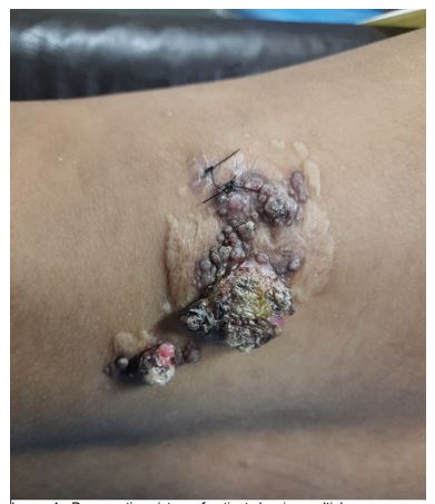
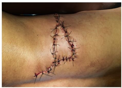
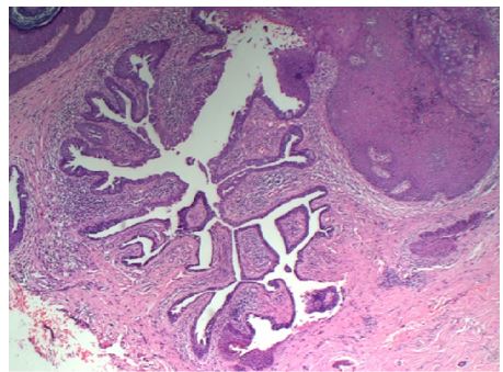
A case of 15year old female presented with lesion over back since childhood, with occasional bleeding and oozing from lesion without any associated systemic complaints. There were multiple verrucous coalescing papules forming plaque with overlying erosion present over left lower back- diagnosed provisionally as angiokeratoma circumscriptum and was biopsied. Histopathology revealed findings consistent with Syringocystadenoma papilliferum. Surgical excision was done and closed with rotation flap. Syringocystadenoma is benign cutaneous adnexal tumor presenting clinically with many morphologies such as warty papules, nodules, plaques with oozing of serous material. Lesion is usually seen in head and neck area in most cases however can also occur on extremities, buttocks, anogenital region. It is characterized by multiple invaginations from skin surface in association with hair follicles lined by cuboidal to columnar epithelium on luminal aspect and myoepithelial cells on outside. There is papillary architecture and dermal ductal component.
Skin appendageal tumours are rare tumours which either arise from or differentiate towards pilosebaceous apparatus, eccrine or apocrine morphology. These are more predominantly found in scalp and anogenital region given more density of pilosebaceous apparatus there. Pilar component predominates over scalp whereas glandular component tends to predominate over face. These present with vague clinical features and diagnosed only after histopathological characterization.
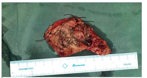
15year old female came with complaint of lesion over back since childhood. It was asymptomatic and pea sized to begin with, gradually increased in size over last 10-12 years. There is history of occasional bleeding and oozing from lesion. There are no systemic symptoms. On examination, patient had multiple skin-coloured papules coalescing to form plaque that was well circumscribed and had few eroded areas on lower left back. Few areas showed signs of spontaneous resorption. She was provisionally diagnosed as? angiokeratoma circumscriptum? lymphangioma circumscriptum. She was investigated further – here hemogram and blood biochemistry were unremarkable. Punch biopsy was taken and histopathological examination showed crateriform invaginations connected with surface epidermis that are lined by double layer of epithelium – luminal epithelium being apocrine and basal being cuboidal. On the floor of invagination, there were many papillary projections forming solid areas. Surrounding dermal tissue showed mixed infiltrate of lymphocytes, plasma cells and neutrophils. Thus histopathological diagnosis of Syringocystadenoma papilliferum was made and excision was planned in coordination with plastic surgery department. Lesion was excised and wound was closed with triangular rotation flap. The excised lesion measured 5X3X1 cm and on histopathology, confirmed diagnosis of Syringocystadenoma papilliferum without any cancerous changes.
Syringocystadenoma papilliferum is a benign adnexal tumor that arises from glandular ductal epithelium. Its origin is uncertain- it’s considered to arise from pluripotent appendageal cells or apoeccrine glands [1]. It can occur as an isolated tumor or as a secondary growth arising in Nevus sebaceous [2]. It occurs commonly in children and adolescents as acquired lesion, but can be congenital in rare cases [3]. In 75
Dear Editorial Team, Clinical Medical Reviews and Reports. My experience with the journal was highly positive. The peer-review process was rigorous, constructive, and completed in a timely manner. The reviewers provided valuable comments that helped improve the quality and clarity of our manuscript. The editorial office was professional, responsive, and supportive throughout all stages of the publication process. Communication was clear and efficient, and any questions were addressed promptly. Overall, I found the journal to maintain high scientific standards and an excellent publication workflow. I would be pleased to consider submitting future work to this journal. Best wishes from, Elena Popa.

It was my pleasure to submit my testimonial concerning the Reviewer Board of our Scientific Journal “Brain and Neurological Disorders”. The Reviewers focused on some modifications and their contribution was helpful. The ladies of our Editorial Office were also supported my efforts. It was my honor to have such a co-operation and I am looking forward for more collaboration.

Dear Grace Pierce, Editorial Coordinator of Journal of Clinical Research and Reports, Thank you for the speedy and efficient peer review process. I appreciate the fact that your peer reviewers do not take months to respond like with some other journals. I would also like to thank the editorial office for responding quickly to my questions. It is an excellent journal. I plan to submit more manuscripts in the future. Best wishes from, Robert W. McGee

Dear Grace Pierce, Editorial Coordinator of Journal of Clinical Research and Reports, Working with you and your team on our recent publication in JCRR has been a truly wonderful and enjoyable experience. The responses were prompt, and the reviewers were patient, constructive, and highly professional. One reviewer in particular gave me the feeling that a professor was carefully reading and commenting on my coursework, which was deeply touching. The entire process was straightforward and hassle‑free, with no tedious online forms to complete. I highly recommend this journal. Best wishes from, DR Aibing Rao, Head of R&D

I Appreciate the Opportunity to Share my Experience with the Journal of Clinical Research and Reports. The peer review process was timely and constructive, and the feedback provided helped improve the quality of our manuscript. The editorial office was professional, responsive, and supportive throughout the process, ensuring smooth communication and efficient handling of the submission. Overall, it was a positive experience collaborating with your team.

Dear Mercy Grace, Editorial Coordinator of Obstetrics Gynecology and Reproductive Sciences, We would like to express our gratitude for your help at all stages of publishing and editing the article. The editors of the magazine answer all the necessary questions and help at every stage. We will definitely continue to cooperate and publish other works in the Obstetrics Gynecology and Reproductive Sciences! Best wishes from, Alla Konstantinovna Politova,
