
Research Article | DOI: https://doi.org/10.31579/2690-4861/586
Shanghai Jahwa United Co., Ltd., Shanghai, 200082, China.
*Corresponding Author: Chen Mo and Zhao Ya, Shanghai Jahwa United Co., Ltd., Shanghai, 200082, China.
Citation: Yan Xinyu, Sun Yi, Chen Mo, Zhu Le, Zhao Ya, (2024), Study on the anti-aging effect of Polygonatum cyrtonema Hua on human skin fibroblasts, International Journal of Clinical Case Reports and Reviews, 19(5); DOI:10.31579/2690-4861/586
Copyright: © 2024, Chen Mo and Zhao Ya. This is an open-access article distributed under the terms of the Creative Commons Attribution License, which permits unrestricted use, distribution, and reproduction in any medium, provided the original author and source are credited.
Received: 21 October 2024 | Accepted: 12 November 2024 | Published: 20 November 2024
Keywords: polygonatum cyrtonema hua; human skin fibroblasts; anti-aging; SASPs
Background: Polygonatum cyrtonema Hua (PCH) is a traditional Chinese medicine, a plant with the same origin of medicine and food, and has high edible value. However, whether Polygonatum cyrtonema Hua has skin care efficacy remains to be studied.
Aims: To explore the anti-aging effect of Polygonatum cyrtonema Hua extract on human skin fibroblasts (FB).
Materials and Methods: An aging model of FB cells induced by H2O2 plus passage was established in vitro. The effect of PCH extract on β - galactosidase, a key index of cell aging, was detected by staining method; The quantitative real-time polymerase chain reaction (qRT-PCR) was used to detect the mRNA expression level of p21 in cells treated with PCH extract; ELISA was used to detect the effects of IL-6, IL-8 inflammatory factors, matrix metalloproteinase-1 (MMP-1) and MCP chemokines in senescent cells; Immunofluorescence detection of DNA damage related protein γH2A.X in cells treated with PCH extract.
Results: Compared with the blank control group, PCH extract can reduce the activity of β - galactosidase in FB cells, reduce the mRNA expression level of p21, inhibit the content of IL-6, IL-8, MMP1, MCP, and inhibit the expression of γH2A.X.
Conclusions :PCH extract can fight against cell aging by reducing SASPs related indicators after cell aging, and can be used as a potential natural active substance for skin anti (inflammatory) aging.
Anti-aging has been an eternal topic since ancient times. People have been trying to find effective and safe anti-aging ingredients. Therefore, we prefer to add pure natural anti-aging ingredients to anti-aging skin care products to achieve anti-aging effects [1]. China is rich in herbal resources, and good natural anti-aging actives may be found.
Polygonatum belongs to the genus Polygonatum in the Liliaceae family. It belongs to the homology of medicine and food [2]. The ancients used Polygonatum as a food for health preservation. The rhizomes are thick and rich in nutrients, including starch, polysaccharides, proteins, etc., as well as rich in essential amino acids and vitamins for the human body. At the same time, the history of medicinal use of Polygonatum has a long history in the records of Chinese herbal medicine. The "Shenxian Zhicao Jing [3]" records: " Polygonatum can widen the middle and replenish qi, harmonize the five internal organs, strengthen the muscles, strengthen the bone marrow, double the strength, make the body not grow old for many years, the color is bright, the whitening becomes darker, and the teeth are regenerated." All of these show that Polygonatum has anti-aging health benefits.
There are three sources of Polygonatum recorded in the 2015 edition of the Chinese Pharmacopoeia, namely Polygonatum sibiricum Red., Polygonatum kingianum Coll. et Hemsl., and Polygonatum cyrtonema Hua (PCH). Compared with the first two types, there are fewer studies on PCH. PCH is mainly distributed in the southern provinces of China, and its rhizomes also have medicinal and edible values [4]. It is reported that PCH has anti-fatigue [5], antioxidant [6], anti-tumor [7] and other effects. There are reports in the literature that the polysaccharide, the main active ingredient of PCH, can extend the lifespan of Caenorhabditis elegans. However, these anti-aging studies on Polygonatum sibiricum mainly focus on oral administration, and there is relatively little research on its external skin effects. Although researchers have previously explored its anti-aging effects on mouse skin [8], it remains to be seen whether it has the same effect on human skin cells.
In this study, we have chosen Polygonatum cyrtonema Hua (PCH), a lesser-studied species, as our research subject, with the intention of developing a natural anti-aging ingredient that can be applied in skincare products.
It is known that the accumulation of senescent cells can cause the aging of tissues or organs. Studies have shown that the number of senescent fibroblasts increases with age, triggering skin aging and accompanied by changes in some aging markers [9]. It is reported that there are many aging-related indicators in skin aging, such as increased SA-β-gal activity [10]; changes in the expression of cyclin-dependent kinase inhibitors (CDKIs) (such as p21, etc.) [11]; elevated expression of senescence-associated secretory phenotype (SASP) and increased DNA damage, etc [12]. This article established a human skin fibroblast aging model using H2O2, and evaluated the anti-bacterial activity of PCH extract through SA-β-gal staining, expression of p21, aging-related secretory phenotype SASPs and expression of DNA damage marker γH2A.X.
1. Materials
Polygonatum cyrtonema Hua (produced in Enshi, Hubei), human fibroblasts (Shaanxi Biocell General Testing Technology Co., Ltd.,Fb19052002), low-sugar DMEM culture medium (Gibco, 11885084), PBS (Gibco, 14040117), H2O2 (Sigma, 386790-M), IL-6 ELISA kit (Abcam, ab178013), IL -8 ELISA Kit (Abcam, ab214030), MCP-1 ELISA Kit (Proteintech, KE00091), Collagen Ι ELISA Kit (Abcam, ab285250), MMP-1 ELISA Kit (Abcam, ab215083), TGF-β1 (Peprotech, 100-21-10UG), dexamethasone (DXMS) (Sigma, D1756), RNAiso Plus (Takara, 9108), reverse transcription kit (Takara, RR037), TB Green Premix Ex Taq (Takara, RR42WR), γH2A.X antibody (Abcam,ab81299), cell senescence β-galactosidase staining kit (Beyotime,C0602), Hochest 33342(Sigma, B2261)
2.Preparation of rhizome extract of PCH [13]
Take appropriate amount of rhizome of PCH and soak it in deionized water at 90℃~100℃ for 1 hour. After that, filter and concentrate to 1 times the volume of the original herb. Add ethanol to a final concentration of 70%. After thorough stirring, the mixture was left to stand overnight at room temperature. The next day the extract was again filtered and concentrated to one times the volume of the original herb. The extract was thus obtained.
3.Establishment of H2O2-induced fibroblast senescence model [14]
D0: Inoculate fibroblasts with cell batch number Fb19052002 into a 6-well plate at a seeding density of 1×10⁵/well; D1: Add serum-free medium containing 400 μM H2O2 and stimulate for 2 hours; discard the solution and wash three times with PBS; add Incubate for 24 hours in the normal culture medium of the sample working solution; D2: discard the liquid and wash three times with PBS; add serum-free medium containing 400 μM H2O2 and stimulate for 2 hours; discard the liquid and wash three times with PBS; after adding the normal culture medium containing the sample working solution Incubate for 24 hours; D3: Repeat the work of D2; D4: Digest with 0.25% trypsin-EDTA for 3 minutes, add the normal medium of the sample working solution for subculture. After 5 generations of continuous passage, cells are seeded into a 6-well plate or a 24-well plate at a plating rate of 80% to 90%, and samples are collected the next day.
4.ELISA test [15]
1) Inoculation: Inoculate cells into a 6-well plate at a plating rate of 80% to 90%, and incubate in an incubator (37°C, 5% CO2) for 24 hours. 2) ELISA detection: After incubation for 24 hours, collect the supernatant and perform detection according to the instructions of the ELISA kit.
5.SA-β-gal staining
Cell senescence β-galactosidase staining kit (Beyotime,C0602) was used according the vendor's protocol.
6.Real-time fluorescence quantitative polymerization chain reaction (q-RT-PCR) [16]
1) Inoculation: Inoculate cells into a 6-well plate at a plating rate of 80% to 90%, and incubate in an incubator (37°C, 5% CO2) for 24 hours. 2) Sample collection: After incubation for 24 hours, discard the supernatant, wash twice with 1 mL/well PBS, add 1 mL RNAiso Plus to each well, pipette to lyse the cells, and collect the sample. 3) Detection: RNA is extracted, reverse transcribed into cDNA, and fluorescent quantitative PCR detection is performed. The 2-△△CT method is used to calculate the results. q-RT-PCR primer information: p21-F: TCTCTGTGTTAGGGGTATATGATGG; p21-R: GAAGGTCGCTGGACGATTTG
7. Immunofluorescence tests [17]
1) Inoculation: Inoculate cells into a 24-well plate at a plating rate of 80% to 90%, and incubate in an incubator (37°C, 5% CO2) for 24 hours. 2) Sample collection: After incubation for 24 hours, discard the solution, wash three times with 1 mL/well PBS, add 1 mL 4% paraformaldehyde to each well and fix at room temperature for 15 min. 4) Blocking: Add 200 μL goat serum dropwise per well and block at room temperature for 60 minutes. 5) Incubate the primary antibody: Discard the goat serum blocking solution, add an appropriate proportion of diluted primary antibody (200 μL/well) (goat serum dilution), and incubate overnight at 4°C. 6) Incubate the secondary antibody: wash with PBS 3 times/5 min, drop into the appropriate diluted fluorescent secondary antibody (200 μL/well), and incubate for 1 h at room temperature in the dark. 7) Nuclear staining: wash with PBS 3 times/5 min, add Hochest 33342 (200 μL/well) dropwise, and incubate at room temperature for 5 min. 8) Cover the slide: Use a needle to pick out the slide, add a drop of anti-fade agent on the slide and place the slide on the slide. 9) Observe and take pictures: Take pictures under a fluorescence microscope within 24 hours.
8. Statistical analysis
All experimental data are expressed as mean ± standard error (± SEM), and all P values are tested by unpaired two-tailed Student t.test in excel. The LSD test was used for comparison between groups; P≤0.05 (two-sided) for various tests indicates significant differences and statistical significance.
1.Effect of PCH extract on aging markers SA-β-gal and p21
Previous research has shown that H2O2 can cause the aging of fibroblasts [18]. In order to explore whether PCH extract has anti-aging effects, we conducted SA-β-gal staining experiments [10]. The results showed that compared with untreated fibroblasts (BC group), H2O2-treated cells (NC group) The proportion of SA-β-gal positive cells increased significantly. Compared with the NC group, the proportion of SA-β-gal positive cells was significantly reduced after incubation with PCH extract at 0.1 % concentration and 1 % concentration, which were reduced by 95.6 % and 97.7 % respectively, even lower than that in the BC group (Figure 1 and Table 1). At the same time, we performed qRT-PCR detection of the aging marker p21 and found that the expression of p21 was reduced by 87.5 % and 90.7 % after incubation with PCH extract at 0.1 % concentration and 1 % concentration (Figure 2). The above results show that PCH extract has anti-aging effects.


Figure 1: SA-β-gal staining of H2O2-induced fibroblasts.
a. White-field images of SA-β-gal staining. BC: blank control group treated with DMEM; NC: negative control group treated with 400uM H2O2; PC-TGF-β1: positive control group treated with 400uM H2O2 and 100 ng/mL TGF-β1; PCH: Polygonatum cyrtonema Hua. Scale bar: 50 μm. Arrow: SA-β-gal positive cells. b. Quantification for the a figure experiments. n=3 each. P values are based on two-tail t-test, and all error bars indicate standard errors. Compared with BC, p < 0> % concentration compares with PCH extract at 1 % concentration, p < 0>
| Sample | Mean | SD |
| BC | 5.30 % | 0.82 % |
| NC | 80.31 % | 3.03 % |
| PC-TGF-β1 | 0.96 % | 0.03 % |
| PCH-0.1 % | 3.50 % | 0.19 % |
| PCH-1 % | 1.85 % | 0.25 % |
Table 1: The proportion of SA-β-gal positive cells.

Figure 2: p21 mRNA expression of H2O2-induced fibroblasts. n=3 each.
2.Effect of PCH extract on the expression of SASP factors
SASP (senescence-associated secretory phenotype) is a typical feature of cellular senescence. IL-6 and IL-8 are one of the main cytokines in SASP signaling and play a direct mediating role in inflammation [19, 20]. MCP-1 is a member of the chemokine CC subfamily, which mediates inflammatory responses and can also induce the production of inflammatory mediators [21]. MMP-1 is a protease that promotes cell invasion and metastasis [22]. At the same time, MMP-1 is involved in the degradation process of collagen [23]. We used ELISA to detect IL-6, IL-8, and MCP-1 and MMP-1. The results showed that 0.1 % concentration of PCH extract can significantly inhibit the increase of IL-6, IL-8, MCP-1 and MMP-1 caused by H2O2. The inhibition rates are 12.13 %, 11.43 %, 80.45 %, and 8.18 % respectively; the PCH extract at a concentration of 1 % can also significantly inhibit the increase in IL-6, IL-8, MCP-1 and MMP-1 caused by H2O2. The inhibition rates are 34.2 %, 29.43 %, 83.54 %, and 38.06 % respectively (Figure 3), indicating that PCH extract can inhibit the expression of SASP-related factors.
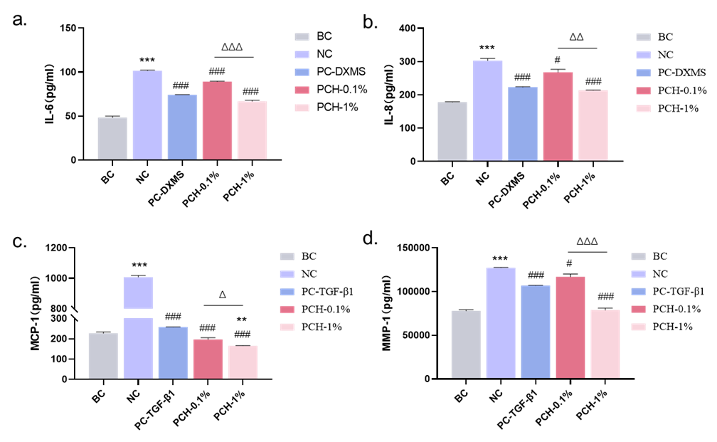
Figure 3a-d: The effect of PCH extract on content of IL-6 IL-8 MCP-1 and MMP-1 in H2O2-induced fibroblasts was determined by ELISA. n=3 each.
3.Effect of PCH extract on DNA damage repair
DNA damage is also one of the signs of cellular aging. As age increases, DNA damage continues to increase, and γH2A.X is a sensitive molecular
marker of DNA damage and repair, used to monitor the occurrence and regression of DNA damage [24]. We performed immunofluorescence staining of γH2A.X on fibroblasts and found that compared with the BC group, PCH extract can significantly reduce the expression of γH2A.X.
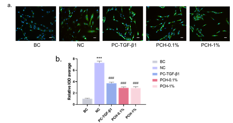
Figure 4: Effect of PCH extract on γH2A.X protein expression in H2O2-induced fibroblasts. (a): Immunofluorescence staining results of γH2A.X in each group, Scale bar: 50 μm; (b): Relative IOD average of γH2A.X in each group. n=3 each.
The skin is the largest organ of the body and plays an important protective role. Senescent cells have been found to accumulate with age and may contribute to age-related skin changes. Like other tissues, skin aging is affected not only by external factors caused by the environment, but also by endogenous changes. Studies have shown that the activity of catalase in aging skin is significantly reduced, and the content of H2O2 is significantly increased [25]. H2O2 will cause an increase in the content of reactive oxygen species ROS in the skin, and ROS is one of the main contributors to aging [26]. On the one hand, excess ROS will activate the NF-κB pathway, thereby activating SASP factors, such as tumor necrosis factor (TNF)-α, interleukin (IL)-1β, IL-6 and other molecules expression, thereby inducing an inflammatory response [27]; on the other hand, ROS can cause DNA damage [28] and induce aging. Therefore, H2O2 was used as an inducer of skin fibroblasts in this study to simulate the aging phenomenon caused by the oxidative process during skin aging in vivo [18].
SA-β-gal [10] and p21[11] are the classical markers of cell senescence. In this study, we found that PCH extract was effective in reducing the expression of SA-β-gal and p21 in H2O2-induced human skin fibroblasts, suggesting the potential of PCH extract in anti-aging of human skin cells. Meanwhile, PCH extract can reduce the expression of inflammation-related factors such as IL-6, IL-8, MCP-1 and MMP-1[19-22] in fibroblasts. It also suggests a potential role of PCH extract in anti-inflammatory aging, which needs to be followed up with further
demonstration. In addition, DNA damage is one of the important factors in skin aging, and in this study, we also found that PCH extract had a significant reduction of the DNA damage marker γH2A.X protein [24], and had a better effect as the concentration increased. Perhaps in the future, the active ingredients can be further enriched through the improvement of the extraction process, in order to enhance the efficacy of PCH extracts in skin anti-aging.
Although some interesting findings were obtained in this study, there are still some limitations. For example, the mechanism of action of PCH extract was not thoroughly investigated in this study, and further studies are needed to elucidate the specific biological effects and molecular mechanisms. Meanwhile, the anti-aging gene Sirtuin 1[29] is critical to the aging process of the skin and the prevention of various chronic diseases. Whether PCH is a Sirtuin 1 activator and prevents the actions of Sirtuin 1 inhibitors with relevance to skin aging is a question worthy of further exploration.
In summary, we found the anti-aging effect of PCH extract on human skin fibroblasts, which is expected to be used as a natural, safe and effective anti-aging ingredient in skin care products in the future.
This study has found that the extract of Polygonatum cyrtonema Hua has anti-aging effects on human skin fibroblasts and can be added to skincare products as a natural anti-aging active ingredient.
Dear Editorial Team, Clinical Medical Reviews and Reports. My experience with the journal was highly positive. The peer-review process was rigorous, constructive, and completed in a timely manner. The reviewers provided valuable comments that helped improve the quality and clarity of our manuscript. The editorial office was professional, responsive, and supportive throughout all stages of the publication process. Communication was clear and efficient, and any questions were addressed promptly. Overall, I found the journal to maintain high scientific standards and an excellent publication workflow. I would be pleased to consider submitting future work to this journal. Best wishes from, Elena Popa.

It was my pleasure to submit my testimonial concerning the Reviewer Board of our Scientific Journal “Brain and Neurological Disorders”. The Reviewers focused on some modifications and their contribution was helpful. The ladies of our Editorial Office were also supported my efforts. It was my honor to have such a co-operation and I am looking forward for more collaboration.

Dear Grace Pierce, Editorial Coordinator of Journal of Clinical Research and Reports, Thank you for the speedy and efficient peer review process. I appreciate the fact that your peer reviewers do not take months to respond like with some other journals. I would also like to thank the editorial office for responding quickly to my questions. It is an excellent journal. I plan to submit more manuscripts in the future. Best wishes from, Robert W. McGee

Dear Grace Pierce, Editorial Coordinator of Journal of Clinical Research and Reports, Working with you and your team on our recent publication in JCRR has been a truly wonderful and enjoyable experience. The responses were prompt, and the reviewers were patient, constructive, and highly professional. One reviewer in particular gave me the feeling that a professor was carefully reading and commenting on my coursework, which was deeply touching. The entire process was straightforward and hassle‑free, with no tedious online forms to complete. I highly recommend this journal. Best wishes from, DR Aibing Rao, Head of R&D

I Appreciate the Opportunity to Share my Experience with the Journal of Clinical Research and Reports. The peer review process was timely and constructive, and the feedback provided helped improve the quality of our manuscript. The editorial office was professional, responsive, and supportive throughout the process, ensuring smooth communication and efficient handling of the submission. Overall, it was a positive experience collaborating with your team.

Dear Mercy Grace, Editorial Coordinator of Obstetrics Gynecology and Reproductive Sciences, We would like to express our gratitude for your help at all stages of publishing and editing the article. The editors of the magazine answer all the necessary questions and help at every stage. We will definitely continue to cooperate and publish other works in the Obstetrics Gynecology and Reproductive Sciences! Best wishes from, Alla Konstantinovna Politova,
