

Case Report | DOI: https://doi.org/10.31579/2694-0248/010
1University hospital of lung diseases "Shefqet Ndroqi", public Pharmacy Service, Odontology Service Tirana, Albania.
*Corresponding Author: Fadil Gradica, University hospital of lung diseases
Citation: F Gradica. (2020) It Just Takes One Phone Call: Simultaneous Activation to Reduce Referral Times. Clinical Orthopaedics and Trauma Care. 2(2); DOI:10.31579/2694-0248/010
Copyright: ©2020 Fadil Gradica, This is an open-access article distributed under the terms of the Creative Commons Attribution License, which permits unrestricted use, distribution, and reproduction in any medium, provided the original author and source are credited.
Received: 11 August 2020 | Accepted: 16 October 2020 | Published: 23 October 2020
Keywords: COVID-19; pneumonia; coronavirus; pneumotorax; computed tomography; x-ray
Coronavirus caused an epidemic in China in December 2019 at an animal market where live and dead animals were sold in Wuhan, China. In a short time, this epidemic spread to different continents.
This virus has been called the 2019 new coronavirus (2019-nCoV) by the World Health Organization. Unlike both MERS-CoV and SARS-CoV, Covid-19 is the seventh member of the coronavirus family that infects humans. It is characteristic for Covid-19 pneumonia that there are subpleural localized ground glass opacities and numerous irregular areas of consolidation in both lungs and especially in the lower lobes.In this case study, we aimed to present a Covid-19 positive in 58 years old man patient with cough, high fever, beginning breathlessness and chest pain on the left, accompanied by pneumothorax in the left hemithorax, after covid -19 diagnosis.
Radiographic imaging of the patient revealed pneumothorax on the left and ground-glass opacities in the bilateral lower lobes. The patient underwent tube thoracostomy from the left hemithorax lateral.
It should be kept in mind that in patients with Covid-19 pneumonia, it may develop in pneumotoacies secondary to lung parenchymal damage. Mortality rates can be reduced in patients with early diagnosis and treatment.
A new case of Coronavirus infection, occurred in Wuhan, in China, in December 2019. On January 2020, this corona virus identified, was called 2019-nCoV [1].
Coronaviruses are enveloped RNA viruses that cause respiratory, enteric, hepatic, and neurological diseases that are common among birds, humans, and other mammals [2].
Up to now, are known six coronavirus species to cause human disease. The four common types cause common cold symptoms. The other two types of severe acute respiratory syndrome coronavirus (SARS-CoV) and Middle East respiratory syndrome coronavirus (MERS-CoV) are of zoonotic origin and can cause fatal diseases [3].
Although the mortality of Covid-19 is lower than that of SARS-CoV and MERS-CoV, the number of confirmed Covid-19 cases has increased significantly [4].
As of August 9, 2020, a total of Coronavirus Cases 19,918,709 and 731,718 deaths were reported worldwide [5, 6].
Radiological imaging plays an important role in the diagnosis and treatment of pneumonia, which is the clinical presentation of Covid-19 [7]. Chest radiographs show low-density pneumonia foci (viral pneumonia), which mostly involve bilateral mid-lower zones in this disease.
Case Report
A 58-year-old male patient was admitted in July to the emergency department with complaints of fever, cough, shortness of breath and increased pain in the left hemithorax. His general condition was not good, inconscious, noncooperative and nonorientated with diabetes with insulinotherapy since 3 years and hypertension equilibrated. Blood pressure arterial 150/90 mmHg, pulse 92/min, respiratory rate 20/min, fever 38.5°C, oxygen saturation (SpO2) with finger probe was 50%.
In blood tests WBC: 13.06 103/uL, NEU lymphocyte: 1.59 103/uL, HGB: 14.4 g/dL, HCT: 42.4%, PLT 188 103/uL, fasting blood sugar: 137.7 mg/dL, urea: 34.3 mg/dL, Creatinine: 0.91 mg/dL, ALT (SGPT): 13 U/L, AST (SGOT): 11 U/L, LDH: 150 U/L.
On radiological examination of the patient, consolidation and ground glass images were observed in the bilateral lower lobes and the accompanying left pneumothorax (Figure 1, 2, 3, 4).
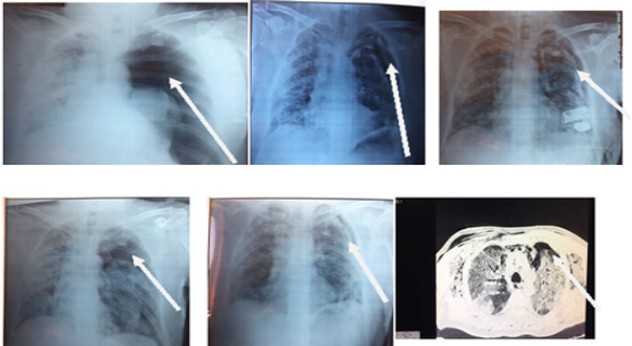
The patient was admitted in the intensive care unit and the ts situation was improving day by day.but 4 days after the hospitalisation a left pneumothorax occured
Covid-19 positive was detected in the polymerase chain reaction analysis (PCR).
Other vital signs were observed stably. There was no reproduction in the blood culture of the patient.
The control arterial blood gas was pH 7.32, PCO2 60.4, PO2 78.6. Vital findings remained stable. Six after the hospitalisation a left pneumothorax occured.In the linea axillaris medioanterior, space 4-5, with local anesthesia, a pleural dranaige was performermed.
Treatment and follow-up of the patient continues.
Discussion
The new coronavirus was identified and isolated by three groups of Chinese scientists. A consortium coordinated by W. Tan from the China Centers for Disease Control and Prevention (China CDC) achieved eight complete viral genome sequences by sequencing from RNA isolation and bronchoalveolar lavage fluid (BALF) in nine patients [8].
The China Centers for Disease Control and Prevention (China CDC) identified 44 672 Covid-19 positive patients on February 14. 965 of them (2.2%) were under 20 and the mortality rate in this age group was 0.1%. 77.8% of the patients reported that they were between the ages of 30 and 69
[9].
Our patient was a 24-year-old young healthy man with no additional disease, previous operation and history of pneumothorax. In his complaints, he had high fever, cough, shortness of breath, and chest pain on the left.
Currently, the RT-PCR test is used as a standard in the definitive diagnosis of Covid-19 infection despite false negativity rates.
Sana S et al. They investigated imaging findings in 919 Covid-19 positive patients and detected ground-glass densities in 88% of cases. 87.5% of parenchymal attitudes were observed to be bilateral. None of them encountered pneumothorax [10].
Conclusion:
In this study, we present a 58-year-old man patient with Covid-19 pneumonia and pneumothorax. We discussed the patient and his radiological findings in the light of the literature. Pneumothorax may develop in Covid-19 pneumonia due to alveolar damage. This can cause increased mortality and morbidity. For this reason, pneumothorax should be kept in mind in the treatment and follow-up of Covid-19 infection.
Conflict of Interest
All authors declare that they have no conflict of interest.
Financial Support
No financial support was recieved for study.
Author Contributions
All authors contributed equally.
Dear Editorial Team, Clinical Medical Reviews and Reports. My experience with the journal was highly positive. The peer-review process was rigorous, constructive, and completed in a timely manner. The reviewers provided valuable comments that helped improve the quality and clarity of our manuscript. The editorial office was professional, responsive, and supportive throughout all stages of the publication process. Communication was clear and efficient, and any questions were addressed promptly. Overall, I found the journal to maintain high scientific standards and an excellent publication workflow. I would be pleased to consider submitting future work to this journal. Best wishes from, Elena Popa.

It was my pleasure to submit my testimonial concerning the Reviewer Board of our Scientific Journal “Brain and Neurological Disorders”. The Reviewers focused on some modifications and their contribution was helpful. The ladies of our Editorial Office were also supported my efforts. It was my honor to have such a co-operation and I am looking forward for more collaboration.

Dear Grace Pierce, Editorial Coordinator of Journal of Clinical Research and Reports, Thank you for the speedy and efficient peer review process. I appreciate the fact that your peer reviewers do not take months to respond like with some other journals. I would also like to thank the editorial office for responding quickly to my questions. It is an excellent journal. I plan to submit more manuscripts in the future. Best wishes from, Robert W. McGee

Dear Grace Pierce, Editorial Coordinator of Journal of Clinical Research and Reports, Working with you and your team on our recent publication in JCRR has been a truly wonderful and enjoyable experience. The responses were prompt, and the reviewers were patient, constructive, and highly professional. One reviewer in particular gave me the feeling that a professor was carefully reading and commenting on my coursework, which was deeply touching. The entire process was straightforward and hassle‑free, with no tedious online forms to complete. I highly recommend this journal. Best wishes from, DR Aibing Rao, Head of R&D

I Appreciate the Opportunity to Share my Experience with the Journal of Clinical Research and Reports. The peer review process was timely and constructive, and the feedback provided helped improve the quality of our manuscript. The editorial office was professional, responsive, and supportive throughout the process, ensuring smooth communication and efficient handling of the submission. Overall, it was a positive experience collaborating with your team.

Dear Mercy Grace, Editorial Coordinator of Obstetrics Gynecology and Reproductive Sciences, We would like to express our gratitude for your help at all stages of publishing and editing the article. The editors of the magazine answer all the necessary questions and help at every stage. We will definitely continue to cooperate and publish other works in the Obstetrics Gynecology and Reproductive Sciences! Best wishes from, Alla Konstantinovna Politova,
