
Case Report | DOI: https://doi.org/10.31579/2690-8808/134
1 First Degree General Surgery Specialist. Auxiliary Professor
2 Second Degree General Surgery Specialist. Ph.D.
3 First Degree Orthopedics and Traumatology Specialist.
4 First Degree General Surgery Specialist Assistant Professor
*Corresponding Author: Juana Teresa Santiago Pérez, Second Degree General Surgery Specialist. Ph.D. e.mail: teresasp@infomed.sld.cu.
Citation: José Miguel González Bárcenas, Juana Teresa Santiago Pérez, Ernesto Miguel Rodríguez Santiago, José Rodríguez Santiago and Asunción Rodríguez Morris. (2022). Splenic peliosis. Presentation of a case. Journal of Clinical Case Reports and Studies 3(7); DOI: 10.31579/2690-8808/134
Copyright: © 2022 Jose Luis Turabian, This is an open-access article distributed under the terms of the Creative Commons Attribution License, which permits unrestricted use, distribution, and reproduction in any medium, provided the original author and source are credited.
Received: 21 May 2022 | Accepted: 03 June 2022 | Published: 28 June 2022
Keywords: peliosis; splenomegaly; rheumatoid arthritis
Peliosis is a very rare benign condition, characterized by sinusoidal lakes or cysts, filled with blood, with or without hyperplasia of the remaining tissue, presenting itself mainly in solid organs of the economy.
It has also been described in other organs such as lungs, pleura, kidneys, adrenal glands and stomach.
Its precise etiology is ignored until today, but associations have been made with other entities such as tumors, viral infections, systemic diseases where they are mentioned, lupus erythematosus, and rheumatoid arthritis, In addition, prolonged therapeutics are noted with Anabolics, Steroids, Erythropoietin, Acetyl-Salicylic Acid, Paracetamol and Vitamin A. among others.
The present case is a 51-year-old female, born of preterm birth and carrier of bronchial asthma, so it deliberately ingests steroids and Vitamin A.
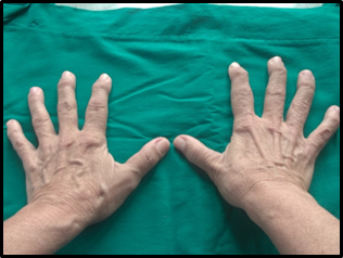
Thus, she goes to the doctor for pain in left hypochondrium, which to the physical examination attracts attention the presence of splenomegaly of more than 4cm. It is also evident the deformity that presents in the third phalanx of both hands that makes you think of physical signs of arthritis rheumatoid.
Peliosis is a very rare benign entity, characterized by cavities filled with blood within solid organs, mainly in the reticuloendothelial system (mainly liver and spleen) [1-2]. Peliosis can also affect other organs such as lungs, pleura, kidneys, adrenal glands, and stomach. Lesions can be single or multiple and of variable size. The etiology is unknown, but it has been related to the ingestion or contact with drugs and various toxins such as steroids and oral contraceptives, being associated with hematological tumor diseases and infectious diseases, particularly tuberculosis; in some cases, it is idiopathic [1-3].
Wagner first described Peliosis in 1861 [4]. In 1916 Schoenlak et al. [5] introduced the term peliosis which means "purple" or "brown", due to the color observed in the tissue. In 1950, Zak [6] described this entity for the first time in the English literature. It took 103 years for Caroli et al [7]. Made the diagnosis in a living patient. In 1971, Drut and Pawlow [8] described the first case of hepatosplenic peliosis in Argentina.
A 51-year-old MML patient with a history of having been born prematurely, in addition to bronchial asthma from the first years of life and from the age of 35, debuted with rheumatoid arthritis; Due to these antecedents, in the course of life she is a high consumer of Vitamin A and D, without a medical prescription, also associated with the deliberate use of steroids due to bronchial asthma, which she still uses sporadically.
In June 2020, he went to the doctor for pain in the left hypochondrium, which he describes as dull, continuous pain without radiation. In addition, a feeling of early fullness. She refers to weakness and a slight loss of appetite. Denies other symptoms.
Physical Exam
Skin paleness - discreet mucosa and deformity of the third phalanx of all the fingers of both hands. (Figure 1)
Abdomen: voluminous, discreetly distended with splenomegaly of 4 cm, below the left costal margin, with regular borders, being painful on palpation.
Blood pressure: 130/80 mmHg. Heart rate: 84 bpm.Rest of the exam without alterations
Complementary Exams
Hemoglobin in 12.1 gr. / l Hematocrit in 39% Erythrosedimentation in 44 mm.,
Coagulation 7 min. Bleeding 1 min
Peripheral lamina: Adequate platelets, mild leukocytosis, neutrophilia.
Red blood cells: normochromic normocytic and reticulocyte count in 1.2%.
Rest of the rigorous exams, without alterations.
Abdominal Ultrasound: Occluded image in the region of the splenic pedicle, measuring around 10x10cms, in addition to verifying the splenomegaly found in the physical examination.
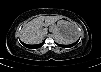
Contrast multisite helical tomography of the abdomen with MPR and MPI reconstruction, showing a liver of normal size and configuration. Spleen showing a hypo dense, septate image, with irregular contours, with low densities in its interior, said lesion slightly compresses and displaces the left kidney, downwards and backwards. The lesion did not capture contrast. (Figure 2)
After the studies carried out, it is discussed in the group of the Surgery Service and her surgical intervention is decided, performing total splenectomy, by conventional route, the patient evolves favorably and is discharged, one week later.
Anatomopathological study
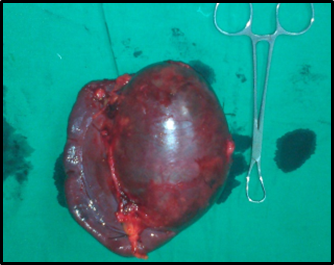
The macroscopic study showed: Surgical specimen from total splenectomy, weighing 500gms. In addition, it measures 15x12x5 cm. presenting a smooth, shiny capsular surface, of a purplish brown color, that bulges and makes relief, in the pedicle region: of relentless consistency, corresponding to the cut, with a cystic, multinodular area, entirely occupied by abundant blood, and measuring 10x7cms. Rest of the splenic parenchyma of friable consistency, reddish in color, with a whitish stippling. Figure 3.
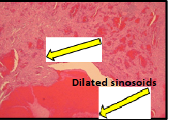
The microscopic study concluded: Splenic tissue with normal histological limits, which alternates with cystic and sinusoidal areas, with thick walls, which are occupied by blood. Figure 4,Histopathological diagnosis: Peliosis Splenica
The term peliosis refers to the histopathological appearance found in the affected organ and is given by the formation of cystic cavities filled with blood in the parenchyma of said organ, which may or may not coincide with the hyperplasia of the remaining tissue [9]. Hepatic localization is the most frequent, most of the time in immune deficient patients, chronically treated with steroids or in seropositive patients with bacterial infections. Cases of peliosis in the liver, spleen, lymph nodes, spinal cord, stomach, ileum and lung [9-11].
Splenic peliosis is an exceptional entity, with less than 100 cases published in the literature, most of the diagnoses being in post-mortem studies, because although it is a benign condition, its management is delayed due to its infrequent presentation. It is associated with tumors, infectious diseases, autoimmune disorders, drug use, transplants, radiation therapy, and splenic vein thrombosis [11].
Hemoperitoneum due to spontaneous splenic rupture or secondary to minimal trauma is a frequent complication [12]. The etiopathogenesis could be related to congenital, toxic factors, or immune complex deposits. Elevation of VEGF (endothelial growth factor) concentrations in patients with splenic peliosis has been argued [10,11].
Diagnosis is difficult, although this entity must be taken into account in cases of hypersplenism in which imaging tests show nodulations or cysts in the splenic parenchyma. In cases of suspected hemoperitoneum, abdominal tomography is the imaging technique of choice [12-14].
Peliosis is very little documented in the reviewed literature. Its identification requires a high index of suspicion, in patients with prolonged treatment with drugs or known toxins capable of producing the disease and who present persistent abdominal pain or suggestive images. The diagnosis is incidental and on other occasions of exclusion. Treatment depends on the underlying cause.
Splenic peliosis is an exceptional medical entity; therefore, the expertise for its diagnosis and subsequent treatment lies precisely in improving knowledge about the disease and medical performance in the consequent action.
The authors declare no conflict of interest
José Miguel González Bárcenas: Data collection, analysis, and statistical processing. Juana Teresa Santiago: She drafted the manuscript and participated in data collection. Ernesto Miguel Rodriguez Santiago: Participated in data collection. Asunción Rodríguez Morris: Participated in literature search. José Rodríguez Santiago: Participated in literature search.
Dear Editorial Team, Clinical Medical Reviews and Reports. My experience with the journal was highly positive. The peer-review process was rigorous, constructive, and completed in a timely manner. The reviewers provided valuable comments that helped improve the quality and clarity of our manuscript. The editorial office was professional, responsive, and supportive throughout all stages of the publication process. Communication was clear and efficient, and any questions were addressed promptly. Overall, I found the journal to maintain high scientific standards and an excellent publication workflow. I would be pleased to consider submitting future work to this journal. Best wishes from, Elena Popa.

It was my pleasure to submit my testimonial concerning the Reviewer Board of our Scientific Journal “Brain and Neurological Disorders”. The Reviewers focused on some modifications and their contribution was helpful. The ladies of our Editorial Office were also supported my efforts. It was my honor to have such a co-operation and I am looking forward for more collaboration.

Dear Grace Pierce, Editorial Coordinator of Journal of Clinical Research and Reports, Thank you for the speedy and efficient peer review process. I appreciate the fact that your peer reviewers do not take months to respond like with some other journals. I would also like to thank the editorial office for responding quickly to my questions. It is an excellent journal. I plan to submit more manuscripts in the future. Best wishes from, Robert W. McGee

Dear Grace Pierce, Editorial Coordinator of Journal of Clinical Research and Reports, Working with you and your team on our recent publication in JCRR has been a truly wonderful and enjoyable experience. The responses were prompt, and the reviewers were patient, constructive, and highly professional. One reviewer in particular gave me the feeling that a professor was carefully reading and commenting on my coursework, which was deeply touching. The entire process was straightforward and hassle‑free, with no tedious online forms to complete. I highly recommend this journal. Best wishes from, DR Aibing Rao, Head of R&D

I Appreciate the Opportunity to Share my Experience with the Journal of Clinical Research and Reports. The peer review process was timely and constructive, and the feedback provided helped improve the quality of our manuscript. The editorial office was professional, responsive, and supportive throughout the process, ensuring smooth communication and efficient handling of the submission. Overall, it was a positive experience collaborating with your team.

Dear Mercy Grace, Editorial Coordinator of Obstetrics Gynecology and Reproductive Sciences, We would like to express our gratitude for your help at all stages of publishing and editing the article. The editors of the magazine answer all the necessary questions and help at every stage. We will definitely continue to cooperate and publish other works in the Obstetrics Gynecology and Reproductive Sciences! Best wishes from, Alla Konstantinovna Politova,
