
Research Article | DOI: https://doi.org/10.31579/2641-5194/011
1. Department of Pediatrics, University of Oklahoma Health Sciences Center, Oklahoma City, Oklahoma.
2 Nishtar Medical College, Nishtar Road, Multan, Pakistan.
3 Department of Pathology, University of Oklahoma Health Sciences Center, Oklahoma City, Oklahoma.
4 Department of Radiology, University of Oklahoma Health Sciences Center, Oklahoma City, Oklahoma.
*Corresponding Author: Zhongxin Yu, Department of Pathology, University of Oklahoma Health Sciences Center, Oklahoma City, Oklahoma. E-mail: Zhongxin-Yu@ouhsc.edu
Citation: Muhammad Shaukat , Muhammad Mahmood , Sepideh Asadbeigi , Rachel Conrad , Issam M Halabi , Ayesha Fatima , Charles Lawrence, Zhongxin Yu. Solid Pseudopapillary Neoplasm of the Pancreas: Clinical-Radiological-Pathological Characteristics of Four Pediatric Cases, J. Gastroenterology Pancreatology and Hepatobilary Disorders, 4(1): DOI: 10.31579/2641-5194/011
Copyright: © 2020 Zhongxin Yu. This is an open-access article distributed under the terms of The Creative Commons Attribution License, which permits unrestricted use, distribution, and reproduction in any medium, provided the original author and source are credited.
Received: 09 March 2019 | Accepted: 13 March 2020 | Published: 17 March 2020
Keywords: solid pseudopapillary neoplasm of the pancreas; SPN; pediatrics; pancreas; Beta-catenin
Solid Pseudopapillary Neoplasm (SPN) of the pancreas represents 1-3 % of all exocrine pancreatic tumors and is uncommon in children. We report four pediatric patients with SPN where each patient posed a unique diagnostic and therapeutic challenge. We describe these four cases with detailed clinical-radiological-pathological correlations. All patients are female, with a median age 13.5 years. Two patients presented with abdominal pain, one with jaundice and one with an incidental pancreatic mass on abdominal CT scan. Radiological studies included abdominal ultrasound, CT scan and MRI of abdomen. Pancreaticoduodenectomy was performed in three patients and laparoscopic distal pancreatectomy in one patient. Mean tumor size was 4.5 cm (ranged from 1.9 to 11.5 cm). All SPNs were benign on histological exam. One patient developed pancreatic insufficiency post-surgery. No tumor recurrence was observed over a mean follow up period of 1 year. We conclude that diagnosis of SPN in pediatric population can be challenging due to non-specific clinical findings, and surgical removal of the tumor is usually required for definitive histologic diagnosis and treatment. Most tumors are benign and recurrence is very rare.
Solid pseudopapillary neoplasm of the pancreas (SPN) is an uncommon tumor of pancreas, accounting for 1-3 % of all exocrine pancreatic tumors [1-3]. It was first described by Frantz in 1959 [4, 5]. Since then it has been known by different names such as solid and papillary epithelial neoplasm, papillary cystic neoplasm, solid and cystic papillary neoplasm, but in 1996, the World Health Organization (WHO) designated it as SPN [6, 7]. As a result of broader use of cross-sectional imaging and better recognition of tumor, there has been a 7-fold increase in the number of SPN cases since 2000 [8,9]. These tumors are mostly of benign nature but 5-15% of SPNs are malignant and liver is the most common site of metastasis [10-14]. Although tumor can occur in all races, Asian and African-American female in their second or third decade of life (mean age 22 to 36.5 years) are most commonly affected with female to male ratio of 10:1 [4, 6, 10, 15]. A typical SPN is a well-encapsulated mass with solid and cystic components due to varying degrees of internal hemorrhage and necrosis [5, 7]. They are predominantly located in the tail (43%) followed by the head (25%) and the body (13%) of pancreas [16, 17]. These tumors are either found incidentally on imaging studies or present with vague abdominal symptoms or mass [1, 4]. Surgical resection is the mainstay of treatment [3]. Overall 5-year survival rate after resection is 90-98%, including patients with metastatic disease [17, 18]. Rarely, patients receive adjuvant chemotherapy for very large or malignant tumors [17]. We are reporting four pediatric cases of SPN where each patient had a distinct clinical presentation and posed a diagnostic challenge.
Method:
We retrospectively reviewed our institutional database to identify patients with SPN from January 2010 to December 2018 and correlated demographic, clinical, radiological, pathological and surgical details.
A previously healthy 14-year-old Hispanic female was admitted with one week of constant, non-radiating, stabbing right upper quadrant abdominal pain (RUQ) with a severity of 6-7/10. For the past few months, she had sour taste in mouth associated with nausea 3 to 4 times per week but no vomiting. Stools were regular and brown. Physical examination showed RUQ and epigastric tenderness with no palpable mass or hepatosplenomegaly. Initial abdominal ultrasound (US) was concerning for "a mass in her stomach" and elevated liver enzymes. She was then referred to our hospital for further management. Her laboratory testing results were summarized in table 1.
AST, Aspartate aminotransferase; ALT, Alanine aminotransferase; GGT, Gamma-glutamyl transferase; INR, International Normalized Ratio; N/A, not available
Abdominal computed tomography (CT) and magnetic resonance imaging (MRI) with contrast showed a large retroperitoneal mass. Because of the uncertainty of the origin of the mass, open laparotomy and biopsy of the mass without excision was performed followed by a trial of Gemcitabine with the intention to shrink the tumor. She underwent modified Whipple procedure (pancreaticoduodenectomy) after no significant decrease in size after 2 cycles of Gemcitabine by repeat abdominal CT. The surgery was uneventful and she was discharged home on post-operative day 7. She developed chronic pancreatic insufficiency 3 years post-surgery, for which she was started on pancreatic enzyme replacement therapy, Aquadeks, and Vitamin D, otherwise, she had no recurrence or metastasis of the tumor with annual abdominal CT for 5 years.
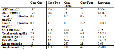
Radiological features: Initial abdomen and pelvis CT showed a large complex, non-invading, well-circumscribed right retroperitoneal mass with areas of enhancement and nodularity within its rim. There was a mass effect on the adjacent duodenum, kidney, liver and portal vein without obstruction. A few small lymph nodes were also seen in the retroperitoneum. Origin of the mass was not very clear on CT scan but was most concerning for cystic pancreatic mass. To better delineate the mass, MRI of abdomen and pelvis with contrast was obtained which also showed a cystic and peripherally solid mass (11.5 x 11.4 x 4.4 cm) with late peripheral nodular enhancement and multiple blood-blood fluid levels of differing stages (Figure 1A-1C).
The exact epicenter of this mass was difficult to locate; however, it was thought to arise from the potential Morison's pouch, which would suggest a sarcoma. However, intimate association with the adjacent pancreatic head also raised the concern for a solid pseudopapillary neoplasm, pancreatoblastoma, or exophytic fibrolamellar hepatocellular carcinoma.
Pathological findings: The initial biopsy showed sheets of cohesive tumor cells arranged around fibrovascular cores, forming a pseudopapillary architecture. The tumor cells were bland with a moderate amount of eosinophilic cytoplasm and relatively uniform nuclei, fine chromatin, and inconspicuous nucleoli (Figures 1D-1E). There was no mitosis or necrosis. The tumor cells were strongly positive for beta-catenin (diffuse) (Figure 1F), CD10 (diffuse) (Figure 1G), anti-alpha 1 trypsin (focal), and weakly positive for synaptophysin. The overall features were consistent with a diagnosis of SPN. The resected tumor was located in the pancreatic head, measuring 10.5 x 10.5 x 10.5 cm. It was well-circumscribed, hemorrhagic, with multiple irregular cystic spaces and necrotic debris on cut surfaces. The histopathological features of the resected tumor was similar to that seen in the initial biopsy, except there were signs suggestive of treatment effect, including fibrosis, various degrees of vessel hyalinization, and hemosiderin. Although the tumor grossly appeared to be well-circumscribed, microscopically, it showed an infiltrative border with the surrounding pancreatic tissue.

A: Axial T2 MRI image shows a mixed signal mass with small peripheral cystic structures involving the head of the pancreas. There is coexistent compression of the gallbladder and the right kidney. B: A coronal post-contrast CT image of the abdomen shows a hypodense mass involving the pancreatic head. There is additional compression of the portal vein, gallbladder, and bowel loops. C: Coronal post-contrast T1 MRI image of the abdomen shows a hypoenhancing mass in the pancreatic head. There is concomitant compression of the adjacent bowel loops. D: Microscopic picture of tumor (H&E stain): The tumor shows pseudopapillae with fibrovascular cores, lined by several layers of bland epithelial cells with eosinophilic cytoplasm. E: Microscopic picture of tumor (H&E stain): The cells are bland with a moderate amount of eosinophilic cytoplasm and relatively uniform nuclei, fine chromatin, and inconspicuous nucleoli. F: Microscopic picture of tumor (beta-catenin immunohistochemical stain): Diffusely positive nuclear and cytoplasmic staining. G: Microscopic picture of tumor (CD10 immunohistochemical stain): Diffusely positive nuclear and cytoplasmic staining.
An 11-year-old, previously healthy African-American female was observed to have jaundice by her dentist during a routine dental examination. She subsequently come to our hospital for further care. She recalled that she did have mild upper abdominal discomfort, dark brown urine, and light tan stools for a few days; otherwise, she had no itching, nausea, vomiting, dysphagia, heartburning, constipation, diarrhea, and weight loss. She denied taking any medications. Physical examination showed jaundice but no hepatosplenomegaly. Her laboratory testing results are summarized in table 1. Abdominal US, CT, and MRI showed a large mass at the pancreatic head. Clinical differential diagnoses included rhabdomyosarcoma, SPN, and others. The patient underwent surgery and had a pancreaticoduodenectomy after intraoperative frozen section ruled out rhabdomyosarcoma that would need neoadjuvant therapy instead of first stage resection. She had no perioperative complications and was discharged home on post-operative day 6. On a follow up visit 2 weeks later, she recovered well with resolution of her jaundice and abdominal discomfort. She subsequently moved to another state and was lost to follow up.
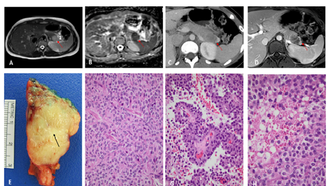
Radiological features: CT abdomen revealed a well-defined (5.3 cm x 6.2 cm x 5.6 cm) mass with mixed solid and cystic components in the region of the head of the pancreas with a thin peripheral rind of tissue and no calcifications. The mass lesion was causing obstruction of the distal common bile duct (CBD) with dilation of the intra and extra-hepatic ducts as well as the gallbladder with layering sludge. MRI of abdomen also revealed a large heterogeneous rounded mass in the region of the head of the pancreas/proximal duodenum with T1 signal intensity suggesting a small amount of blood products. Delayed imaging showed mild progressive enhancement suggesting a fibrous or solid/soft tissue component. Again, compression and dilation of CBD and main pancreatic duct was seen. Mild compression of portal vein and inferior vena cava (IVC) without obstruction was also visualized. There were few scattered mesenteric and periaortic lymph nodes (Figure 2A-2B). Differential diagnosis included intraductal papillary mucinous neoplasm (IPMN), serous or mucinous cystic neoplasm, and solid pseudopapillary neoplasm.
Pathological findings: The intraoperative frozen section showed an epithelioid tumor with papillary structure, suggestive for SPN. The resected tumor was well-circumscribed and partially encapsulated. It was located in the pancreatic head, measuring 6.5 x 6.5 x 4.5 cm. Sectioning revealed a red-tan, solid and cystic tumor with hemorrhage and edematous changes (Figures 2C). Microscopically, the tumor showed groups of discohesive tumor cells arranged in a pseudopapillary fashion with prominent hemorrhage and cystic changes (2D). The tumor cells had a moderate amount of eosinophilic cytoplasm and relatively uniform nuclei, fine chromatin, and inconspicuous nucleoli; some of the tumor cells show clear cell changes (Figure 2E). There was no mitosis or necrosis. Multifocal perineural and lymphovascular invasion was observed, but no definitive invasion into the muscular vessels was identified (Figure 2G-2H). Immunohistochemical stains showed strongly positivity for beta-catenin (diffuse), CD10 (focal), and alpha-1 antitrypsin (rare cells). The overall findings were diagnostic for SPN.

A: Axial T2 image of the upper abdomen shows a mixed signal intensity mass arising from the head of the pancreas. There is additional dilation of the pancreatic duct. B: Axial T1 postcontrast MRI image shows an irregularly enhancing mass at the head of the pancreas. There is compression of the IVC and the portal vein at the level of the porta hepatis. C: Gross picture of the pancreatic head containing the tumor that measures 6.5 x 6.5 x 4.5 cm. The residual pancreas can be seen (Stars indicating benign residual pancreatic tissue). D: Microscopic picture of tumor (H&E stain): The tumor shows groups of discohesive tumor cells arranged in a pseudopapillary fashion with prominent hemorrhage and cystic changes. E: Microscopic picture of tumor (H&E stain): Some of the tumor cells show clear cell changes.
A 13-year-old Caucasian female developed right lower quadrant abdominal pain. Abdominal CT showed an inflamed appendix, which was removed laparoscopically at an outside hospital. CT also showed a distal pancreatic mass, for which she was referred to our hospital for further evaluation after appendectomy. She had no abdominal pain or other symptoms during her initial evaluation at our hospital, and her physical exam was unremarkable. Laboratory results are summarized in table 1. She underwent laparoscopic distal pancreatectomy with an uneventful post-operative course and was discharged home on post-operative day 3. Follow up MRI of abdomen 8 months later did not reveal any recurrence of the tumor, and her lipase and amylase normalized.
Radiological features: CT and MRI of abdomen revealed low density, hypo-enhancing mass in the area of pancreatic tail with no significant mass effect on surrounding structures. Axial ADC map of the mass showed a low signal intensity reflecting tumor hypercellularity and a high suspicion of SPN (Figure 3A-D).
Pathological findings: The distal pancreatectomy showed a well-circumscribed, non-encapsulated, solid mass measuring 2.2 x 2.0 x 1.9 cm. It had a lobulated, yellow tan cut surface with an apparent central scar. The tumor was located in the tail of pancreas (Figure 3E). Microscopically, the tumor was composed predominantly of solid areas with focal pseudopapillary structures (Figures 3F-3G). There were pale areas composed of cells with clear cytoplasm and mucinous secretions. Intracytoplasmic hyaline globules were also appreciated in multiple areas (Figure 3H). The distal residual pancreatic parenchyma showed atrophic changes with diminished exocrine acini, but preserved islets of Langerhans. The tumor invaded into the pancreatic parenchyma and showed perineural invasion. The tumor cells were positive for beta-catenin (diffuse), CD10 (diffuse), alpha-1 antitrypsin (multifocal focal), and vimentin (diffuse) and negative for chromogranin, synaptophysin, and pan-cytokeratin. The overall findings were diagnostic for SPN.
A: Axial T2 MRI image shows a mildly hyperintense mass in the pancreatic tail. B: Axial ADC map at the level of the pancreas shows a low signal intensity pancreatic tail mass reflecting tumor hypercellularity. C: Axial post-contrast CT image of the upper abdomen shows a low-density pancreatic tail mass. D: Axial postcontrast T1 MRI image of the upper abdomen shows a hypoenhancing pancreatic tail mass. E: Gross picture of the pancreatic tail containing the tumor that measures 2.2 x 2.0 x 1.9 cm. The tumor has lobulated, yellow-tan cut surfaces with an apparent central scar (arrow indicating the central scar). F: Microscopic picture of tumor (H&E stain): Solid nests of poorly cohesive cells are seen. G: Microscopic picture of tumor (H&E stain): Sheets of cohesive tumor cells arranged around a fibrovascular core forming pseudopapillary architecture. H: Microscopic picture of tumor (H&E stain): Intracytoplasmic hyaline globules are seen in multiple areas (arrows are indicating the hyaline globules).
A 14 year-old- Caucasian female presented with sudden onset of epigastric pain and non-bilious, non-bloody vomiting after she was “elbowed in the abdomen” during a soccer game. She had a significant clinical history of a similar blunt abdominal trauma during a basketball game with gradually worsening epigastric pain and vomiting two years earlier, for which she had a CT scan suspected for pseudo cyst of the pancreatic head at an outside hospital and received drainage by interventional radiology; the aspirate yielded a small volume of bloody aspirate (no tissue was obtained for histopathological examination at the time) with subsequent reduction in the size of the mass and improvement in her abdominal pain. She was discharged with a diagnosis of questionable intra-abdominal hematoma, most likely duodenal hematoma. She was followed by serial abdominal ultrasounds at outside hospital, which reportedly showed gradual reduction in the size of the mass, seemed to be expected in a patient with a resolving hematoma. She had no abdominal symptoms over the following two years, until current presentation. At current presentation, physical examination revealed a sick-looking patient in moderate pain. Her abdomen was severely tender to palpation in the epigastric area with hypoactive bowel sounds. Abdominal US, CT scan, and MRI revealed heterogeneous mass with scattered calcification between the pancreatic head and proximal duodenum, raising suspicions for a pancreatic tumor at the same location with similar radiological features as seen two years ago. Her laboratory testing results at this admission are summarized in table 1. She had fine needle aspiration (FNA) revealed 20 ml of serosanguineous fluid and a core tissue biopsy. Subsequently, she underwent a pancreaticoduodenectomy. Post-operatively, she suffered intra-abdominal fluid collections that required drainage and antibiotics. Repeat MRI 6 months later did not show recurrence of the tumor.
Radiological features: Abdominal US revealed a heterogeneous mass in RUQ measuring 9 x 5 x 7 cm, that increased in size from the ultrasound 2 years ago. MRI of the abdomen also showed a prominent heterogeneous mass with areas of hemorrhage and cystic change with some calcifications, interposed in the region between the pancreatic head and proximal duodenum with a mass effect upon the pancreatic head, proximal duodenum, upper abdominal IVC and the extrahepatic portal vein (Figure 4A-4B). The epicenter of the mass was difficult to discern. There was mild dilatation of the CBD. Differential considerations included SPN or a complex hematoma arising from duodenum or the groove between the pancreatic head and the duodenum.
Pathological findings: FNA showed few large clusters of epithelioid cells with rare associated fibro-vascular structures (Figure 4C-4D). The core biopsy also showed fibro-connective tissue with a neoplastic proliferation consisting of fibro-vascular cores, with a small amount of associated myxoid stromal changes, surrounded by the oval epithelioid cells. Intracytoplasmic hyaline globules were also appreciated and were highlighted by Periodic Acid-Schiff stain after diastase treatment (PASD) (Figures 4E-4F). The cells were positive for beta-catenin (focal) and CD10 (diffuse). These features were diagnostic for SPN. The resected specimen showed a well-circumscribed mass located in the head of the pancreas, measuring 6.5 x 4.5 x 4.3 cm. It was solid and cystic with hemorrhage and calcification on cut surfaces (Figure 4G). Microscopic examination showed similar histological features and consistent with the diagnosis of SPN. There were prominent hemorrhagic cystic changes as well as calcification and hemosiderin deposition. Perineural invasion was present, but lymph-vascular invasion was absent. The uninvolved residual distal pancreatic parenchyma showed atrophic changes with fibrosis and diminished exocrine glands.
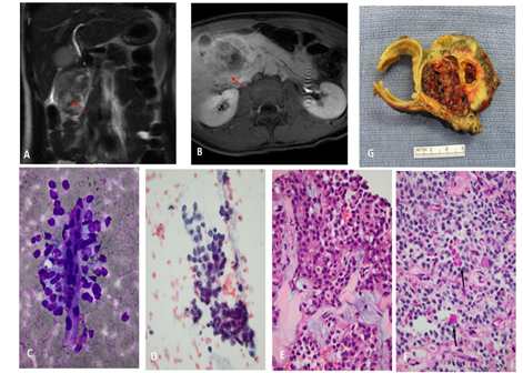
A: Coronal T2 MRI image shows a mixed signal intensity mass in the pancreatic head creating mild dilation of the common duct. B: Axial post-contrast T1 image of the upper abdomen shows a modestly enhancing mass in the pancreatic head with compression of the duodenum and the gallbladder. C: Microscopic picture of tumor (Diff-Quick stain): Clusters of epithelioid cells with rare associated fibro-vascular structures. D: Microscopic picture of tumor (PAP stain): Tumor cells with eosinophilic cytoplasm with large and uniform nuclei, inconspicuous nucleoli and rare grooves. D: Microscopic picture of tumor (H&E stain): Tumor with fibro-vascular cores. Intracytoplasmic hyaline globules are seen. F: Microscopic picture of tumor (PASD stain): Intracytoplasmic hyaline globules are highlighted by Periodic Acid-Schiff stain after diastase treatment (PASD) and are indicated with arrows. G: Gross picture of the pancreatic head containing the tumor that measures 6.5 x 4.5 x 4.3 cm.
Our report focuses on the variable clinical presentation of SPN, which can be very challenging to diagnose at times, and reviews the radiological and pathological differentials of SPN. SPN comprises 1-3% of all pancreatic tumors and affects young female with a mean age of 22-36.5 years (2-74 years). Asian and African-American races show higher incidence of the disease, with a female to male ratio of 10:1. Consistent with the epidemiologic data, our reported cases are all female teenagers in their second decade.
The pathogenesis and origin of SPN is still unclear. A somatic point mutation has been noticed in CTNNB1, which is the gene encoding for beta-catenin in Wnt singling pathway. This mutation leads to condensation of beta-catenin and cyclin-D1 in the nucleus [19]. The loss of E-cadherin expression on cellular membranes of neoplastic cells has also been attributed to this gene mutation, leading to discohesiveness of tumor cells and the resulting pseudopapillary architecture [20].
SPNs can present in a variety of ways. Most SPNs are found incidentally on routine imaging studies or physical examination as an abdominal mass in an otherwise asymptomatic individual. Sometimes, patients present with severe abdominal pain after suffering blunt abdominal trauma, and tumor is found on imaging [14]. Other symptoms can happen due to compression of the surrounding structures by the tumor. Jaundice is rare but sometimes SPN of head of pancreas can cause jaundice and elevated liver enzymes [10]. Of note, every patient in our report presented with different chief complaint, which highlights the variable clinical expression of this tumor.
SPN can be detected by abdominal US, CT, MRI or PET scan. On CT, SPN appear as a well-defined, heterogeneous, hypoattenuating mass with variable solid and cystic components4. MRI, however, can better differentiate between solid and cystic components [7]. Sometimes, areas of calcification may be present at the periphery. Cystic components are mostly centrally located. Endoscopic ultrasound guided fine needle aspiration (EUS-FNA) is another modality to obtain preoperative diagnosis for solid and cystic pancreatic lesions [21]. Aspirated fluid shows low amylase and low CEA (carcinoembryonic antigen), which can be helpful in differentiating it from other cystic structures [22]. Biopsy diagnosis of SPN is confirmed by curative resection.
On gross examination, SPN appears as well circumscribed, encapsulated solid mass with varying degrees of central necrosis and hemorrhage that corresponds to cystic component on imaging [15,23]. Histologic findings demonstrate uniform polyhedral cells lining small-hyalinized fibrovascular stalks. Between the pseudopapillae, degenerative changes, foamy histiocytes, and erythrocytes may be seen, along with hyalinized and focally calcified connective tissue and cholesterol crystals. Round to oval nuclei with occasional grooves and convolutions are typical. Nucleoli are faint, and mitoses are infrequent. Intracellular and extracellular metachromatic hyaline globules may also be seen [24]. Special stains demonstrate diffuse positivity for nuclear and cytoplasmic beta-catenin and neuron specific enolase (NSE) and vimentin, with focal intense small clusters or single cell staining for alpha-1-antitrypsin and alpha-1-antichymotrypsin [25]. CD10, vimentin, and DOG1 (discovered on gastrointestinal stromal tumor 1) are often positive, suggesting a possible centroacinar cell origin [26]. CD117, CD56, and galectin-3 may be expressed, as well as progesterone receptors, alternately suggesting derivation from genital ridge/ovarian anlage related cells attached during embryogenesis to the pancreas [27]. Various other epithelial markers, neuroendocrine markers, pancreatic enzymes, islet cell hormones, CEA, and CA19.9 have been described, prompting a third theory of origin from primitive pancreatic cells with potential for endocrine and exocrine differentiation [28].
The differential diagnosis for SPN includes pancreatoblastoma, pancreatic endocrine neoplasm, pancreatic pseudocyst, serous cystadenoma, mucinous cystic neoplasm [29]. Pancreatoblastoma is a very uncommon pediatric pancreatic neoplasm, usually occurs in children younger than 5 years old. Histologically, it consists of solid sheets and nests of uniform epithelial cells mixed with occasional acinar and ductal structures and characteristic squamoid corpuscles. A 50% cure rate with excision is seen in infants, although prognosis is worse in adults [30]. Pancreatic endocrine neoplasms occur at any age but are rare in childhood. It is often associated with multiple endocrine neoplasia syndrome type 1 (MEN1). Some tumors may secrete pancreatic hormones, which are referred as functional tumors. Microscopically, the tumors are comprised of nests of polygonal cells with salt-and-pepper chromatin. Positive staining may be noted for chromogranin, synaptophysin, peptides, insulin, glucagon, somatostatin, pancreatic polypeptide, gastrin, vasoactive intestinal polypeptide, or CEA, but nuclear staining for beta-catenin is negative. Pancreatic psuedocysts are the most common type of pancreatic cystic lesion and are more frequent in alcoholic males. Clinically, the patients may have high amylase and low CEA [22]. FNA biopsy reveals predominately necrotic debris, macrophages, rare epithelial cells, and absent mucin. Histologically, it is easily to be differentiated from SPN by its lacking epithelial linings. Serous cystadenoma is a benign neoplasm presenting as multiple (>6), small (<2cm>
Although most SPN are of benign nature, about 5-10% exhibit malignant potential with metastasis and happens commonly in male patients [32]. Malignant tumors tend to be larger in diameter (>10cm) and contain larger solid portion as compared to benign SPN. Some other characteristic features of malignant SPN include pancreatic tissue infiltration, focal capsular invasion, perineural, vascular or adjacent organ invasion, and metastasis [16]. The most common sites of metastasis are liver, spleen, regional lymph nodes and mesentery [11]. Some rare sites of metastasis are lung and stomach [12].
Curative resection of tumor is the mainstay of treatment, even in patients with malignant and metastatic SPN [6]. Different surgical procedures have been performed depending upon the location of the tumor. Pancreaticoduodenectomy or Whipple procedure is used to resect SPN on head of pancreas, and distal pancreatectomy is done for tumors limited to the tail of pancreas. Prophylactic splenectomy has also been performed in cases where tumor invades the splenic vasculature [3]. Post-surgical complications may include chronic pancreatic insufficiency, pancreatic fistula, intra-abdominal abscess, and gastric fistula [33]. Enucleation of the tumor is also an option, has much lower morbidity than pancreaticoduodenectomy, and offers a chance to preserve of endocrine and exocrine function [9]. Although, their role in overall prognosis is not yet well established, certain chemotherapeutic agents like cisplatin, 5-FU, and gemcitabine have been used in an attempt to shrink very large tumors or tumors with malignant features [17].
The post-operative recurrence is 10-15% and certain high risk features (tumor size > 5cm, lympho-vascular invasion, lymph node metastasis, synchronous metastasis and positive tumor margin) are associated with postoperative recurrence of SPN [3, 8, 33]. Most of the relapses occur more than 5 years after resection; therefore, follow-up should be continued > 5 years in patients with high risk features [8]. Overall 5-year survival rate after resection is >95%, even in patients with metastatic disease [34].
In conclusion, SPN is a rare tumor in pediatric patients and can pose certain diagnostic and therapeutic challenges due to its location and risk of morbidity with surgical removal. It has very low malignant potential with excellent survival rate after resection.
The authors report no conflict of interest.
Dear Editorial Team, Clinical Medical Reviews and Reports. My experience with the journal was highly positive. The peer-review process was rigorous, constructive, and completed in a timely manner. The reviewers provided valuable comments that helped improve the quality and clarity of our manuscript. The editorial office was professional, responsive, and supportive throughout all stages of the publication process. Communication was clear and efficient, and any questions were addressed promptly. Overall, I found the journal to maintain high scientific standards and an excellent publication workflow. I would be pleased to consider submitting future work to this journal. Best wishes from, Elena Popa.

It was my pleasure to submit my testimonial concerning the Reviewer Board of our Scientific Journal “Brain and Neurological Disorders”. The Reviewers focused on some modifications and their contribution was helpful. The ladies of our Editorial Office were also supported my efforts. It was my honor to have such a co-operation and I am looking forward for more collaboration.

Dear Grace Pierce, Editorial Coordinator of Journal of Clinical Research and Reports, Thank you for the speedy and efficient peer review process. I appreciate the fact that your peer reviewers do not take months to respond like with some other journals. I would also like to thank the editorial office for responding quickly to my questions. It is an excellent journal. I plan to submit more manuscripts in the future. Best wishes from, Robert W. McGee

Dear Grace Pierce, Editorial Coordinator of Journal of Clinical Research and Reports, Working with you and your team on our recent publication in JCRR has been a truly wonderful and enjoyable experience. The responses were prompt, and the reviewers were patient, constructive, and highly professional. One reviewer in particular gave me the feeling that a professor was carefully reading and commenting on my coursework, which was deeply touching. The entire process was straightforward and hassle‑free, with no tedious online forms to complete. I highly recommend this journal. Best wishes from, DR Aibing Rao, Head of R&D

I Appreciate the Opportunity to Share my Experience with the Journal of Clinical Research and Reports. The peer review process was timely and constructive, and the feedback provided helped improve the quality of our manuscript. The editorial office was professional, responsive, and supportive throughout the process, ensuring smooth communication and efficient handling of the submission. Overall, it was a positive experience collaborating with your team.

Dear Mercy Grace, Editorial Coordinator of Obstetrics Gynecology and Reproductive Sciences, We would like to express our gratitude for your help at all stages of publishing and editing the article. The editors of the magazine answer all the necessary questions and help at every stage. We will definitely continue to cooperate and publish other works in the Obstetrics Gynecology and Reproductive Sciences! Best wishes from, Alla Konstantinovna Politova,
