
Case Reports | DOI: https://doi.org/10.31579/2690-8794/024
*Corresponding Author: Selma Benkirane, Department of Dermatology, University Hospital Hassan II, Fez, Morocco
Citation: Selma Benkirane (2020) Skin metastases revealing lung cancer, J. Clinical Medical Reviews and Reports, 2(4), DOI: 10.31579/2690-8794/024
Copyright: © 2020, Selma Benkirane. This is an open access article distributed under the Creative Commons Attribution License, which permits unrestricted use, distribution, and reproduction in any medium, provided the original work is properly cited.
Received: 27 May 2020 | Accepted: 22 June 2020 | Published: 25 June 2020
Keywords: Keywords
Abstract
Skin metastases represent an infrequent secondary location of deep cancers. They represent 0.6 to 10% of metastases from solid tumors [1]. We are reporting a case.
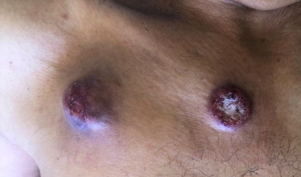
60-year-old patient, chronic smoking for 30 years not weaned, consulted in dermatology for painless lesions in the trunk gradually increasing in size. On clinical examination, these were two cutaneous nodules of the angiomatous type, measuring for the largest 3cm in diameter with a firm and painless character sitting at the high level of the trunk [Figure 1] without tendency to spontaneous regression of lesions. Dermoscopic examination revealed a milky red area, tortuous vessels, and a rainbow appearance [Figure 2].The whole evolving in a context of apyrexia and deterioration of the general state. The rest of the examination was normal, especially the lymph nodes. The patient underwent a biopsy with a histological result in favor of a secondary location. A thoracic CT scan was performed showing a right para-hilar tissue process with confirmation of a bronchial adenocarcinoma by fibroscopy. The extension assessment, notably a bone scan and a cerebral, abdominal and pelvic computed tomography complement, was without particularities. The patient was referred to oncology for possible polychemotherapy and additional management.

Cutaneous metastases represent about 2% of all skin tumors [2] and are usually associated with a poor prognosis, being a hallmark of widely spread visceral disease. Moreover, they can be the first sign of an asymptomatic, yet occult malignancy [3]. Several series have found that melanoma is the most frequent primary neoplasm leading to cutaneous metastases [4]. After melanoma, lung cancer is the most frequent origin of cutaneous metastases in men and breast cancer in women [5]. Lung cancer is often diagnosed at a metastatic stage, with a predilection for lymph node, pleural, contralateral pulmonary, cerebral, bone and adrenal localizations. Skin metastases are rare with an incidence of 2.9-5.3% for all cancers and 1-12% for lung cancer. The lesions are often made of a single nodule with several immobile hard subcutaneous with a size varying from (0.5 to 10 cm), the skin metastases of pulmonary origin are often of the adenocarcinoma type followed by the epidermal carcinoma as well as the carcinoma pulmonary with small cells, our patient presented two subcutaneous nodules of which one ulcerated.
Careful clinical examination, the site of the lesions and the sex of the patient point to skin metastases due to solid cancer. However, histological and immunohistochemical examination remains the key to the diagnosis.
The authors have declared that they have no conflict of interest.
Dear Editorial Team, Clinical Medical Reviews and Reports. My experience with the journal was highly positive. The peer-review process was rigorous, constructive, and completed in a timely manner. The reviewers provided valuable comments that helped improve the quality and clarity of our manuscript. The editorial office was professional, responsive, and supportive throughout all stages of the publication process. Communication was clear and efficient, and any questions were addressed promptly. Overall, I found the journal to maintain high scientific standards and an excellent publication workflow. I would be pleased to consider submitting future work to this journal. Best wishes from, Elena Popa.

It was my pleasure to submit my testimonial concerning the Reviewer Board of our Scientific Journal “Brain and Neurological Disorders”. The Reviewers focused on some modifications and their contribution was helpful. The ladies of our Editorial Office were also supported my efforts. It was my honor to have such a co-operation and I am looking forward for more collaboration.

Dear Grace Pierce, Editorial Coordinator of Journal of Clinical Research and Reports, Thank you for the speedy and efficient peer review process. I appreciate the fact that your peer reviewers do not take months to respond like with some other journals. I would also like to thank the editorial office for responding quickly to my questions. It is an excellent journal. I plan to submit more manuscripts in the future. Best wishes from, Robert W. McGee

Dear Grace Pierce, Editorial Coordinator of Journal of Clinical Research and Reports, Working with you and your team on our recent publication in JCRR has been a truly wonderful and enjoyable experience. The responses were prompt, and the reviewers were patient, constructive, and highly professional. One reviewer in particular gave me the feeling that a professor was carefully reading and commenting on my coursework, which was deeply touching. The entire process was straightforward and hassle‑free, with no tedious online forms to complete. I highly recommend this journal. Best wishes from, DR Aibing Rao, Head of R&D

I Appreciate the Opportunity to Share my Experience with the Journal of Clinical Research and Reports. The peer review process was timely and constructive, and the feedback provided helped improve the quality of our manuscript. The editorial office was professional, responsive, and supportive throughout the process, ensuring smooth communication and efficient handling of the submission. Overall, it was a positive experience collaborating with your team.

Dear Mercy Grace, Editorial Coordinator of Obstetrics Gynecology and Reproductive Sciences, We would like to express our gratitude for your help at all stages of publishing and editing the article. The editors of the magazine answer all the necessary questions and help at every stage. We will definitely continue to cooperate and publish other works in the Obstetrics Gynecology and Reproductive Sciences! Best wishes from, Alla Konstantinovna Politova,
