
Research Article | DOI: https://doi.org/10.31579/2641-0419/010
1 Center for Diagnostic Nanosystems, Marshall University, Huntington, WV, USA.
2 Department of Pharmaceutical Science and Research, School of Pharmacy, Marshall University, Huntington, WV, USA.
3 Department of Internal Medicine, Joan C. Edwards School of Medicine, Marshall University, Huntington, WV, USA.
4 Biotechnology Graduate Program West Virginia State University, Institute, WV.
5 Department of Public Heath, Marshall University, Huntington, WV, USA.
6 Department of Health and Human Service, School of Kinesiology, Marshall University, Huntington, WV.
7 Department of Pharmacology, Physiology and Toxicology, Joan C. Edwards School of Medicine, Marshall University, Huntington, WV, USA.
8 Department of Cardiology, Joan C. Edwards School of Medicine, Marshall University, Huntington, WV, USA.
*Corresponding Author: Kevin M. Rice, Center for Diagnostic Nanosystems, Marshall University, USA
Citation: Kevin M. Rice, Short Communication: The Effects Chronic Acetaminophen Treatment on Age-Associated Alterations of Cardiac Function in the Female F344xBN Heart, J.Clinical Cardiology and Cardiovascular Interventions. 1(2);Doi: 10.31579/2641-0419/010
Copyright: © 2018. Kevin M. Rice This is an open-access article distributed under the terms of the Creative Commons Attribution License, which permits unrestricted use, distribution, and reproduction in any medium, provided the original author and source are credited.
Received: 09 September 2018 | Accepted: 10 October 2018 | Published: 15 October 2018
Keywords: Cardiac Function; Chronic Acetaminophen;Echocardiographic procedures
Background
Although several studies have investigated the age-associated changes in male and female F344 as well as male Fischer 344/NNiaHSD x Brown Norway/BiNia F1 (F344xBN), no study to our knowledge has examined the age-associated changes in structure and function in the female F344xBN heart using echocardiographic measures. This information is crucial in determining whether or not the female F344xBN is an appropriate aging model. Additional studies have also suggested that age-associated increases in levels of oxidative stress may cause cardiac dysfunction and that chronic acetaminophen (APAP) ingestion may be protective against increased oxidative stress. On the basis of these studies we examined the function and structure of the aged female F344xBN heart in the absence and presence of chronic APAP treatment.
Methods
To investigate if the aging-related changes in cardiovascular structure and function can be attenuated with APAP treatment, aged (22-month old) female F344xBN rats were administered APAP (30 mg/kg body weight/day) for 8 months in drinking water and echocardiograms assessments were performed.
Results
Aging was associated with evidence of diastolic (impaired left ventricle relaxation time) and systolic dysfunction (fractional shortening and end systolic volume). The incidence of arrhythmias was not different with age. However, valvular dysfunction was increased. Chronic APAP treatment did not attenuate the age-associated changes in cardiac structure and function in the female F344xBN. However the occurrence of valve dysfunction with aging was significantly lowered in the APAP treated 30-month female hearts.
The American Heart Association estimates that 80 million Americans have one or more types of cardiovascular disease (CVD). It is estimated that 38.1 million of those afflicted are aged 60 or older. As a person ages he or she has a higher risk of developing myocardial infarction, stroke, atherosclerosis, peripheral occlusive disease, diabetes, hypertension [1-3].Thepotential bmechanism of increase risk of disease with age is that there is an increase of reactive oxygen species (ROS) which leads to oxidative stress [4]. Recently acetaminophen (APAP), an antipyretic and analgesic, has shown to be protective against oxidative stress in the heart and the vasculature of the brain due to its antioxidant properties [5-7].
Due to these antioxidant effects APAP is considered to be cardioprotective during ischemia/reperfusion, hypoxia/reoxygenation, exogenous peroxynitrite administration, and experimentally induced myocardial infarction due to its ability to block or reduce the development of mitochondrial permeability transition pores, mitochondrial swelling, cytochrome c release, late stage apoptosis, protein oxidation, production of hydroxyl radicals, arrhythmias, and peroxynitrite induced dysfunction in hearts [5, 8-16].
The Fischer 344/NNiaHSD x Brown Norway/BiNia F1 (F344xBN) has been recommended by the National Institute of Aging as a rat model of aging due to its increased life span and fewer age-associated pathologies compared to the standard model of aging – the Fischer 344 rat. Although several studies have investigated the age-associated changes in male and female F344 rats as well as male F344xBN rats [17-21], no study to our knowledge has examined the age-associated changes in structure and function in the female F344xBN heart using echocardiographic measures.
This information is crucial in determining whether or not the female F344xBN is an appropriate aging model to study the development of cardiovascular disease. Due to evidence that age-associated increase in levels of oxidative stress may cause cardiac dysfunction, we also determined the function and structure of the aged female F344xBN heart with chronic APAP treatment. The purpose of this study was to determine the age-associated changes in the female F344xBN cardiac structure and function and whether chronic APAP treatment would attenuate these effects.
Materials and Methods
All procedures were performed in accordance with the Guide for the Care and Use of Laboratory Animals as approved by the Council of the American Physiological Society and the Animal Use Review Board of Marshall University. All procedures were conducted in strict accordance with the Public Health Service animal welfare policy. Young (6-month), adult (22-month), aged (26-month), and very aged (30-month) female F344xBN rats were obtained from the National Institute for Aging were housed two per cage in an Association for Assessment and Accreditation of Laboratory Animal Care International (AAALAC) approved vivarium. Housing conditions consisted of a 12 h-12 h light-dark cycle with a temperature maintained at 22 ± 2˚C. Animals were provided with food and water ad libitum. Rats were allowed to recover from shipment for at least two weeks before experimentation. During this time the animals were carefully observed and weighed weekly. Any rat showing signs of failure to thrive, such as precipitous weight loss, disinterest in the environment, or unexpected gait alterations were excluded from the study. Six Female F344xBN rats were randomly assigned to each group (6C – 6-month animal; 22C – 22-month animal; 26C – 26-month animal; 26T – 26-month APAP-treated animal; 30C – 30-month animal; 30T – 30-month APAP-treated animal).
A therapeutic dose of APAP (30 mg/kg/day) was administered through the drinking water for treated animals at 22-months of age. Changes in body weight and water intake were monitored throughout the study to maintain a therapeutic dose. The 6-, 22-, 26-, and 30-month age-matched control groups received tap water and were maintained under the same conditions as the APAP treated groups.
Echocardiographic procedures
Control (6-, 22-, 26-, and 30-month) and APAP treated (26- and 30-month) rats were anesthetized with an intraperitoneal (ip) injection of ketamine (40 mg/kg) and xyaline (10 kg/mg). The ventral thoraxes where shaved, and the rats were placed either on their backs or left side and covered with ultrasonic transmission gel for adequate sonic transference. A Phillips 5500 ECHO system with a 12 MHz transducer was used to take two-dimensional ECHO measurements, two-dimensional guided M-mode, Doppler M-mode, and other recordings from parasternal long- and short-axis views. Parasternal long- and short axis views were used to determine two-dimensional cardiac structural measurements. The echocardiographic views were then used to position the M-mode echocardiographic line. In order to determine the presence of valve dysfunctions, valvular blood flow velocities were evaluated using pulse wave Doppler with the probe toward the apex (x-axis) and the depth along the y-axis (long axis procedure). Ejection fraction and fractional shortening during systole was calculated using the evaluation of wall structure from short-axis procedures with the probe oriented toward the left ventricle and across the heart. M-mode displays were analyzed by a digital echocardiographic analysis system.
The following measurements were selected for each assessment of cardiac structure and function. The structural parameters included diastolic (IVSd) and systolic (IVSs) left ventricular septal thickness, diastolic (LVIDd) and systolic (LVIDs) left ventricular internal dimension, diastiolic (LVPWd) and systolic (LVPWs) left ventricular posterior wall thickness, and right ventricular diastolic internal dimension (RVd). Functional measurement included ejection fraction (EF) and left ventricular fractional shortening during systole (FS).
Additional echocardiographic measurements included mitral valve deceleration (MV decel time), left ventricular mass (LVM), end-systolic volume (ESV), end-diastolic volume (EDV), peak velocity of the A wave (Amax), and peak velocity of the E wave (Emax) were used to evaluate systolic function. Mitral valve deceleration, Emax, Amax, and MV E/A ratio were used to evaluate diastolic function.
Electrocardiogram (EKG) measurements and Heart Collection
After completion of treatment and echocardiographic procedures, animals were anesthetized with an ip injection of ketamine (40 mg/kg) and xyaline (10 mg/kg) and supplemented as necessary for reflexive responses. Before heart collection, electrocardiograms (EKGs) with leads I, II, and III were measured in age-matched control and treated animals using the Biopac Student Lab software (BIOPAC Systems, Inc., Microsoft). After completion of EKG measurements, the heart was removed after a midline laparotomy and was then placed in Krebs-Ringer bicarbonate buffer (KRB) containing the following: 118 mM NaCl, 4.7 mM KCl, 2.5 mM CaCl2, 1.2 mM KH2PO4, 1.2 mM MgSO4, 24.2 mM NaHCO3, and 10 mM a-D-glucose (pH 7.4) equilibrated with 5% CO2/95% O2 and maintained at 37° C. Isolated hearts were quickly massaged to remove any blood from the ventricles, cleaned of connective tissue, weighed, and immediately snap frozen in liquid nitrogen for further analysis.
Results were reported as mean ± SEM. Statistical analyses were performed using Sigma Stat 3.5 statistical software (Systat Software, Inc). Age comparisons between ECHO structural, functional parameters, and morphologic indices were evaluated by one-way ANOVA or Kruskal-Wallis one-way Analysis of Variance on Ranks with the Student-Newman-Keuls or Dunn's methods as the post hoc test, respectively. Differences between age-matched control and treated animals were determined by Student’s T-test. The level of significance accepted a priori was P < 0.05.
Echocardiographic Structural Parameters
Echocardiographic evaluation of cardiac structural parameters in female F344xBN rats are compared in Table 1. Left ventricular septal thickness (IVS) during systole (lVSs) significantly increased at 26-months (0.253 ± 0.003 cm) compared to 6-months (0.193 ± 0.008 cm; P < 0.05); however, no age-associated changes during diastole (lVSd) were found. Left ventricular internal dimension during systole (LVIDs) significantly increased at 22- (0.340 ± 0.006 cm) and 26-months (0.331 ± 0.009 cm) compared to 6-month (0.378 ± 0.017 cm; P < 0.05). Treatment with APAP significantly decreased LVIDs compared to control animals at 30-months (0.344 ± 0.015 vs 0.356 ± 0.011 cm, respectively; P = 0.004). During diastole LVID did not change with age but significantly increased with APAP treatment at 26-months (0.630 ± 0.009 cm) when compared to control (0.593 ± 0.009 cm; P < 0.001). Left ventricular posterior wall thickness (LVPW) during systole (LVPWs) and diastole (LVPWd) was significantly increased at 22- (0.273 ± 0.004 and 0.183 ± 0.004 cm, respectively) and 30-months (0.288 ± 0.009 and 0.189 ± 0.008 cm, respectively) compared to 6-months (0.193 ± 0.002 and 0.153 ± 0.010 cm, respectively; P < 0.05). APAP treatment increased both LVPWs and LVPWd thickness at 26-months compared to control (0.314 ± 0.007 and 0.185 ± 0.003 cm, respectively; P = 0.002). No changes were found in right ventricular dimension during diastole or left ventricular mass with age or APAP treatment.
0.340 ± 0.006a
0.273 ± 0.004a
0.183 ± 0.004a
0.253 ± 0.003a
0.331 ± 0.009a
0.630 ± 0.009d
0.314 ± 0.007d
0.185 ± 0.003d
0.288 ± 0.009a
0.189 ± 0.008a
0.334 ± 0.015d
Table1:Echocardiographic evaluation of cardiac structural parameters in female F344xBN rats (mean ± SEM; n = ## rats/group). 6C – 6-month animal; 22C – 22-month animal; 26C – 26-month animal; 26T – 26-month APAP-treated animal; 30C – 30-month animal; 30T – 30-month APAP-treated animal; aP < 0.05 significant difference from 6-month control animal; dP < 0.05 age-matched control vs. APAP treated animal.
Echocardiographic Functional Parameters
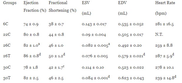
Echocardiographic evaluation of cardiac functional parameters in female F344xBN rats can be found in Table 2 and 3. As described in Table 2, ejection fraction was significantly increased at 26-months (82 ± 1.0%) compared to 6-months (74 ± 0.9%; P < 0.05). APAP treatment at 26-months (86 ± 0.8%) also significantly increased the ejection fraction compared to control (P = 0.009). Fractional shortening significantly decreased at 30-months (41.5 ± 1.7%) compared to 26-months (45.6 ± 1.0%; P < 0.05). APAP treatment at 26-months (49.8 ± 1.2%) significantly increased the fractional shortening when compared to control (P = 0.007). End systolic volume (ESV) was significantly decreased at 26-months (0.082 ± 0.005 mL) when compared to 6-months (0.143 ± 0.017 mL; P < 0.05). APAP treatment significantly decreased ESV at 30-months (0.084 ± 0.001 mL) when compared to control (0.114 ± 0.10 mL; P=0.034). APAP treatment at 26-months (0.579 ± 0.021 mL) significantly increased end diastolic volume when compared to control (0.492 ± 0.20 mL; P = 0.005). Heart rate did not change with age but significantly increased with APAP treatment at 26- (286.6 ± 5.9 bpm; P = 0.015) and decreased at 30-months (238.7 ± 14.8 bpm; P = 0.029) when compared to controls.
Table 2. Echocardiographic evaluation of cardiac functional parameters in female F344xBN rats with and without APAP (mean ± SEM; n = ## rats/group).
6C – 6-month animal; 22C – 22-month animal; 26C – 26-month animal; 26T – 26-month APAP-treated animal; 30C – 30-month animal; 30T – 30-month APAP-treated animal; aP < 0.05 significant difference from 6-month control animal;cP < 0.05 significant difference from 26-month control animal; dP < 0.05 age-matched control vs. treated animal; N.T. – not tested.
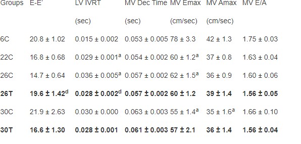
As described in Table 3, no age-associated changes in E-E’ ratio was found in the female F344xBN rats. APAP treatment increased the E-E’ ratio at 26-months (19.61 ± 1.42) compared to control (14.73 ± 0.64; P = 0.017). Left ventricular isovolumetric relaxation time (LV IVRT) was significantly increased at 22- (0.029 ± 0.001 sec) and 26-months (0.036 ± 0.005 sec) compared to 6-months (0.015 ± 0.002 sec; P < 0.05). Treatment with APAP at 26-months (0.028 ± 0.002 sec) significantly decreased LV IVRT compared to control (P < 0.001). There were no changes in mitral valve deceleration (MV Dec) time with age or APAP treatment. With age mitral valve peak velocity of the E wave (MV Emax) significantly decreased at 22- (59.8 ± 1.2 cm/sec), 26- (61.5 ± 1.5 cm/sec), and 30-months (55.4 ± 1.4 cm/sec) when compared to 6-months (78.0 ± 3.3 cm/sec; P < 0.05). Aging also significantly decreased peak velocity of the A wave (MV Amax) at 30-months (35.1 ± 1.6 cm/sec) when compared to 6-months (41.5 ± 1.3 cm/sec; P < 0.05). No change in MV Emax or Amax was found with APAP treatment. No changes were found in MV E/A ratio with age or APAP treatment.
Table 3. Echocardiographic evaluation of cardiac functional parameters in female F344xBN rats with and without APAP (mean ± SEM; n = ## rats/group).
6C – 6-month animal; 22C – 22-month animal; 26C – 26-month animal; 26T – 26-month APAP-treated animal; 30C – 30-month animal; 30T – 30-month APAP-treated animal; aP < 0.05 significant difference from 6-month control animal;bP < 0.05 significant difference from 22-month control animal; cP < 0.05 significant difference from 26-month control animal; dP < 0.05 age-matched control vs. treated animal.
Table 4. Total body weight (BW) and heart weight (HW) in female F344xBN rats at 6-, 22-, 26-, and 30-months of age with or without APAP treatment (means ± SEM; n = ## rats/group).
6C – 6-month animal; 22C – 22-month animal; 26C – 26-month animal; 26T – 26-month APAP-treated animal; 30C – 30-month animal; 30T – 30-month APAP-treated animal; aP < 0.05 significant difference from 6-month control animal;bP < 0.05 significant difference from 22-month control animal; cP < 0.05 significant difference from 26-month control animal; dP < 0.05 age-matched control vs. treated animal.
Ejection Fraction (%)
Fractional Shortening (%)
0.082 ± 0.005a
0.579 ± 0.021d
0.084 ± 0.001d
0.029 ± 0.001a
0.036 ± 0.005a
0.028 ± 0.002d
Tricuspid valve dysfunction was higher in 22-, 26-, and 30-month (52.4%, 21.7%, 33.3%) female hearts relative to 6-month. APAP treatment at 26-months (33.3%) had a higher percentage of female rats with tricuspid dysfunction compared to 26-month control group. However, with APAP treatment at 30-months the percentage of female rats with tricuspid dysfunction (33.3%) was the same when relative to the 6-month group. The percentage of female rats with mitral dysfunction also was higher with age (22C: 19.0%; 26C: 17.4%; 30C: 13.3%) relative to 6-month rats. APAP treatment at 26-months had a similar percentage of mitral dysfunction compared to control; however, APAP treatment at 30-months had a lower percentage when compared to the 30-month control group. The presence of aortic dysfunction with age was only seen in the 30-month control group (20%). At 26-months with APAP treatment (26T: 16.7%; 30T: 15.0%) the percentage of female rats with aortic dysfunction was higher compared to control groups (26C: 0%; 30C: 20.0%). The percentage of aortic dysfunction in 30-month APAP rats was lower (20.0%) than the 30-month control rats (15%). Female rats with pulmonary valve dysfunction were higher but this percentage was similar or lower with APAP treatment at 26- and 30-months (26T: 77.8%; 30T: 45.0%) when compared to control groups (26C: 78.3%; 30C: 66.7%).
Electrocardiogram Analysis
The presence of arrhythmias was not found in the vast majority of female F344xBN rats, and therefore the incidence was not different between control and APAP treatment groups. In the 30-month APAP treated group one, premature ventricular contraction was observed (data not shown).
Heart Weight, Body Weight, and Heart Weight to Body Weight Ratio
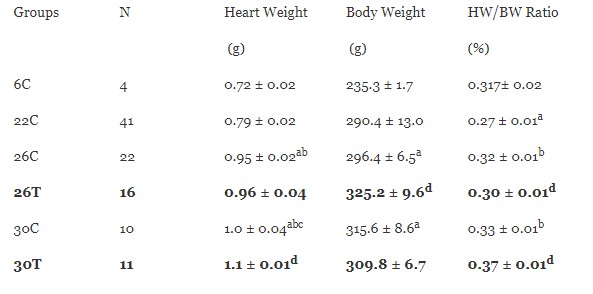
Female F344xBN heart weights and body weights were compared in Table 4. Heart weight increased significantly with age at 26-months (0.954 ± 0.002 g) compared to 6- and 22-month groups (6C: 0.721 ± 0.002 g; 22C: 0.787 ± 0.02 g; P < 0.001). At 30-months (1.04 ± 0.04 g) heart weight significantly increased compared to all age groups (P < 0.05). APAP treatment significantly increased heart weight at 30-months when compared to control (P = 0.029). Body weight increased significantly at 26- (296.4 ± 6.5 g) and 30-months (315.6 ± 8.6 g) when compared to 6-month (235.3 ± 1.7 g; P < 0.05). When normalized to body weight, heart weights were significantly decreased at 22-months (0.272 ± 0.01%) compared to 6-months (0.307 ± 0.02%; P < 0.05). However, normalized heart weights at 26- (0.321 ± 0.01%) and 30-month (0.327 ± 0.01%) rats was significantly increased compared to 6-months (P < 0.05). APAP treatment significantly decreased heart weight after normalization to body weight at 26-months (0.295 ± 0.01%; P = 0.048) but significantly increased at 30-months (0.367 ± 0.01%; P = 0.002) compared to controls.
The present study was conducted to obtain the cardiac function reference values for the aging female F344xBN rats. The F344xBN rat, according to the National Institute of Aging, is the preferred animal model to study age-associated pathophysiological changes due to its longer life span and low occurrence of pathologies [22]. The 6-, 22-, 26-, and 30-month age groups were chosen based off survivability curves from the National Institute of Aging to represent females in the third, seventh, and eighth decade of life [22, 23]. These reference values are critical in order to compare the effects of aging on systolic and diastolic function in order to establish if the female F344XBN rat is an appropriate model for aging cardiac dysfunction.
Previous work in our laboratory has indicated that with aging there is an increase in oxidative-nitrosative stress which may responsible for the age-associated changes in cardiac structure and function [24-26]. This age-associated increase in oxidative-nitrosative stress is attenuated with chronic APAP treatment [20] due to its antioxidative properties. In order to investigate the role of age-associated oxidative-nitrosative stress on cardiac structure and function, APAP was administered to the treatment groups in order to see if it would attenuate these age-associated changes in cardiac function.
Increased risk of diastolic dysfunction with aging appears to occur more often in women compared to men [27]. These diastolic dysfunctions oftentimes precede the development of systolic dysfunction [27]. Diastolic dysfunction in humans is defined by prolonged deceleration time, a high A wave velocity, a low E wave velocity, and prolonged isovolumetric relaxation [27]. In the present study of the female F344xBN heart, we observed age-related increases in left ventricular relaxation time, decrease in E`, and a trend in mitral valve deceleration time. Nonetheless, we did not find any changes in the E/A ratio with age nor did APAP treatment produce a change (Table 3). In contrast, Boluyt et al. show that aged female F344 animals have increased LV IVRT, decreased E wave, and increased A wave velocity [28]. In our study, as in the Boluyt et al. study with age there was a longer left ventricular relaxation time, a reduced E’ wave velocity, a trend toward an increased mitral valve deceleration time, and no change in the E/A ratio [28]. Taken together, these data suggest that aging in female F344 and F344xBN rats, much like that seen in humans, is characterized by changes in diastolic function. APAP treatment had no effect on this age related change.
As previously observed in F344 rats, systolic function in female F344xBN rats did not appear to be significantly impaired with aging (Table 2) [28, 29]. Similar to the F344 rats, we found a slight increase in 26-month ejection fraction; this parameter, however, must be interpreted carefully as there was also increased valvular regurgitation at this measurement time [28]. Forman et al. find that F344 male rats have higher incidence of mitral regurgitation (MR) relative to female rats [29]. This may explain differences in age related functional changes between male and female rats. More investigation is needed to determine the mechanism of age related differences in cardiac function in male and female F344xBN rats. Nonetheless, our data are consistent with the notion that APAP significantly improved the ejection fraction (and therefore increased the EDV and reduced the ESV) at 26 months and there was trend towards improvement at 30 months (note that the ESV was significantly lower in treated compared to age matched controls at this time point).
Unlike our previous data regarding the presence of age associated arrhythmias in the male F334XBN [17], the aging female F344XBN failed to demonstrate age associated arrhythmias. However, APAP treatment attenuated the age associated incidents of valvular dysfunction.
These data suggest that aging in the female F344xBN rat heart is associated with changes in cardiac structure (Table 1) and function (Tables 2 and 3) and that chronic APAP treatment may diminish the incidence of valvular dysfunction in most situations, but increase the incidence in others (e.g., increased tricuspid dysfunction at 26 months). APAP appears to result in beneficial changes in ejection fraction (Table 2), but a variable effect on heart rate (i.e., increased at 26 months, but decreased at 30 months compared to control). This study provides reference values for cardiac structure and function for adult female F344XBN rats. Further investigation regarding other parameters of cardiac function is currently underway.
The American Heart Association estimates that 80 million Americans have one or more types of cardiovascular disease (CVD). It is estimated that 38.1 million of those afflicted are aged 60 or older. As a person ages he or she has a higher risk of developing myocardial infarction, stroke, atherosclerosis, peripheral occlusive disease, diabetes, hypertension [1-3].Thepotential bmechanism of increase risk of disease with age is that there is an increase of reactive oxygen species (ROS) which leads to oxidative stress [4]. Recently acetaminophen (APAP), an antipyretic and analgesic, has shown to be protective against oxidative stress in the heart and the vasculature of the brain due to its antioxidant properties [5-7].
Due to these antioxidant effects APAP is considered to be cardioprotective during ischemia/reperfusion, hypoxia/reoxygenation, exogenous peroxynitrite administration, and experimentally induced myocardial infarction due to its ability to block or reduce the development of mitochondrial permeability transition pores, mitochondrial swelling, cytochrome c release, late stage apoptosis, protein oxidation, production of hydroxyl radicals, arrhythmias, and peroxynitrite induced dysfunction in hearts [5, 8-16].
The Fischer 344/NNiaHSD x Brown Norway/BiNia F1 (F344xBN) has been recommended by the National Institute of Aging as a rat model of aging due to its increased life span and fewer age-associated pathologies compared to the standard model of aging – the Fischer 344 rat. Although several studies have investigated the age-associated changes in male and female F344 rats as well as male F344xBN rats [17-21], no study to our knowledge has examined the age-associated changes in structure and function in the female F344xBN heart using echocardiographic measures.
This information is crucial in determining whether or not the female F344xBN is an appropriate aging model to study the development of cardiovascular disease. Due to evidence that age-associated increase in levels of oxidative stress may cause cardiac dysfunction, we also determined the function and structure of the aged female F344xBN heart with chronic APAP treatment. The purpose of this study was to determine the age-associated changes in the female F344xBN cardiac structure and function and whether chronic APAP treatment would attenuate these effects.
All procedures were performed in accordance with the Guide for the Care and Use of Laboratory Animals as approved by the Council of the American Physiological Society and the Animal Use Review Board of Marshall University. All procedures were conducted in strict accordance with the Public Health Service animal welfare policy. Young (6-month), adult (22-month), aged (26-month), and very aged (30-month) female F344xBN rats were obtained from the National Institute for Aging were housed two per cage in an Association for Assessment and Accreditation of Laboratory Animal Care International (AAALAC) approved vivarium. Housing conditions consisted of a 12 h-12 h light-dark cycle with a temperature maintained at 22 ± 2˚C. Animals were provided with food and water ad libitum. Rats were allowed to recover from shipment for at least two weeks before experimentation. During this time the animals were carefully observed and weighed weekly. Any rat showing signs of failure to thrive, such as precipitous weight loss, disinterest in the environment, or unexpected gait alterations were excluded from the study. Six Female F344xBN rats were randomly assigned to each group (6C – 6-month animal; 22C – 22-month animal; 26C – 26-month animal; 26T – 26-month APAP-treated animal; 30C – 30-month animal; 30T – 30-month APAP-treated animal).
A therapeutic dose of APAP (30 mg/kg/day) was administered through the drinking water for treated animals at 22-months of age. Changes in body weight and water intake were monitored throughout the study to maintain a therapeutic dose. The 6-, 22-, 26-, and 30-month age-matched control groups received tap water and were maintained under the same conditions as the APAP treated groups.
Control (6-, 22-, 26-, and 30-month) and APAP treated (26- and 30-month) rats were anesthetized with an intraperitoneal (ip) injection of ketamine (40 mg/kg) and xyaline (10 kg/mg). The ventral thoraxes where shaved, and the rats were placed either on their backs or left side and covered with ultrasonic transmission gel for adequate sonic transference. A Phillips 5500 ECHO system with a 12 MHz transducer was used to take two-dimensional ECHO measurements, two-dimensional guided M-mode, Doppler M-mode, and other recordings from parasternal long- and short-axis views. Parasternal long- and short axis views were used to determine two-dimensional cardiac structural measurements. The echocardiographic views were then used to position the M-mode echocardiographic line. In order to determine the presence of valve dysfunctions, valvular blood flow velocities were evaluated using pulse wave Doppler with the probe toward the apex (x-axis) and the depth along the y-axis (long axis procedure). Ejection fraction and fractional shortening during systole was calculated using the evaluation of wall structure from short-axis procedures with the probe oriented toward the left ventricle and across the heart. M-mode displays were analyzed by a digital echocardiographic analysis system.
The following measurements were selected for each assessment of cardiac structure and function. The structural parameters included diastolic (IVSd) and systolic (IVSs) left ventricular septal thickness, diastolic (LVIDd) and systolic (LVIDs) left ventricular internal dimension, diastiolic (LVPWd) and systolic (LVPWs) left ventricular posterior wall thickness, and right ventricular diastolic internal dimension (RVd). Functional measurement included ejection fraction (EF) and left ventricular fractional shortening during systole (FS).
Additional echocardiographic measurements included mitral valve deceleration (MV decel time), left ventricular mass (LVM), end-systolic volume (ESV), end-diastolic volume (EDV), peak velocity of the A wave (Amax), and peak velocity of the E wave (Emax) were used to evaluate systolic function. Mitral valve deceleration, Emax, Amax, and MV E/A ratio were used to evaluate diastolic function.
After completion of treatment and echocardiographic procedures, animals were anesthetized with an ip injection of ketamine (40 mg/kg) and xyaline (10 mg/kg) and supplemented as necessary for reflexive responses. Before heart collection, electrocardiograms (EKGs) with leads I, II, and III were measured in age-matched control and treated animals using the Biopac Student Lab software (BIOPAC Systems, Inc., Microsoft). After completion of EKG measurements, the heart was removed after a midline laparotomy and was then placed in Krebs-Ringer bicarbonate buffer (KRB) containing the following: 118 mM NaCl, 4.7 mM KCl, 2.5 mM CaCl2, 1.2 mM KH2PO4, 1.2 mM MgSO4, 24.2 mM NaHCO3, and 10 mM a-D-glucose (pH 7.4) equilibrated with 5% CO2/95% O2 and maintained at 37° C. Isolated hearts were quickly massaged to remove any blood from the ventricles, cleaned of connective tissue, weighed, and immediately snap frozen in liquid nitrogen for further analysis.
Results were reported as mean ± SEM. Statistical analyses were performed using Sigma Stat 3.5 statistical software (Systat Software, Inc). Age comparisons between ECHO structural, functional parameters, and morphologic indices were evaluated by one-way ANOVA or Kruskal-Wallis one-way Analysis of Variance on Ranks with the Student-Newman-Keuls or Dunn's methods as the post hoc test, respectively. Differences between age-matched control and treated animals were determined by Student’s T-test. The level of significance accepted a priori was P < 0.05.
Echocardiographic evaluation of cardiac structural parameters in female F344xBN rats are compared in Table 1. Left ventricular septal thickness (IVS) during systole (lVSs) significantly increased at 26-months (0.253 ± 0.003 cm) compared to 6-months (0.193 ± 0.008 cm; P < 0.05); however, no age-associated changes during diastole (lVSd) were found. Left ventricular internal dimension during systole (LVIDs) significantly increased at 22- (0.340 ± 0.006 cm) and 26-months (0.331 ± 0.009 cm) compared to 6-month (0.378 ± 0.017 cm; P < 0.05). Treatment with APAP significantly decreased LVIDs compared to control animals at 30-months (0.344 ± 0.015 vs 0.356 ± 0.011 cm, respectively; P = 0.004). During diastole LVID did not change with age but significantly increased with APAP treatment at 26-months (0.630 ± 0.009 cm) when compared to control (0.593 ± 0.009 cm; P < 0.001). Left ventricular posterior wall thickness (LVPW) during systole (LVPWs) and diastole (LVPWd) was significantly increased at 22- (0.273 ± 0.004 and 0.183 ± 0.004 cm, respectively) and 30-months (0.288 ± 0.009 and 0.189 ± 0.008 cm, respectively) compared to 6-months (0.193 ± 0.002 and 0.153 ± 0.010 cm, respectively; P < 0.05). APAP treatment increased both LVPWs and LVPWd thickness at 26-months compared to control (0.314 ± 0.007 and 0.185 ± 0.003 cm, respectively; P = 0.002). No changes were found in right ventricular dimension during diastole or left ventricular mass with age or APAP treatment.

Echocardiographic evaluation of cardiac functional parameters in female F344xBN rats can be found in Table 2 and 3. As described in Table 2, ejection fraction was significantly increased at 26-months (82 ± 1.0%) compared to 6-months (74 ± 0.9%; P < 0.05). APAP treatment at 26-months (86 ± 0.8%) also significantly increased the ejection fraction compared to control (P = 0.009). Fractional shortening significantly decreased at 30-months (41.5 ± 1.7%) compared to 26-months (45.6 ± 1.0%; P < 0.05). APAP treatment at 26-months (49.8 ± 1.2%) significantly increased the fractional shortening when compared to control (P = 0.007). End systolic volume (ESV) was significantly decreased at 26-months (0.082 ± 0.005 mL) when compared to 6-months (0.143 ± 0.017 mL; P < 0.05). APAP treatment significantly decreased ESV at 30-months (0.084 ± 0.001 mL) when compared to control (0.114 ± 0.10 mL; P=0.034). APAP treatment at 26-months (0.579 ± 0.021 mL) significantly increased end diastolic volume when compared to control (0.492 ± 0.20 mL; P = 0.005). Heart rate did not change with age but significantly increased with APAP treatment at 26- (286.6 ± 5.9 bpm; P = 0.015) and decreased at 30-months (238.7 ± 14.8 bpm; P = 0.029) when compared to controls.
6C – 6-month animal; 22C – 22-month animal; 26C – 26-month animal; 26T – 26-month APAP-treated animal; 30C – 30-month animal; 30T – 30-month APAP-treated animal; aP < 0.05 significant difference from 6-month control animal;cP < 0.05 significant difference from 26-month control animal; dP < 0.05 age-matched control vs. treated animal; N.T. – not tested.
As described in Table 3, no age-associated changes in E-E’ ratio was found in the female F344xBN rats. APAP treatment increased the E-E’ ratio at 26-months (19.61 ± 1.42) compared to control (14.73 ± 0.64; P = 0.017). Left ventricular isovolumetric relaxation time (LV IVRT) was significantly increased at 22- (0.029 ± 0.001 sec) and 26-months (0.036 ± 0.005 sec) compared to 6-months (0.015 ± 0.002 sec; P < 0.05). Treatment with APAP at 26-months (0.028 ± 0.002 sec) significantly decreased LV IVRT compared to control (P < 0.001). There were no changes in mitral valve deceleration (MV Dec) time with age or APAP treatment. With age mitral valve peak velocity of the E wave (MV Emax) significantly decreased at 22- (59.8 ± 1.2 cm/sec), 26- (61.5 ± 1.5 cm/sec), and 30-months (55.4 ± 1.4 cm/sec) when compared to 6-months (78.0 ± 3.3 cm/sec; P < 0.05). Aging also significantly decreased peak velocity of the A wave (MV Amax) at 30-months (35.1 ± 1.6 cm/sec) when compared to 6-months (41.5 ± 1.3 cm/sec; P < 0.05). No change in MV Emax or Amax was found with APAP treatment. No changes were found in MV E/A ratio with age or APAP treatment.
6C – 6-month animal; 22C – 22-month animal; 26C – 26-month animal; 26T – 26-month APAP-treated animal; 30C – 30-month animal; 30T – 30-month APAP-treated animal; aP < 0.05 significant difference from 6-month control animal;bP < 0.05 significant difference from 22-month control animal; cP < 0.05 significant difference from 26-month control animal; dP < 0.05 age-matched control vs. treated animal.
Tricuspid valve dysfunction was higher in 22-, 26-, and 30-month (52.4%, 21.7%, 33.3%) female hearts relative to 6-month. APAP treatment at 26-months (33.3%) had a higher percentage of female rats with tricuspid dysfunction compared to 26-month control group. However, with APAP treatment at 30-months the percentage of female rats with tricuspid dysfunction (33.3%) was the same when relative to the 6-month group. The percentage of female rats with mitral dysfunction also was higher with age (22C: 19.0%; 26C: 17.4%; 30C: 13.3%) relative to 6-month rats. APAP treatment at 26-months had a similar percentage of mitral dysfunction compared to control; however, APAP treatment at 30-months had a lower percentage when compared to the 30-month control group. The presence of aortic dysfunction with age was only seen in the 30-month control group (20%). At 26-months with APAP treatment (26T: 16.7%; 30T: 15.0%) the percentage of female rats with aortic dysfunction was higher compared to control groups (26C: 0%; 30C: 20.0%). The percentage of aortic dysfunction in 30-month APAP rats was lower (20.0%) than the 30-month control rats (15%). Female rats with pulmonary valve dysfunction were higher but this percentage was similar or lower with APAP treatment at 26- and 30-months (26T: 77.8%; 30T: 45.0%) when compared to control groups (26C: 78.3%; 30C: 66.7%).
The presence of arrhythmias was not found in the vast majority of female F344xBN rats, and therefore the incidence was not different between control and APAP treatment groups. In the 30-month APAP treated group one, premature ventricular contraction was observed (data not shown).
Female F344xBN heart weights and body weights were compared in Table 4. Heart weight increased significantly with age at 26-months (0.954 ± 0.002 g) compared to 6- and 22-month groups (6C: 0.721 ± 0.002 g; 22C: 0.787 ± 0.02 g; P < 0.001). At 30-months (1.04 ± 0.04 g) heart weight significantly increased compared to all age groups (P < 0.05). APAP treatment significantly increased heart weight at 30-months when compared to control (P = 0.029). Body weight increased significantly at 26- (296.4 ± 6.5 g) and 30-months (315.6 ± 8.6 g) when compared to 6-month (235.3 ± 1.7 g; P < 0.05). When normalized to body weight, heart weights were significantly decreased at 22-months (0.272 ± 0.01%) compared to 6-months (0.307 ± 0.02%; P < 0.05). However, normalized heart weights at 26- (0.321 ± 0.01%) and 30-month (0.327 ± 0.01%) rats was significantly increased compared to 6-months (P < 0.05). APAP treatment significantly decreased heart weight after normalization to body weight at 26-months (0.295 ± 0.01%; P = 0.048) but significantly increased at 30-months (0.367 ± 0.01%; P = 0.002) compared to controls.
6C – 6-month animal; 22C – 22-month animal; 26C – 26-month animal; 26T – 26-month APAP-treated animal; 30C – 30-month animal; 30T – 30-month APAP-treated animal; aP < 0.05 significant difference from 6-month control animal;bP < 0.05 significant difference from 22-month control animal; cP < 0.05 significant difference from 26-month control animal; dP < 0.05 age-matched control vs. treated animal.
The present study was conducted to obtain the cardiac function reference values for the aging female F344xBN rats. The F344xBN rat, according to the National Institute of Aging, is the preferred animal model to study age-associated pathophysiological changes due to its longer life span and low occurrence of pathologies [22]. The 6-, 22-, 26-, and 30-month age groups were chosen based off survivability curves from the National Institute of Aging to represent females in the third, seventh, and eighth decade of life [22, 23]. These reference values are critical in order to compare the effects of aging on systolic and diastolic function in order to establish if the female F344XBN rat is an appropriate model for aging cardiac dysfunction.
Previous work in our laboratory has indicated that with aging there is an increase in oxidative-nitrosative stress which may responsible for the age-associated changes in cardiac structure and function [24-26]. This age-associated increase in oxidative-nitrosative stress is attenuated with chronic APAP treatment [20] due to its antioxidative properties. In order to investigate the role of age-associated oxidative-nitrosative stress on cardiac structure and function, APAP was administered to the treatment groups in order to see if it would attenuate these age-associated changes in cardiac function.
Increased risk of diastolic dysfunction with aging appears to occur more often in women compared to men [27]. These diastolic dysfunctions oftentimes precede the development of systolic dysfunction [27]. Diastolic dysfunction in humans is defined by prolonged deceleration time, a high A wave velocity, a low E wave velocity, and prolonged isovolumetric relaxation [27]. In the present study of the female F344xBN heart, we observed age-related increases in left ventricular relaxation time, decrease in E`, and a trend in mitral valve deceleration time. Nonetheless, we did not find any changes in the E/A ratio with age nor did APAP treatment produce a change (Table 3). In contrast, Boluyt et al. show that aged female F344 animals have increased LV IVRT, decreased E wave, and increased A wave velocity [28]. In our study, as in the Boluyt et al. study with age there was a longer left ventricular relaxation time, a reduced E’ wave velocity, a trend toward an increased mitral valve deceleration time, and no change in the E/A ratio [28]. Taken together, these data suggest that aging in female F344 and F344xBN rats, much like that seen in humans, is characterized by changes in diastolic function. APAP treatment had no effect on this age related change.
As previously observed in F344 rats, systolic function in female F344xBN rats did not appear to be significantly impaired with aging (Table 2) [28, 29]. Similar to the F344 rats, we found a slight increase in 26-month ejection fraction; this parameter, however, must be interpreted carefully as there was also increased valvular regurgitation at this measurement time [28]. Forman et al. find that F344 male rats have higher incidence of mitral regurgitation (MR) relative to female rats [29]. This may explain differences in age related functional changes between male and female rats. More investigation is needed to determine the mechanism of age related differences in cardiac function in male and female F344xBN rats. Nonetheless, our data are consistent with the notion that APAP significantly improved the ejection fraction (and therefore increased the EDV and reduced the ESV) at 26 months and there was trend towards improvement at 30 months (note that the ESV was significantly lower in treated compared to age matched controls at this time point).
Unlike our previous data regarding the presence of age associated arrhythmias in the male F334XBN [17], the aging female F344XBN failed to demonstrate age associated arrhythmias. However, APAP treatment attenuated the age associated incidents of valvular dysfunction.
These data suggest that aging in the female F344xBN rat heart is associated with changes in cardiac structure (Table 1) and function (Tables 2 and 3) and that chronic APAP treatment may diminish the incidence of valvular dysfunction in most situations, but increase the incidence in others (e.g., increased tricuspid dysfunction at 26 months). APAP appears to result in beneficial changes in ejection fraction (Table 2), but a variable effect on heart rate (i.e., increased at 26 months, but decreased at 30 months compared to control). This study provides reference values for cardiac structure and function for adult female F344XBN rats. Further investigation regarding other parameters of cardiac function is currently underway.
Dear Editorial Team, Clinical Medical Reviews and Reports. My experience with the journal was highly positive. The peer-review process was rigorous, constructive, and completed in a timely manner. The reviewers provided valuable comments that helped improve the quality and clarity of our manuscript. The editorial office was professional, responsive, and supportive throughout all stages of the publication process. Communication was clear and efficient, and any questions were addressed promptly. Overall, I found the journal to maintain high scientific standards and an excellent publication workflow. I would be pleased to consider submitting future work to this journal. Best wishes from, Elena Popa.

It was my pleasure to submit my testimonial concerning the Reviewer Board of our Scientific Journal “Brain and Neurological Disorders”. The Reviewers focused on some modifications and their contribution was helpful. The ladies of our Editorial Office were also supported my efforts. It was my honor to have such a co-operation and I am looking forward for more collaboration.

Dear Grace Pierce, Editorial Coordinator of Journal of Clinical Research and Reports, Thank you for the speedy and efficient peer review process. I appreciate the fact that your peer reviewers do not take months to respond like with some other journals. I would also like to thank the editorial office for responding quickly to my questions. It is an excellent journal. I plan to submit more manuscripts in the future. Best wishes from, Robert W. McGee

Dear Grace Pierce, Editorial Coordinator of Journal of Clinical Research and Reports, Working with you and your team on our recent publication in JCRR has been a truly wonderful and enjoyable experience. The responses were prompt, and the reviewers were patient, constructive, and highly professional. One reviewer in particular gave me the feeling that a professor was carefully reading and commenting on my coursework, which was deeply touching. The entire process was straightforward and hassle‑free, with no tedious online forms to complete. I highly recommend this journal. Best wishes from, DR Aibing Rao, Head of R&D

I Appreciate the Opportunity to Share my Experience with the Journal of Clinical Research and Reports. The peer review process was timely and constructive, and the feedback provided helped improve the quality of our manuscript. The editorial office was professional, responsive, and supportive throughout the process, ensuring smooth communication and efficient handling of the submission. Overall, it was a positive experience collaborating with your team.

Dear Mercy Grace, Editorial Coordinator of Obstetrics Gynecology and Reproductive Sciences, We would like to express our gratitude for your help at all stages of publishing and editing the article. The editors of the magazine answer all the necessary questions and help at every stage. We will definitely continue to cooperate and publish other works in the Obstetrics Gynecology and Reproductive Sciences! Best wishes from, Alla Konstantinovna Politova,
