AUCTORES
Globalize your Research

Research Article | DOI: https://doi.org/10.31579/2641-0419/417
1Heart and Brain Center of Clinical Excellence, Pleven, Bulgaria.
2Bulgarian Cardiac Institute.
3Medical University, Pleven, Bulgaria.
4Heart and Brain, Burgas, Bulgaria.
5Specialized hospital for active cardiology treatment, Varna, Bulgaria.
*Corresponding Author: А. Ivanova, Heart and Brain Center of Clinical Excellence, Pleven, Bulgaria.
Citation: А. Ivanova, I. Simova, Т. Vekov, V. Коrnovski, J. Кrasnaliev, (2025), Use of Streptokinase for intravenous thrombolysis in pulmonary embolism: Practice and results (Data from the pulmonary embolism registry of Bogodogo University Hospital Center in Burkina Faso (PER/UHC Bogodogo-BF), J Clinical Cardiology and Cardiovascular Interventions, 8(2); DOI: 10.31579/2641-0419/417
Copyright: © 2025, А. Ivanova. This is an open access article distributed under the Creative Commons Attribution License, which permits unrestricted use, distribution, and reproduction in any medium, provided the original work is properly cited.
Received: 13 January 2025 | Accepted: 29 January 2025 | Published: 06 February 2025
Keywords: familial hypercholesterolemia; artificial intelligence; statinе; ezetimibe; PCSK9 inhibitors; xanthomas; corneal arcus
Introduction: Familial hypercholesterolemia (FH) is an autosomal dominant genetic manifestation with early, aggressive, and recurrent cardiovascular events. According to literature data, this is seen in one in 200 individuals in the general population and is interpreted as the most common hereditary cardiovascular disease. Following the current trends in modern medicine, we used the artificial intelligence (AI) method to achieve greater efficiency in the recognition of FH patients.
Objective: Our primary goal was to create, with the help of AI, an algorithm for FH screening in patients, admitted to the Clinics of the Bulgarian Cardiac Institute. A secondary task was to optimize the therapy of patients, according to the latest recommendations for behavior in dyslipidemias of the European Society of Cardiology.
Methods: For a period of one year, we conducted a prospective study in which all hospitalized patients for any atherosclerotic event requiring treatment were followed in three hospitals in Bulgaria - Pleven, Burgas and Varna, covering Cardiology, Cardiac Surgery, Neurology and Vascular Surgery clinics. The electronic records were stored in the hospital's Gamma Cod Master (GCM) program. The algorithm for patient inclusion and data collection uses AI, through which, according to set parameters, patients are selected according to age, diagnosis, LDL-C value, type, and dose of antilipemic therapy. Each indicator gives certain points that are summed up. For each patient with a final score of ≥4, an alarm signal is created in the GSM, which can be easily seen by the attending physician. Despite AI, the main task is the attending physician to check the accuracy of the integrated data, to specify the physical status for the presence of stigmata such as xanthomas and corneal arcus at an early age, to collect additional family history for early atherosclerosis, to additionally recalculate the points of the DLCN score, and to optimize the therapy according to the recommendations of the European Society of Cardiology for dyslipidemias.
Results: We prospectively followed 438 patients signaled by AI. The largest number of electronic files were analyzed in the cardiology clinics, with the most patients being screened for FH in the Cardiology Clinic in Pleven – 174, of which 79 patients (46%) had probable FH. In 49% of the total number of patients (438), we found a family history of an early atherosclerotic event, and 32% of the patients had a previous cardiovascular event. Only 1.5% of those hospitalized were on combined statin and ezetimibe therapy, and 15% of the total number of patients were completely missing lipid- lowering therapy. Through AI, patient therapy was optimized, also increasing the number of PCSK9 inhibitor prescriptions 3-to 4.5-fold (depending on the region).
Conclusion: The development, implementation and use of AI for FH screening is possible and easily applicable. Despite the use of AI, many FH patients remain undiagnosed. This confirms the need for active participation of the attending physician, whose role in correct diagnosis and treatment, as well as in improving the patient's prognosis, remains leading.
Familial hypercholesterolemia (FH) is an autosomal-dominant genetic disease associated with early, aggressive and recurrent atherosclerotic cardiovascular events. It is caused by an allelic mutation in a gene responsible for lipid metabolism, which is associated with persistence of high levels of low-density lipoprotein (LDL-C). According to literature data, the disease is observed in one in 200 individuals in the general population and is interpreted as the most common hereditary cardiovascular disease (1,2,3).
FH has two forms: homozygous and heterozygous, the latter being widespread and having one mutated allele in the following genes: LDLR, ApoB, PCSK9, or LDLRAP1 (1, 4, 8). In the homozygous form of FH, carriers have two pathogenic mutations (homozygotes, i.e. with two identical mutations or compound heterozygotes, i.e. with different mutations). The condition of homozygous FH is very rare (1:160,000 to 1:250,000) and results in a high incidence of acute myocardial infarction (AMI) or other atherosclerotic events in childhood or adolescence (1).
Patients suspected of having genetic dyslipidemia can be recognized by extremely high LDL-C levels, which correlate with a family history of premature atherosclerotic cardiovascular disease (3, 4).
According to the latest recommendations of the European Society of Cardiology (ESC) for dyslipidemias (5, 6), LDL-C target values, in very high and high-risk individuals, should be <1>
| Treatment | Average reduction of LDL-C |
| Statinе with a moderate intensity | ~30 % |
| Statinе withhigh intensity | ~50 % |
| Statinе withhigh intensity + ezetimibe | ~65 % |
| PCSK9 inhibitors | ~60 % |
| PCSK9 inhibitors + statin withhigh intensity | ~75 % |
| PCSK9 inhibitors + statin with high intensity + ezetimibe | ~85% |
Table 1: Expected reduction of LDL-C, with combined therapies:
2021 ESC Guidelines on cardiovascular disease prevention in clinical practice
Despite the well-known behavior algorithm for hypercholesterolemia, the lipid profile is often underestimated by clinicians and not correctly interpreted according to the patient's risk profile. Assessment of physical status for the presence of xanthomas, arcus corneal, and xanthelasma should be a guiding criterion and presumptive factor for FH (Figs. 1, 2, 3).
Components of the diagnosis: Identification of patients with FH is a multifaceted process, including family history of dyslipidemia and early cardiovascular events, physical status stigmata: tendon xanthomas, corneal arcus, manifestation of early atherosclerotic cardiovascular disease, LDL-C values, as well as genetic testing. In recent years, Lipoprotein(a) – for short Lp (a), called the “genetic” lipoprotein, has also found informative value and significance (4, 5, 7).
In the process of determining the causes of hypercholesterolemia, other conditions associated with elevated LDL-C levels, such as thyroid disease, cholelithiasis, kidney disease, hyperglycemia, and hyperalbuminemia, must be excluded (8, 9).
Use of imaging methods for early identification of cardiovascular disease such as 12-lead electrocardiogram, echocardiography, computed tomography angiography scan, selective coronary angiography with intravascular ultrasound (IVUS) and Fractional Flow Reserve (FFR) would also benefit and support the diagnosis (figure 3).
A diagnosis of FH can be made by genetic testing, which is rarely applied in practice, and/or by using the readily available criteria of the Dutch Lipid Clinical Network (DLCN) score (Table 2)
| Category | Characteristic marks | Points |
| Family history | First-degree relatives with known early coronary and/orvascular disease (males<55> First-degree relatives with tendinous xanthomas and/orpresence of cornealarcus, or children aged <18> | 1
2 |
| Clinical history | Patients withearly coronary disease, (men <55> | 2 |
| Patients with early cerebrovascular or peripheral vascular disease (men <55> | 1 | |
| Physical examination | Tendinous xanthomata Corneal arcus before 45 years of age | 6 4 |
| Levels of LDL-C | >8.5 mmol/l 6.5-8.4 mmol/l 5.0-6.4 mmol/l 4.0-4.9 | 8 5 3 1 |
| Genetic analysis | Functional mutations in the genes of LDL-R, APOB, PCSK9 | 8 |
| Genetic analysis | Functional mutations in the genesof LDL-R, APOB, PCSK9 | 8 |
Table 2: Criteria for the diagnosis of Heterozygous FH, according to the Dutch Lipid Clinical Network.
Dutch Lipid Clinic Network criteria for Familial Hypercholesterolemia (FH) Last edited 26 Jan 2018. Last reviewed 28 Nov 2023 <3>8 points: definite FH
According to national registries, Bulgaria is among the countries with the highest number of cardiovascular diseases. Many patients do not suspect that they have FH, and in most cases the disease is discovered after the realization of an acute vascular accident (acute myocardial infarction or stroke at a young age, men <55>
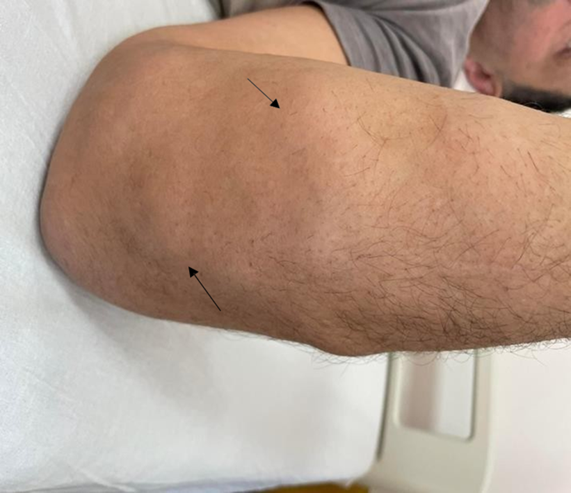
Figure 1: Xanthomas on a 47-year-old patient with newly diagnosed three-vessel coronary disease and newly established hypercholesterolemia. Upon admission to the Pleven "Heart and Brain" Hospital with LDL 8 mmol/l, total cholesterol 9.7 mmol/l, emergency cardiac surgical treatment was performed due to unstable angina pectoris. The family history of early ischemic heart disease.Personal archive, the patient has given consent to publish the material.

Figure 2: Xanthelasma of a 46-year-old patient treated at MBAL "Heart and Brain" Pleven with established hypercholesterolemia, multivessel coronary artery disease, in which aorto-coronary bypass was performed. LDL-C 6 mmol/l, total cholesterol 12 mmol/l at diagnosis, untreated until hospitalization for acute coronary syndrome. Family history: Father died at the age of 47 from acute myocardial infarction, sister with severe dyslipidemia. Personal archive, the patient has given consent to publish the material

Figure 3: Arcus corneal and xanthelasma in a 54-year-old patient hospitalized for a second time with myocardial infarction. First vascular accident of a 50-year-old age. With chronic arterial occlusive disease of the lower extremities. Ineffectively treated dyslipidemia, LDL-C 3.9 mmol/l upon admission against the background of rosuvastatin 10 mg/per day. Personal archive, photo provided for publication with patient consent.
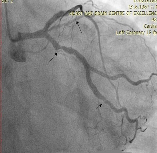
Figure 4: Selective coronary angiography of a 55-year-old woman with established ischemic heart disease, LDL 4 mmol/l, total cholesterol 6 mmol/l, on therapy with rosuvastatin 20 mg/per day. and ezetemibe 10 mg/per day, in which we started treatment with a PCSK9 inhibitor.
For a period of one year, we conducted a prospective study, including all patients (men < 65>
The analysis used data from electronic records of patients treated in a specialized cardiology hospital in Varna and two large multi-specialty hospitals in Pleven and Burgas, where departments of Cardiology, Cardiac Surgery, Neurology and Vascular Surgery were covered. Electronic records were stored and generated using the hospital's Gamma Cod Master (GCM) program.
The follow-up period was one year, starting in the month of July 2021 and ending in July 2022. The algorithm for patient inclusion and data collection uses AI and runs in the following sequence:
I. Patients under 65 years of age for men and under 70 years of age for women are selected.
II.Distribution of points according to diagnosis
a.Each patient with any of the following diagnoses receives 2 points
I20.0 Unstable angina pectoris
I20.1 Angina with documented spasm I20.8 Other types of anginas
I20.9 Angina, unspecified
I21.0 Acute anterior wall transmural myocardial infarction I21.1 Acute transmural myocardial infarction of inferior wall
I21.2 Acute transmural myocardial infarction with other specified locations I21.3 Acute transmural myocardial infarction of unspecified location
I21.4 Acute subendocardial myocardial infarction I21.9 Acute myocardial infarction, unspecified I22.0 Recurrent anterior wall myocardial infarction I22.1 Recurrent inferior wall myocardial infarction
I22.8 Recurrent myocardial infarction with other specified location I22.9 Recurrent myocardial infarction of unspecified location
I23.0 Hemopericardium as an ongoing complication of acute myocardial infarction I23.1 Interatrial defect as an ongoing complication of acute myocardial infarction
I23.2 Interventricular defect as an ongoing complication of acute myocardial infarction
I23.3 Rupture of the heart wall without hemopericardium as an ongoing complication of acute myocardial infarction
I23.4 Rupture of tendon chord as an ongoing complication of acute myocardial infarction
I23.5 Papillary muscle rupture as an ongoing complication of acute myocardial infarction
I23.6 Atrial, atrial auricle, or chamber thrombosis as an ongoing complication of acute myocardial infarction
I23.8 Other ongoing complications of acute myocardial infarction I24.0 Coronary thrombosis not leading to myocardial infarction I24.1 Dressler syndrome.
I24.8 Other forms of acute ischemic heart disease I24.9 Acute ischemic heart disease, unspecified
I25.0 Atherosclerotic cardiovascular disease - described as such I25.1 Atherosclerotic heart disease.
I25.2 Old myocardial infarction I25.3 Cardiac aneurysm
I25.4 Coronary artery aneurysm I25.5 Ischemic cardiomyopathy
I25.6 Asymptomatic myocardial ischemia
I25.8 Other forms of chronic ischemic heart disease I25.9 Chronic ischemic heart disease, unspecified
b.Each patient with your diagnosis receives 1 of the following points: I63.0 Cerebral infarction caused by thrombosis of precerebral arteries I63.1 Cerebral infarction caused by embolism of precerebral arteries.
I63.2 Cerebral infarction caused by unspecified occlusion or stenosis of precerebral arteries.
I63.3 Cerebral infarction caused by thrombosis of cerebral arteries I63.4 Cerebral infarction caused by embolism of cerebral arteries.
I63.5 Cerebral infarction caused by unspecified occlusion or stenosis of cerebral arteries.
I63.6 Cerebral infarction caused by cerebral vein thrombosis - non-pyogenic I63.8 Another cerebral infarction.
I63.9 Cerebral infarction, unspecified
I65.0 Occlusion and stenosis of vertebral artery I65.1 Occlusion and stenosis of basilar artery I65.2 Occlusion and stenosis of carotid artery
I65.3 Occlusion and stenosis of multiple and bilateral precerebral arteries
I65.8 Occlusion and stenosis of other precerebral arteries
I65.9 Occlusion and stenosis of unspecified precerebral artery I66.0 Occlusion and stenosis of middle cerebral artery
I66.1 Occlusion and stenosis of anterior cerebral artery I66.2 Posterior cerebral artery occlusion and stenosis I66.3 Occlusion and stenosis of cerebellar arteries
I66.4 Occlusion and stenosis of multiple and bilateral cerebral arteries I66.8 Occlusion and stenosis of another cerebral artery
I66.9 Occlusion and stenosis of unspecified cerebral artery I67.0 Dissection of cerebral arteries without rupture
I67.1 Unruptured cerebral aneurysm I67.2 Cerebral atherosclerosis
I69.3 Sequelae of cerebral infarction
I69.4 Sequelae of stroke, unspecified as cerebral hemorrhage or infarction I69.8 Sequelae of other and unspecified cerebrovascular diseases
I70.0 Atherosclerosis of aorta
I70.1 Atherosclerosis of renal artery
I70.2 Atherosclerosis of arteries of extremities I70.8 Atherosclerosis of other arteries
I70.9 Generalized and unspecified atherosclerosis I74.0 Embolism and thrombosis of abdominal aorta
I74.1 Embolism and thrombosis of other and unspecified parts of aorta I74.2 Embolism and thrombosis of arteries of upper extremities
I74.3 Embolism and thrombosis of arteries of lower extremities
I74.4 Embolism and thrombosis of arteries of extremities, unspecified I74.5 Embolism and thrombosis of iliac artery
I74.8 Embolism and thrombosis of other arteries I74.9 Embolism and thrombosis of unspecified artery Z95.1 Presence of aortic coronary shunt graft
Z95.5 Presence of coronary angioplasty implant and graft
Z95.8 Presence of other cardiac and vascular implants and transplants
Note: according to category II a patient can receive either 1 or 2 points. Consider the higher points. The maximum number of points in this category is 2.
III.Distribution of points according to LDL cholesterol
a.The LDL cholesterol value on admission is obtained
b.It is determined whether this value is achieved on the background of statin therapy and at what dose
For this purpose, in the History section, all possible trade names of statins (according to the attached list) are searched for, creating an 80% margin of coincidence with the correct spelling (due to the frequently observed cases of incorrect spelling). In addition, we made it possible to write the name of the statin in both Cyrillic and Latin letters. When a statin is detected in therapy from the anamnesis, the dose in which it was administered is extracted (in the absence of a specified dose, a dose of 10 mg is taken).
c.The trade name of the statin is transferred to the corresponding generic.
The value of LDL-C is recalculated by multiplying by a corresponding coefficient, according to the type and dose of statin taken - see table 3.
The recalculation is done at a certain point in time, i.e. if the patient is hospitalized several times, the algorithm is triggered for each of them. Usually, the values of LDL- C and antilipemic therapy at each hospitalization differ, but the recalculated LDL-C remains approximately the same. If there is a difference with the total number of points, it is usually due to the entry of a new diagnosis in the patient's medical record.
| Antilipemic drug | Coefficient of correction |
| Atorvastatin 10 mg | 1.618123 |
| Atorvastatin 20 mg | 1.763668 |
| Atorvastatin 40 mg | 1.937984 |
| Atorvastatin 80 mg | 2.150538 |
| Rosuvastatin 5 mg | 1.709402 |
| Rosuvastatin 10 mg | 1.872659 |
| Rosuvastatin 20 mg | 2.070393 |
| Rosuvastatin 40 mg | 2.314815 |
| Rosuvastatin 80 mg | 2.624672 |
| Simvastatin 10 mg | 1.37741 |
| Simvastatin 20mg | 1.492537 |
| Simvastatin 40 mg | 1.636661 |
| Simvastatin 80 mg | 1.818182 |
| Ezetimibe 10 mg | 1.236094 |
| Ezetimibe/Simvastatin 10/10 mg | 1.855288 |
| Ezetimibe/Simvastatin 10/20 mg | 2.008032 |
| Ezetimibe/Simvastatin 10/40 mg | 2.252252 |
| Ezetimibe/Simvastatin 10/80 mg | 2.463054 |
| Ezetimibe/Atorvastatin 10/10 mg | 2 |
| Ezetimibe/Atorvastatin 10/20 mg | 2.173913 |
| Ezetimibe/Atorvastatin 10/40 mg | 2.173913 |
| Ezetimibe/Atorvastatin 10/80 mg | 2.5 |
| Ezetimibe/Rosuvastatin 10/10 mg | 2.48139 |
| Ezetimibe/Rosuvastatin 10/20 mg | 2.739726 |
| Ezetimibe/Rosuvastatin 10/40 mg | 3.333333 |
| Pravastatin 10 mg | 1.251564 |
| Pravastatin 20 mg | 1.322751 |
| Pravastatin 40 mg | 1.422475 |
| Others | 1.43 |
Table 3: Coefficient of correction, according to the type and dose of statin
d.The value of LDL cholesterol thus obtained is transferred into a number of points, according to table 4:
| LDL cholesterol (mmol/l) | Points |
| ≥ 8.5 | 8 |
| 6.5 – 8.4 | 5 |
| 5.0 – 6.4 | 3 |
| 4.0 – 4.9 | 1 |
Table 4. Values of LDL cholesterol, transferred into number of points
III. The points of categories II and III are added together and the final number of points is obtained.
IV. For each patient with a cut-off score ≥4, an alarm / output signal is created in the GCM to be easily visible to the attending physician.
Our chosen signaling method is the coloring of the at-risk patient, visible to the medical staff with access to the relevant ward, after the signaling has been activated.
Despite the help of AI, the main task remains for the attending physician to check the accuracy of the integrated data and whether the manifestation of the cardiovascular disease is early, to supplement the information with the data of the physical status in view of the presence or absence of xanthomas and corneal arcus of early age, collect additional family information for early atherosclerosis, further recalculate the DLCN score, and optimize therapy according to the recommendations of the European Society of Cardiology.
Statistical analysis: Statistical analysis was performed using SPSS for Windows version 19.0 statistical software. Continuous variables were presented as mean ± standard deviation (SD). Categorical variables were presented as a percentage. The DLCN score was used to calculate the probability of FH.
Demographic and clinical characteristics of patients were analyzed through electronic records stored in GCM.
Ethical considerations: The clinical trial protocol was reviewed and approved by the local ethics committee. All research procedures adhere to the principles of the Declaration of Helsinki. Consent for FH treatment and personal data collection and analysis was obtained from all patients.
Results: 438 patients hospitalized and treated for any atherosclerotic indication were screened for FH. The prospective study continued from July 2021 to July 2022 and was conducted in seven department in three hospitals of the Bulgarian Cardiac Institute - two multi-specialty hospitals, including cardiology, cardiac surgery, neurology and vascular surgery clinics and one specialized cardiology hospital from three major cities in Bulgaria - Pleven, Varna, Burgas with a large patient flow.
Our primary outcome is to use AI to detect and analyses patients with probable and definite FH. Secondary objectives were to optimize the therapy of patients with DLCN ≥6 points, according to ESC dyslipidemia guideline from 2019 and to demonstrate whether the AI method would improve early recognition of patients with suspected FH.
Of the 438 patients signaled by AI, the largest number of electronic files was analyzed in the Cardiology Clinic of "Heart and Brain", Pleven (174), followed by 149 patients in the Cardiology Clinic in Varna (table 5).
From 174 patients detected by AI in the Cardiology Clinic of "Heart and Brain"- Pleven, the average value of AI points was 6.21 (standard deviation - SD ± 1.59). Of the total number of patients detected by AI, an individually physician-calculated DLCN score in 153 patients. These patients for risk profile, sequence of atherosclerotic disease, family history for early cardiovascular events, physical status with targeted follow-up for presence of corneal arcus and xanthomas. A DLCN score ≥6 points were found in 79 patients (46%), i.e., probable FH. In all hospitalized patients, the therapy was optimized according to the latest recommendations for the treatment of hypercholesterolemia (ESC 2019), in 78 patients (45%) PCSK9 inhibitor was added to the standard therapy with a maximum tolerated dose of statin, with or without ezetimibe.
In the same hospital, in the Department of Cardiac Surgery, the AI-assisted FH algorithm flagged 59 patients with a mean score of 6.37 (SD 1.79), of which 44% (26 patients) had an individual DLCN
score calculated by the attending physician ≥ 6 points, and treatment with monoclonal antibodies was started in 10 patients (17%).
The smallest number of patients were recognized in the Clinic for Vascular Surgery (3), and two of them were suspected of possible FH and the therapy was optimized to reach target LDL-C values according to the profile of the specific patients.
At the Neurology Clinic of "Heart and Brain", Pleven, AI has not reported any patients. Our explanation was for a non-routine study of a lipid profile in hospitalized patients in this department.
In SHAT of Cardiology, Varna, from 149 patients signaled by AI as potential for FH, the system gave an average value of 6.25 points (SD 1.77), of which DLCN score was calculated by the attending physician in 50 patients, 22 patients (14%) were assessed as possible FH, and in 9 patients (6%) an PCSK9-inhibitor was added.
For "Heart and Brain" Medical Center, Burgas, despite the small number of patients in total for the Clinic of Cardiology and Cardiac Surgery (53), a score for FH was calculated by the attending physician for 50 patients, with 23 of them having possible FH (43%), in 21 patients (40%) a PCSK9-inhibitor was added, indicating a good working algorithm and follow-up by the attending physician.
For the previous year without an AI for Pleven region, twenty patients received treatment with a PCSK9 inhibitor, and compared to ninety patients after the screening program was started. For 2019 without the use of AI for the hospitals involved in Burgas and Varna, only ten patients received treatment for FH with the administration of a monoclonal antibody, compared to thirty patients after the program was started (Table 5).
| Clinic | Number of patients, signaled by AI | Number of points by AI (average value ± SD) | Actual number of patients with DLCN score calculated by physician | Patients with probable FH- number (%) | Patients with PCSK9 inhibitor included - number(%) |
| Clinic of Cardiology „Heart and Brain“, Pleven | 174 | 6,21 (1,59) | 153 | 79 (46%) | 78 (45%) |
Cardiac surgery “Heartand Brain“, Pleven | 59 | 6,37 (1,79) | 26 | 26 (44%) | 10 (17%) |
| Clinic of Vascular Surgery „Heart and Brain“, Pleven | 3 | 5,33 (1,15) | 3 | 2 (66%) | 2 (66%) |
| Clinic of Neurology „Heart and Brain“,Pleven | 0 | 0 | 0 | 0 | 0 |
| Clinic of Cardiology and Cardiac surgery,Heart and Brain“,Burgas | 53 | 6,5 (2,01) | 50 | 23 (43%) | 21 (40%) |
| SHAT of Cardiology-Varna | 149 | 6,25 (1,77) | 50 | 22 (14%) | 9 (6%) |
| Total | 438 | 6,13 | 282 (64%) | 152 (35%) | 120 (28%) |
Table 5. Potential patients for FH in Clinics of the Bulgarian Cardiac Institute for a period of 12 months.
Legend: SD-standard deviation, AI-artificial intelligence, PCSK9-inhibitor (proprotein convertase subtilisin- kexin type 9 inhibitor).
Regarding the demographic characteristics (table 6), most of the patients recognized by AI were male – 310 patients (71%)., the majority were in age group 3 (56-70 years), 30% of analyzed patients were smokers, 30% had concomitant type 2 diabetes, 20% with obesity, 66% of patients had accompanying arterial hypertension. A family history of an early atherosclerotic event was found in 215 patients (49%) (Table 6).
| Gender | N | % |
| F | 128 | 29 |
| M | 310 | 71 |
| Age | ||
| <40> | 22 | 5 |
Table 6. Distribution of followed patients by demographic and clinical characteristics.
In 141 patients (32%), the GCM database had information on prior hospitalizations related to an atherosclerotic event (Table 6). This fact gives us reason to believe that antilipemic therapy has not been optimized in these individuals. This was also confirmed when analyzing baseline lipid profile levels (Figure 4) as well as baseline lipid-lowering therapy (Figures 5, 6). As it turns out, only 1.5% of patients were taking combined therapy - statin with ezetimibe. In 15% of patients, lipid-lowering therapy was never given (Figure 6).
According to the available lipid profile, the patients were divided into three groups (Table 4). As can be seen, most patients (74%) had LDL-C levels of 2.05 to 3.95 mmol/l, far from the target values of 1.4 and 1.8 mmol/l for the persons with extremely high and high cardiovascular risk, respectively.
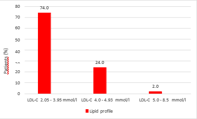
Figure 4: Distribution of patients by LDL-C levels at hospitalization, expressed as percentages:

Figure 5: Incoming lipid-lowering therapy by type of statin, expressed as a percentage.

Figure 6. Patients on ezetimibe monotherapy, combination therapy and no lipid-lowering therapy, expressed as a percentage:
In our prospective study, we demonstrated that with the help of artificial intelligence, the detection rate of patients with probable FH increased significantly and from 3 to 4.5 times patients were stared on PCSK9 inhibitor therapy.
It is noteworthy that in Cardiology clinics, the screening for FH is most successfully carried out. The reason for this may be due to the more frequent discussions of FH in the cardiology community and in cardiology departments compared to surgical specialties such as the Vascular Surgery and Cardiac Surgery Clinics. The non-analyzed patients in the Neurology Clinic appeared to be due to an unscreened lipid profile during hospitalization, which is a shortcoming of our analysis and perhaps needs to be changed as a strategy in this regard.
A disadvantage of our program that we find is that the dataset or AI algorithm is only available for use within our structure at the Hospitals of the Bulgarian Cardiology Institute and is limited to the hospital population level, making independent validation of the data not possible. Also, the presented analyses were conducted in a period when we did not monitor Lp a, as a more reliable marker for verifying patients with probable FH, as well as other markers for optimizing diagnosis. The role of the anti-aging gene, for example, Sirtuin 1, is crucial for the prevention of various chronic diseases such as diabetes and cardiovascular disease. It has been suggested that patients with FH should be evaluated for plasma levels of Sirtuin 1 in relation to insulin resistance and hepatic cholesterol metabolism. Sirtuin 1 activators versus Sirtuin 1 inhibitors could be evaluated in patients with FH (15).
The percentage of patients with concomitant diabetes mellitus type 2 (DM type 2) and hypercholesterolemia (30%) and arterial hypertension (66%) that we found were similar to those cited in the large study Da VINCI (38% for DM type 2 and 81% for arterial hypertension, respectively) associated with the use of lipid-lowering therapy in primary and secondary care in Central and Eastern Europe (CEE) (13). Da VINCI analyzed the proportion of patients from CEE who reached LDL-C targets based on individual cardiovascular risk recommended by the 2016 and 2019 ESC and European Atherosclerosis Society (EAS) guidelines. This study followed a total of 5,888 adults, of whom 2,154 were patients with dyslipidemia and lipid-lowering therapy in a CEE region believed to be undertreated, which is why this region of Europe has the highest cardiovascular mortality.
Patients from 18 European countries were included between June 21, 2017, and November 20, 2018, and for Eastern Europe, the Czech Republic (n=509), Hungary (n=319), Poland (n=460), Romania (n=259), Slovakia (n=123) and Ukraine (n=484). The researchers concluded that more than half of patients in CEE did not reach LDL-C target values, highlighting the significant gap between guidelines and clinical practice for dyslipidemias.
The results we achieved analyzing the patient profiles submitted by AI for FH screening are like those cited in Da VINCI. We also identified late-diagnosed FH patients with already overt atherosclerotic disease, who are untreated for dyslipidemia or insufficient therapy to effectively lower LDL-C (Figure 1, 2, 3). An additional impression, which we found in our work with patients with probable FH, is the lack of sufficient awareness on their part. Sometimes we saw an unjustified negative attitude of some patients and doctors towards the occurrence of side effects from taking statin therapy and the understanding of "adverse effects of high-dose statins". This was a frequent argument put forward by patients, why, despite an acute myocardial infarction, they are not optimal antilipemic therapy and why after optimizing LDL-C levels, they reduce the dose or even stop the drug. According to data from a retrospective study including patients with dyslipidemia followed for ≥3 years in a lipid clinic at a University Hospital in Greece, the frequency of clinically significant elevation of liver enzymes (>3× upper limit of normal (ULN) in 2) and statin-associated muscle symptoms (SAMS) expressed in muscle cramps, increased creatine kinase (CK) > 10 × ULN and rhabdomyolysis, during follow-up is low. Most of these patients were still able to tolerate statin treatment (14).
Unfortunately, in Bulgaria, a lack of criticality was also found among a significant number of doctors, mainly from outpatient practice and general practitioners, towards the pursuit of achieving target LDL-C levels and underestimation of patients with very high and high cardiovascular risk. Also, many patients stop taking statins and/or ezetimibe when treated with a PCSK9 inhibitor, which does not correspond to good clinical practice. These facts indicate the need for a broader discussion about FH, including the participation of both doctors and patients. Keeping a register for FH. We believe that the development of a family screening program for FH is necessary, as well as a rich informational awareness of patients. All this is related to the global change of the existing national strategy for patients with familial hypercholesterolemia.
Our ambition is to continue our research in this direction in the future. We are currently conducting a family screening program for patients identified by AI and treated for FH. We are working to expand the AI system to a larger number of healthcare facilities, with the aim of implementing integration with electronic health records, as well as application in primary care settings.
Unfortunately, potential obstacles that we would encounter are related to resource constraints and training requirements. Our goal is to continue using the program to identify patients with familial hypercholesterolemia in the future to achieve control of cardiovascular events. Our studies in this direction are ongoing. We have also added a routine serum Lp a test to the lipid panel. We are currently conducting a family screening program for patients identified by AI and treated for FH.
For our second outcomes – optimization of therapy, our conclusion is that the primary role is for the attending physician, to assess the patient's risk and follow the recommendations for behavior in dyslipidemias.
All these mentioned efforts, such as AI data analysis, screening, and proper treatment, will give us both an opportunity to recognize FH patients earlier and to implement cascade family screening. By reducing the rate of patients with cardiovascular events, the financial burden on health systems will be reduced and the quality of life of these patients will be improved, which is inextricably linked to a reduction in mortality in this population.
The development, implementation and use of AI for FH screening is workable in clinical practice and is easily applicable. The AI-based method identifies patients with probable FH. However, the role of the treating physician remains essential to make a correct diagnosis and initiate appropriate treatment, as well as to improve the patient's prognosis.
Clearly Auctoresonline and particularly Psychology and Mental Health Care Journal is dedicated to improving health care services for individuals and populations. The editorial boards' ability to efficiently recognize and share the global importance of health literacy with a variety of stakeholders. Auctoresonline publishing platform can be used to facilitate of optimal client-based services and should be added to health care professionals' repertoire of evidence-based health care resources.

Journal of Clinical Cardiology and Cardiovascular Intervention The submission and review process was adequate. However I think that the publication total value should have been enlightened in early fases. Thank you for all.

Journal of Women Health Care and Issues By the present mail, I want to say thank to you and tour colleagues for facilitating my published article. Specially thank you for the peer review process, support from the editorial office. I appreciate positively the quality of your journal.

Journal of Clinical Research and Reports I would be very delighted to submit my testimonial regarding the reviewer board and the editorial office. The reviewer board were accurate and helpful regarding any modifications for my manuscript. And the editorial office were very helpful and supportive in contacting and monitoring with any update and offering help. It was my pleasure to contribute with your promising Journal and I am looking forward for more collaboration.

We would like to thank the Journal of Thoracic Disease and Cardiothoracic Surgery because of the services they provided us for our articles. The peer-review process was done in a very excellent time manner, and the opinions of the reviewers helped us to improve our manuscript further. The editorial office had an outstanding correspondence with us and guided us in many ways. During a hard time of the pandemic that is affecting every one of us tremendously, the editorial office helped us make everything easier for publishing scientific work. Hope for a more scientific relationship with your Journal.

The peer-review process which consisted high quality queries on the paper. I did answer six reviewers’ questions and comments before the paper was accepted. The support from the editorial office is excellent.

Journal of Neuroscience and Neurological Surgery. I had the experience of publishing a research article recently. The whole process was simple from submission to publication. The reviewers made specific and valuable recommendations and corrections that improved the quality of my publication. I strongly recommend this Journal.

Dr. Katarzyna Byczkowska My testimonial covering: "The peer review process is quick and effective. The support from the editorial office is very professional and friendly. Quality of the Clinical Cardiology and Cardiovascular Interventions is scientific and publishes ground-breaking research on cardiology that is useful for other professionals in the field.

Thank you most sincerely, with regard to the support you have given in relation to the reviewing process and the processing of my article entitled "Large Cell Neuroendocrine Carcinoma of The Prostate Gland: A Review and Update" for publication in your esteemed Journal, Journal of Cancer Research and Cellular Therapeutics". The editorial team has been very supportive.

Testimony of Journal of Clinical Otorhinolaryngology: work with your Reviews has been a educational and constructive experience. The editorial office were very helpful and supportive. It was a pleasure to contribute to your Journal.

Dr. Bernard Terkimbi Utoo, I am happy to publish my scientific work in Journal of Women Health Care and Issues (JWHCI). The manuscript submission was seamless and peer review process was top notch. I was amazed that 4 reviewers worked on the manuscript which made it a highly technical, standard and excellent quality paper. I appreciate the format and consideration for the APC as well as the speed of publication. It is my pleasure to continue with this scientific relationship with the esteem JWHCI.

This is an acknowledgment for peer reviewers, editorial board of Journal of Clinical Research and Reports. They show a lot of consideration for us as publishers for our research article “Evaluation of the different factors associated with side effects of COVID-19 vaccination on medical students, Mutah university, Al-Karak, Jordan”, in a very professional and easy way. This journal is one of outstanding medical journal.

Dear Hao Jiang, to Journal of Nutrition and Food Processing We greatly appreciate the efficient, professional and rapid processing of our paper by your team. If there is anything else we should do, please do not hesitate to let us know. On behalf of my co-authors, we would like to express our great appreciation to editor and reviewers.

As an author who has recently published in the journal "Brain and Neurological Disorders". I am delighted to provide a testimonial on the peer review process, editorial office support, and the overall quality of the journal. The peer review process at Brain and Neurological Disorders is rigorous and meticulous, ensuring that only high-quality, evidence-based research is published. The reviewers are experts in their fields, and their comments and suggestions were constructive and helped improve the quality of my manuscript. The review process was timely and efficient, with clear communication from the editorial office at each stage. The support from the editorial office was exceptional throughout the entire process. The editorial staff was responsive, professional, and always willing to help. They provided valuable guidance on formatting, structure, and ethical considerations, making the submission process seamless. Moreover, they kept me informed about the status of my manuscript and provided timely updates, which made the process less stressful. The journal Brain and Neurological Disorders is of the highest quality, with a strong focus on publishing cutting-edge research in the field of neurology. The articles published in this journal are well-researched, rigorously peer-reviewed, and written by experts in the field. The journal maintains high standards, ensuring that readers are provided with the most up-to-date and reliable information on brain and neurological disorders. In conclusion, I had a wonderful experience publishing in Brain and Neurological Disorders. The peer review process was thorough, the editorial office provided exceptional support, and the journal's quality is second to none. I would highly recommend this journal to any researcher working in the field of neurology and brain disorders.
Dear Agrippa Hilda, Journal of Neuroscience and Neurological Surgery, Editorial Coordinator, I trust this message finds you well. I want to extend my appreciation for considering my article for publication in your esteemed journal. I am pleased to provide a testimonial regarding the peer review process and the support received from your editorial office. The peer review process for my paper was carried out in a highly professional and thorough manner. The feedback and comments provided by the authors were constructive and very useful in improving the quality of the manuscript. This rigorous assessment process undoubtedly contributes to the high standards maintained by your journal.

International Journal of Clinical Case Reports and Reviews. I strongly recommend to consider submitting your work to this high-quality journal. The support and availability of the Editorial staff is outstanding and the review process was both efficient and rigorous.

Thank you very much for publishing my Research Article titled “Comparing Treatment Outcome Of Allergic Rhinitis Patients After Using Fluticasone Nasal Spray And Nasal Douching" in the Journal of Clinical Otorhinolaryngology. As Medical Professionals we are immensely benefited from study of various informative Articles and Papers published in this high quality Journal. I look forward to enriching my knowledge by regular study of the Journal and contribute my future work in the field of ENT through the Journal for use by the medical fraternity. The support from the Editorial office was excellent and very prompt. I also welcome the comments received from the readers of my Research Article.

Dear Erica Kelsey, Editorial Coordinator of Cancer Research and Cellular Therapeutics Our team is very satisfied with the processing of our paper by your journal. That was fast, efficient, rigorous, but without unnecessary complications. We appreciated the very short time between the submission of the paper and its publication on line on your site.

I am very glad to say that the peer review process is very successful and fast and support from the Editorial Office. Therefore, I would like to continue our scientific relationship for a long time. And I especially thank you for your kindly attention towards my article. Have a good day!

"We recently published an article entitled “Influence of beta-Cyclodextrins upon the Degradation of Carbofuran Derivatives under Alkaline Conditions" in the Journal of “Pesticides and Biofertilizers” to show that the cyclodextrins protect the carbamates increasing their half-life time in the presence of basic conditions This will be very helpful to understand carbofuran behaviour in the analytical, agro-environmental and food areas. We greatly appreciated the interaction with the editor and the editorial team; we were particularly well accompanied during the course of the revision process, since all various steps towards publication were short and without delay".

I would like to express my gratitude towards you process of article review and submission. I found this to be very fair and expedient. Your follow up has been excellent. I have many publications in national and international journal and your process has been one of the best so far. Keep up the great work.

We are grateful for this opportunity to provide a glowing recommendation to the Journal of Psychiatry and Psychotherapy. We found that the editorial team were very supportive, helpful, kept us abreast of timelines and over all very professional in nature. The peer review process was rigorous, efficient and constructive that really enhanced our article submission. The experience with this journal remains one of our best ever and we look forward to providing future submissions in the near future.

I am very pleased to serve as EBM of the journal, I hope many years of my experience in stem cells can help the journal from one way or another. As we know, stem cells hold great potential for regenerative medicine, which are mostly used to promote the repair response of diseased, dysfunctional or injured tissue using stem cells or their derivatives. I think Stem Cell Research and Therapeutics International is a great platform to publish and share the understanding towards the biology and translational or clinical application of stem cells.

I would like to give my testimony in the support I have got by the peer review process and to support the editorial office where they were of asset to support young author like me to be encouraged to publish their work in your respected journal and globalize and share knowledge across the globe. I really give my great gratitude to your journal and the peer review including the editorial office.

I am delighted to publish our manuscript entitled "A Perspective on Cocaine Induced Stroke - Its Mechanisms and Management" in the Journal of Neuroscience and Neurological Surgery. The peer review process, support from the editorial office, and quality of the journal are excellent. The manuscripts published are of high quality and of excellent scientific value. I recommend this journal very much to colleagues.

Dr.Tania Muñoz, My experience as researcher and author of a review article in The Journal Clinical Cardiology and Interventions has been very enriching and stimulating. The editorial team is excellent, performs its work with absolute responsibility and delivery. They are proactive, dynamic and receptive to all proposals. Supporting at all times the vast universe of authors who choose them as an option for publication. The team of review specialists, members of the editorial board, are brilliant professionals, with remarkable performance in medical research and scientific methodology. Together they form a frontline team that consolidates the JCCI as a magnificent option for the publication and review of high-level medical articles and broad collective interest. I am honored to be able to share my review article and open to receive all your comments.

“The peer review process of JPMHC is quick and effective. Authors are benefited by good and professional reviewers with huge experience in the field of psychology and mental health. The support from the editorial office is very professional. People to contact to are friendly and happy to help and assist any query authors might have. Quality of the Journal is scientific and publishes ground-breaking research on mental health that is useful for other professionals in the field”.

Dear editorial department: On behalf of our team, I hereby certify the reliability and superiority of the International Journal of Clinical Case Reports and Reviews in the peer review process, editorial support, and journal quality. Firstly, the peer review process of the International Journal of Clinical Case Reports and Reviews is rigorous, fair, transparent, fast, and of high quality. The editorial department invites experts from relevant fields as anonymous reviewers to review all submitted manuscripts. These experts have rich academic backgrounds and experience, and can accurately evaluate the academic quality, originality, and suitability of manuscripts. The editorial department is committed to ensuring the rigor of the peer review process, while also making every effort to ensure a fast review cycle to meet the needs of authors and the academic community. Secondly, the editorial team of the International Journal of Clinical Case Reports and Reviews is composed of a group of senior scholars and professionals with rich experience and professional knowledge in related fields. The editorial department is committed to assisting authors in improving their manuscripts, ensuring their academic accuracy, clarity, and completeness. Editors actively collaborate with authors, providing useful suggestions and feedback to promote the improvement and development of the manuscript. We believe that the support of the editorial department is one of the key factors in ensuring the quality of the journal. Finally, the International Journal of Clinical Case Reports and Reviews is renowned for its high- quality articles and strict academic standards. The editorial department is committed to publishing innovative and academically valuable research results to promote the development and progress of related fields. The International Journal of Clinical Case Reports and Reviews is reasonably priced and ensures excellent service and quality ratio, allowing authors to obtain high-level academic publishing opportunities in an affordable manner. I hereby solemnly declare that the International Journal of Clinical Case Reports and Reviews has a high level of credibility and superiority in terms of peer review process, editorial support, reasonable fees, and journal quality. Sincerely, Rui Tao.

Clinical Cardiology and Cardiovascular Interventions I testity the covering of the peer review process, support from the editorial office, and quality of the journal.

Clinical Cardiology and Cardiovascular Interventions, we deeply appreciate the interest shown in our work and its publication. It has been a true pleasure to collaborate with you. The peer review process, as well as the support provided by the editorial office, have been exceptional, and the quality of the journal is very high, which was a determining factor in our decision to publish with you.

The peer reviewers process is quick and effective, the supports from editorial office is excellent, the quality of journal is high. I would like to collabroate with Internatioanl journal of Clinical Case Reports and Reviews journal clinically in the future time.

Clinical Cardiology and Cardiovascular Interventions, I would like to express my sincerest gratitude for the trust placed in our team for the publication in your journal. It has been a true pleasure to collaborate with you on this project. I am pleased to inform you that both the peer review process and the attention from the editorial coordination have been excellent. Your team has worked with dedication and professionalism to ensure that your publication meets the highest standards of quality. We are confident that this collaboration will result in mutual success, and we are eager to see the fruits of this shared effort.

Dear Dr. Jessica Magne, Editorial Coordinator 0f Clinical Cardiology and Cardiovascular Interventions, I hope this message finds you well. I want to express my utmost gratitude for your excellent work and for the dedication and speed in the publication process of my article titled "Navigating Innovation: Qualitative Insights on Using Technology for Health Education in Acute Coronary Syndrome Patients." I am very satisfied with the peer review process, the support from the editorial office, and the quality of the journal. I hope we can maintain our scientific relationship in the long term.

Dear Monica Gissare, - Editorial Coordinator of Nutrition and Food Processing. ¨My testimony with you is truly professional, with a positive response regarding the follow-up of the article and its review, you took into account my qualities and the importance of the topic¨.

Dear Dr. Jessica Magne, Editorial Coordinator 0f Clinical Cardiology and Cardiovascular Interventions, The review process for the article “The Handling of Anti-aggregants and Anticoagulants in the Oncologic Heart Patient Submitted to Surgery” was extremely rigorous and detailed. From the initial submission to the final acceptance, the editorial team at the “Journal of Clinical Cardiology and Cardiovascular Interventions” demonstrated a high level of professionalism and dedication. The reviewers provided constructive and detailed feedback, which was essential for improving the quality of our work. Communication was always clear and efficient, ensuring that all our questions were promptly addressed. The quality of the “Journal of Clinical Cardiology and Cardiovascular Interventions” is undeniable. It is a peer-reviewed, open-access publication dedicated exclusively to disseminating high-quality research in the field of clinical cardiology and cardiovascular interventions. The journal's impact factor is currently under evaluation, and it is indexed in reputable databases, which further reinforces its credibility and relevance in the scientific field. I highly recommend this journal to researchers looking for a reputable platform to publish their studies.

Dear Editorial Coordinator of the Journal of Nutrition and Food Processing! "I would like to thank the Journal of Nutrition and Food Processing for including and publishing my article. The peer review process was very quick, movement and precise. The Editorial Board has done an extremely conscientious job with much help, valuable comments and advices. I find the journal very valuable from a professional point of view, thank you very much for allowing me to be part of it and I would like to participate in the future!”

Dealing with The Journal of Neurology and Neurological Surgery was very smooth and comprehensive. The office staff took time to address my needs and the response from editors and the office was prompt and fair. I certainly hope to publish with this journal again.Their professionalism is apparent and more than satisfactory. Susan Weiner

My Testimonial Covering as fellowing: Lin-Show Chin. The peer reviewers process is quick and effective, the supports from editorial office is excellent, the quality of journal is high. I would like to collabroate with Internatioanl journal of Clinical Case Reports and Reviews.

My experience publishing in Psychology and Mental Health Care was exceptional. The peer review process was rigorous and constructive, with reviewers providing valuable insights that helped enhance the quality of our work. The editorial team was highly supportive and responsive, making the submission process smooth and efficient. The journal's commitment to high standards and academic rigor makes it a respected platform for quality research. I am grateful for the opportunity to publish in such a reputable journal.

My experience publishing in International Journal of Clinical Case Reports and Reviews was exceptional. I Come forth to Provide a Testimonial Covering the Peer Review Process and the editorial office for the Professional and Impartial Evaluation of the Manuscript.

I would like to offer my testimony in the support. I have received through the peer review process and support the editorial office where they are to support young authors like me, encourage them to publish their work in your esteemed journals, and globalize and share knowledge globally. I really appreciate your journal, peer review, and editorial office.

Dear Agrippa Hilda- Editorial Coordinator of Journal of Neuroscience and Neurological Surgery, "The peer review process was very quick and of high quality, which can also be seen in the articles in the journal. The collaboration with the editorial office was very good."

I would like to express my sincere gratitude for the support and efficiency provided by the editorial office throughout the publication process of my article, “Delayed Vulvar Metastases from Rectal Carcinoma: A Case Report.” I greatly appreciate the assistance and guidance I received from your team, which made the entire process smooth and efficient. The peer review process was thorough and constructive, contributing to the overall quality of the final article. I am very grateful for the high level of professionalism and commitment shown by the editorial staff, and I look forward to maintaining a long-term collaboration with the International Journal of Clinical Case Reports and Reviews.

To Dear Erin Aust, I would like to express my heartfelt appreciation for the opportunity to have my work published in this esteemed journal. The entire publication process was smooth and well-organized, and I am extremely satisfied with the final result. The Editorial Team demonstrated the utmost professionalism, providing prompt and insightful feedback throughout the review process. Their clear communication and constructive suggestions were invaluable in enhancing my manuscript, and their meticulous attention to detail and dedication to quality are truly commendable. Additionally, the support from the Editorial Office was exceptional. From the initial submission to the final publication, I was guided through every step of the process with great care and professionalism. The team's responsiveness and assistance made the entire experience both easy and stress-free. I am also deeply impressed by the quality and reputation of the journal. It is an honor to have my research featured in such a respected publication, and I am confident that it will make a meaningful contribution to the field.

"I am grateful for the opportunity of contributing to [International Journal of Clinical Case Reports and Reviews] and for the rigorous review process that enhances the quality of research published in your esteemed journal. I sincerely appreciate the time and effort of your team who have dedicatedly helped me in improvising changes and modifying my manuscript. The insightful comments and constructive feedback provided have been invaluable in refining and strengthening my work".

I thank the ‘Journal of Clinical Research and Reports’ for accepting this article for publication. This is a rigorously peer reviewed journal which is on all major global scientific data bases. I note the review process was prompt, thorough and professionally critical. It gave us an insight into a number of important scientific/statistical issues. The review prompted us to review the relevant literature again and look at the limitations of the study. The peer reviewers were open, clear in the instructions and the editorial team was very prompt in their communication. This journal certainly publishes quality research articles. I would recommend the journal for any future publications.

Dear Jessica Magne, with gratitude for the joint work. Fast process of receiving and processing the submitted scientific materials in “Clinical Cardiology and Cardiovascular Interventions”. High level of competence of the editors with clear and correct recommendations and ideas for enriching the article.

We found the peer review process quick and positive in its input. The support from the editorial officer has been very agile, always with the intention of improving the article and taking into account our subsequent corrections.

My article, titled 'No Way Out of the Smartphone Epidemic Without Considering the Insights of Brain Research,' has been republished in the International Journal of Clinical Case Reports and Reviews. The review process was seamless and professional, with the editors being both friendly and supportive. I am deeply grateful for their efforts.

To Dear Erin Aust – Editorial Coordinator of Journal of General Medicine and Clinical Practice! I declare that I am absolutely satisfied with your work carried out with great competence in following the manuscript during the various stages from its receipt, during the revision process to the final acceptance for publication. Thank Prof. Elvira Farina

Dear Jessica, and the super professional team of the ‘Clinical Cardiology and Cardiovascular Interventions’ I am sincerely grateful to the coordinated work of the journal team for the no problem with the submission of my manuscript: “Cardiometabolic Disorders in A Pregnant Woman with Severe Preeclampsia on the Background of Morbid Obesity (Case Report).” The review process by 5 experts was fast, and the comments were professional, which made it more specific and academic, and the process of publication and presentation of the article was excellent. I recommend that my colleagues publish articles in this journal, and I am interested in further scientific cooperation. Sincerely and best wishes, Dr. Oleg Golyanovskiy.

Dear Ashley Rosa, Editorial Coordinator of the journal - Psychology and Mental Health Care. " The process of obtaining publication of my article in the Psychology and Mental Health Journal was positive in all areas. The peer review process resulted in a number of valuable comments, the editorial process was collaborative and timely, and the quality of this journal has been quickly noticed, resulting in alternative journals contacting me to publish with them." Warm regards, Susan Anne Smith, PhD. Australian Breastfeeding Association.

Dear Jessica Magne, Editorial Coordinator, Clinical Cardiology and Cardiovascular Interventions, Auctores Publishing LLC. I appreciate the journal (JCCI) editorial office support, the entire team leads were always ready to help, not only on technical front but also on thorough process. Also, I should thank dear reviewers’ attention to detail and creative approach to teach me and bring new insights by their comments. Surely, more discussions and introduction of other hemodynamic devices would provide better prevention and management of shock states. Your efforts and dedication in presenting educational materials in this journal are commendable. Best wishes from, Farahnaz Fallahian.

Dear Maria Emerson, Editorial Coordinator, International Journal of Clinical Case Reports and Reviews, Auctores Publishing LLC. I am delighted to have published our manuscript, "Acute Colonic Pseudo-Obstruction (ACPO): A rare but serious complication following caesarean section." I want to thank the editorial team, especially Maria Emerson, for their prompt review of the manuscript, quick responses to queries, and overall support. Yours sincerely Dr. Victor Olagundoye.

Dear Ashley Rosa, Editorial Coordinator, International Journal of Clinical Case Reports and Reviews. Many thanks for publishing this manuscript after I lost confidence the editors were most helpful, more than other journals Best wishes from, Susan Anne Smith, PhD. Australian Breastfeeding Association.

Dear Agrippa Hilda, Editorial Coordinator, Journal of Neuroscience and Neurological Surgery. The entire process including article submission, review, revision, and publication was extremely easy. The journal editor was prompt and helpful, and the reviewers contributed to the quality of the paper. Thank you so much! Eric Nussbaum, MD

Dr Hala Al Shaikh This is to acknowledge that the peer review process for the article ’ A Novel Gnrh1 Gene Mutation in Four Omani Male Siblings, Presentation and Management ’ sent to the International Journal of Clinical Case Reports and Reviews was quick and smooth. The editorial office was prompt with easy communication.

Dear Erin Aust, Editorial Coordinator, Journal of General Medicine and Clinical Practice. We are pleased to share our experience with the “Journal of General Medicine and Clinical Practice”, following the successful publication of our article. The peer review process was thorough and constructive, helping to improve the clarity and quality of the manuscript. We are especially thankful to Ms. Erin Aust, the Editorial Coordinator, for her prompt communication and continuous support throughout the process. Her professionalism ensured a smooth and efficient publication experience. The journal upholds high editorial standards, and we highly recommend it to fellow researchers seeking a credible platform for their work. Best wishes By, Dr. Rakhi Mishra.

Dear Jessica Magne, Editorial Coordinator, Clinical Cardiology and Cardiovascular Interventions, Auctores Publishing LLC. The peer review process of the journal of Clinical Cardiology and Cardiovascular Interventions was excellent and fast, as was the support of the editorial office and the quality of the journal. Kind regards Walter F. Riesen Prof. Dr. Dr. h.c. Walter F. Riesen.

Dear Ashley Rosa, Editorial Coordinator, International Journal of Clinical Case Reports and Reviews, Auctores Publishing LLC. Thank you for publishing our article, Exploring Clozapine's Efficacy in Managing Aggression: A Multiple Single-Case Study in Forensic Psychiatry in the international journal of clinical case reports and reviews. We found the peer review process very professional and efficient. The comments were constructive, and the whole process was efficient. On behalf of the co-authors, I would like to thank you for publishing this article. With regards, Dr. Jelle R. Lettinga.

Dear Clarissa Eric, Editorial Coordinator, Journal of Clinical Case Reports and Studies, I would like to express my deep admiration for the exceptional professionalism demonstrated by your journal. I am thoroughly impressed by the speed of the editorial process, the substantive and insightful reviews, and the meticulous preparation of the manuscript for publication. Additionally, I greatly appreciate the courteous and immediate responses from your editorial office to all my inquiries. Best Regards, Dariusz Ziora

Dear Chrystine Mejia, Editorial Coordinator, Journal of Neurodegeneration and Neurorehabilitation, Auctores Publishing LLC, We would like to thank the editorial team for the smooth and high-quality communication leading up to the publication of our article in the Journal of Neurodegeneration and Neurorehabilitation. The reviewers have extensive knowledge in the field, and their relevant questions helped to add value to our publication. Kind regards, Dr. Ravi Shrivastava.

Dear Clarissa Eric, Editorial Coordinator, Journal of Clinical Case Reports and Studies, Auctores Publishing LLC, USA Office: +1-(302)-520-2644. I would like to express my sincere appreciation for the efficient and professional handling of my case report by the ‘Journal of Clinical Case Reports and Studies’. The peer review process was not only fast but also highly constructive—the reviewers’ comments were clear, relevant, and greatly helped me improve the quality and clarity of my manuscript. I also received excellent support from the editorial office throughout the process. Communication was smooth and timely, and I felt well guided at every stage, from submission to publication. The overall quality and rigor of the journal are truly commendable. I am pleased to have published my work with Journal of Clinical Case Reports and Studies, and I look forward to future opportunities for collaboration. Sincerely, Aline Tollet, UCLouvain.

Dear Ms. Mayra Duenas, Editorial Coordinator, International Journal of Clinical Case Reports and Reviews. “The International Journal of Clinical Case Reports and Reviews represented the “ideal house” to share with the research community a first experience with the use of the Simeox device for speech rehabilitation. High scientific reputation and attractive website communication were first determinants for the selection of this Journal, and the following submission process exceeded expectations: fast but highly professional peer review, great support by the editorial office, elegant graphic layout. Exactly what a dynamic research team - also composed by allied professionals - needs!" From, Chiara Beccaluva, PT - Italy.

Dear Maria Emerson, Editorial Coordinator, we have deeply appreciated the professionalism demonstrated by the International Journal of Clinical Case Reports and Reviews. The reviewers have extensive knowledge of our field and have been very efficient and fast in supporting the process. I am really looking forward to further collaboration. Thanks. Best regards, Dr. Claudio Ligresti

Dear Chrystine Mejia, Editorial Coordinator, Journal of Neurodegeneration and Neurorehabilitation. “The peer review process was efficient and constructive, and the editorial office provided excellent communication and support throughout. The journal ensures scientific rigor and high editorial standards, while also offering a smooth and timely publication process. We sincerely appreciate the work of the editorial team in facilitating the dissemination of innovative approaches such as the Bonori Method.” Best regards, Dr. Matteo Bonori.

I recommend without hesitation submitting relevant papers on medical decision making to the International Journal of Clinical Case Reports and Reviews. I am very grateful to the editorial staff. Maria Emerson was a pleasure to communicate with. The time from submission to publication was an extremely short 3 weeks. The editorial staff submitted the paper to three reviewers. Two of the reviewers commented positively on the value of publishing the paper. The editorial staff quickly recognized the third reviewer’s comments as an unjust attempt to reject the paper. I revised the paper as recommended by the first two reviewers.

Dear Maria Emerson, Editorial Coordinator, Journal of Clinical Research and Reports. Thank you for publishing our case report: "Clinical Case of Effective Fetal Stem Cells Treatment in a Patient with Autism Spectrum Disorder" within the "Journal of Clinical Research and Reports" being submitted by the team of EmCell doctors from Kyiv, Ukraine. We much appreciate a professional and transparent peer-review process from Auctores. All research Doctors are so grateful to your Editorial Office and Auctores Publishing support! I amiably wish our article publication maintained a top quality of your International Scientific Journal. My best wishes for a prosperity of the Journal of Clinical Research and Reports. Hope our scientific relationship and cooperation will remain long lasting. Thank you very much indeed. Kind regards, Dr. Andriy Sinelnyk Cell Therapy Center EmCell

Dear Editorial Team, Clinical Cardiology and Cardiovascular Interventions. It was truly a rewarding experience to work with the journal “Clinical Cardiology and Cardiovascular Interventions”. The peer review process was insightful and encouraging, helping us refine our work to a higher standard. The editorial office offered exceptional support with prompt and thoughtful communication. I highly value the journal’s role in promoting scientific advancement and am honored to be part of it. Best regards, Meng-Jou Lee, MD, Department of Anesthesiology, National Taiwan University Hospital.

Dear Editorial Team, Journal-Clinical Cardiology and Cardiovascular Interventions, “Publishing my article with Clinical Cardiology and Cardiovascular Interventions has been a highly positive experience. The peer-review process was rigorous yet supportive, offering valuable feedback that strengthened my work. The editorial team demonstrated exceptional professionalism, prompt communication, and a genuine commitment to maintaining the highest scientific standards. I am very pleased with the publication quality and proud to be associated with such a reputable journal.” Warm regards, Dr. Mahmoud Kamal Moustafa Ahmed

Dear Maria Emerson, Editorial Coordinator of ‘International Journal of Clinical Case Reports and Reviews’, I appreciate the opportunity to publish my article with your journal. The editorial office provided clear communication during the submission and review process, and I found the overall experience professional and constructive. Best regards, Elena Salvatore.

Dear Mayra Duenas, Editorial Coordinator of ‘International Journal of Clinical Case Reports and Reviews Herewith I confirm an optimal peer review process and a great support of the editorial office of the present journal

Dear Editorial Team, Clinical Cardiology and Cardiovascular Interventions. I am really grateful for the peers review; their feedback gave me the opportunity to reflect on the message and impact of my work and to ameliorate the article. The editors did a great job in addition by encouraging me to continue with the process of publishing.

Dear Cecilia Lilly, Editorial Coordinator, Endocrinology and Disorders, Thank you so much for your quick response regarding reviewing and all process till publishing our manuscript entitled: Prevalence of Pre-Diabetes and its Associated Risk Factors Among Nile College Students, Sudan. Best regards, Dr Mamoun Magzoub.

International Journal of Clinical Case Reports and Reviews is a high quality journal that has a clear and concise submission process. The peer review process was comprehensive and constructive. Support from the editorial office was excellent, since the administrative staff were responsive. The journal provides a fast and timely publication timeline.

Dear Maria Emerson, Editorial Coordinator of International Journal of Clinical Case Reports and Reviews, What distinguishes International Journal of Clinical Case Report and Review is not only the scientific rigor of its publications, but the intellectual climate in which research is evaluated. The submission process is refreshingly free of unnecessary formal barriers and bureaucratic rituals that often complicate academic publishing without adding real value. The peer-review system is demanding yet constructive, guided by genuine scientific dialogue rather than hierarchical or authoritarian attitudes. Reviewers act as collaborators in improving the manuscript, not as gatekeepers imposing arbitrary standards. This journal offers a rare balance: high methodological standards combined with a respectful, transparent, and supportive editorial approach. In an era where publishing can feel more burdensome than research itself, this platform restores the original purpose of peer review — to refine ideas, not to obstruct them Prof. Perlat Kapisyzi, FCCP PULMONOLOGIST AND THORACIC IMAGING.

Dear Mayra Duenas, Editorial Coordinator of the journal IJCCR, I write here a little on my experience as an author submitting to the International Journal of Clinical Case Reports and Reviews (IJCCR). This was my first submission to IJCCR and my manuscript was inherently an outsider’s effort. It attempted to broadly identify and then make some sense of life’s under-appreciated mysteries. I initially had responded to a request for possible submissions. I then contacted IJCCR with a tentative topic for a manuscript. They quickly got back with an approval for the submission, but with a particular requirement that it be medically relevant. I then put together a manuscript and submitted it. After the usual back-and-forth over forms and formality, the manuscript was sent off for reviews. Within 2 weeks I got back 4 reviews which were both helpful and also surprising. Surprising in that the topic was somewhat foreign to medical literature. My subsequent updates in response to the reviewer comments went smoothly and in short order I had a series of proofs to evaluate. All in all, the whole publication process seemed outstanding. It was both helpful in terms of the paper’s content and also in terms of its efficient and friendly communications. Thank you all very much. Sincerely, Ted Christopher, Rochester, NY.

Dear Grace Pierce, Editorial Coordinator of the journal IJCCR, I had a very positive experience with Auctores - Journal throughout the publication process. The Editorial Team was highly responsive, professional, and supportive at every stage. I would like to extend my sincere thanks to the Editor: Grace Pierce, for her guidance and assistance. The peer-review process was smooth and constructive, helping improve the quality of my work. I would gladly recommend Auctores Journal to fellow researchers and authors. Dr. SABITA SINHA, Medical Oncologist, MD (Electro Homeopathy).

Dear Maria Emerson, Editorial Coordinator of - Journal of Clinical Research and Reports. ''I am pleased to provide this testimonial following the publication of our recent case report in this journal. The peer review process was rigorous, constructive, thorough, and conducted in a timely manner. The reviewers’ comments were thoughtful, detailed, and highly constructive, contributing substantially to the refinement, clarity, and scientific robustness of our manuscript. The process was conducted with professionalism and academic integrity throughout. The support provided by the editorial office was exemplary. Communication was consistently prompt, clear, and courteous at all stages of the submission and publication process. The editorial team demonstrated a high level of organization and responsiveness, ensuring that all queries were addressed efficiently and that the process remained transparent and well-coordinated. The overall quality of the journal is reflected in its strong editorial standards, commitment to scientific excellence, and dedication to publishing clinically meaningful research. It has been a privilege to publish our work in this journal, and we would welcome the opportunity to contribute further in the future.'' Best wishes from, Dr. Trogkanis Efstratios, Cardiologist.