
Case Report | DOI: https://doi.org/10.31579/2690-4861/027
*Corresponding Author: Ravi Kumar Chittoria, Department of Plastic Surgery Jawaharlal Institute of Postgraduate Medical Education and Research (JIPMER) Pondicherry India
Citation: Neljo T, Ravi K, Saurabh G, Chirra L R, Padmalakshmi B. (2020) Role of topical insulin in electric burns management. International Journal of Clinical Case Reports and Reviews. 2(3); DOI: 10.31579/2690-4861/027
Copyright: © 2020 Ravi Kumar Chittoria, This is an open-access article distributed under the terms of the Creative Commons Attribution License, which permits unrestricted use, distribution, and reproduction in any medium, provided the original author and source are credited.
Received: 25 May 2020 | Accepted: 12 June 2020 | Published: 18 June 2020
Keywords: insulin therapy, Electric burns
Electric burns are known for non-healing and plastic surgeons face with difficulty in wound management. In chronic wounds there will be a lack of growth factors. Insulin therapy is a method of wound healing by stimulation of growth factors and provide growth factors for wound healing. In this study we have used topical insulin for wound bed preparation of patient’s electric burns wound.
Adult wound healing can be categorized into three stages: inflammatory phase, proliferative phase, and remodelling phase. The three stages have to occur in correct conjunction to result in good wound healing. Wound bed preparation is a novel concept which is used to manage wounds that fail to heal by itself and can be summarized using T.I.M.E with T for tissue: non-viable or deficient. I for infection/inflammation, M for moisture balance. E for epidermis which was changed to E for edge.Recently in literature, we happened to come across use of topical insulin for use in wound bed preparation.
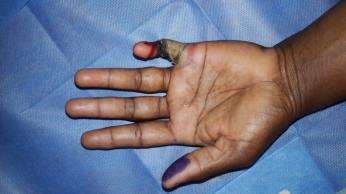
Materials and methods: This study was conducted in the department of Plastic Surgery at tertiary care center after the departmental ethical committee approval was taken. Informed written consent was taken from the patient. The details of the patient in study are as follows: 40 year old female without any known co morbidities with history of accidental electric burns from low voltage source and sustained circumferential 3rd to 4th degree burns over the left little finger with loss of vascularity of the distal area and 2nd degree burns over the medial aspect of the ring finger in the proximal phalanx (figure 1).
Patient underwent little finger disarticulation after 1 week when the line of demarcation was developed and edges were loosely approximated to facilitate healing (figure 2).

Following the procedure patient was dressed regularly. She developed a raw area of 3.5cm2 over the medial aspect of the ring finger which failed to show any evidence of healing. Wound bed preparation was done for the patient with topical insulin therapy as her ulcer did not show any evidence of healing. 0.2ml of regular insulin was injected onto the border and 0.15ml was sprayed on the floor onto 3.5cm2 of the wound when the regular dressing was performed (figure 3).

This was repeated once every 3-5 days when the dressing was changed .The wound was reassessed after 2weeks for evidence of wound healing.
Results: The wound bed showed good granulation tissue after 2weeks (figure 4)

Discussion
Burn injury is a major cause of trauma to the human body, causing death as well as disability, with a long healing period. The mortality rate of burn injury has decreased with new treatment modalities, but secondary infections and prolonged healing periods still affect the mortality rates.For this purpose, different kinds of dressings and pharmacotherapies have been developed, but most are costly, and the mechanisms underlying these therapies have not been fully documented. Many therapeutic methods are available to effect the wound healing such as the topical application of insulin, growth factors, negative pressure assisted wound closure, oxidized regenerated cellulose/collagen, hyaluronic acid conjugated with glycidyl methacrylate or gelatin dressings.
Role of topical insulin in wound healing has been in literature since 1970s [1]. Studies done in animal models and humans have found that topical Insulin therapy exerts its effects through the IGF 1 receptor. Insulin is known to stimulate keratinocytes and also the rate of endothelial proliferation leading to a faster neovascularization and also formation of granulation tissue [2]. Topical insulin application rapidly induces upregulation of the insulin signalling related proteins on wound areas following injury. IRS-1 binds the PI3-kinase, one of the SH2 proteins, through the multiple tyrosine phosphorylated sites [3, 4].
Insulin triggers the keratinocyte migration depending on the dose and time in the chronic wounds. Insulin demonstrates its effect through an insulin receptor dependent but EGF/EGF-R non-dependent way. The fact that it increases the keratinocyte migration on the PI3K-Akt-Rac1 pathway and cause stimulation of the keratinocytes by enabling the production of α3 and LN332 molecules has been proven by the in vitro studies [6,7,8,9]. Insulin is a hormone that also affects collagen production. Insulin selectively and strongly stimulates the collagen production in dermal fibroblasts [10, 11].The cost of insulin ranges from 130-500 INR per vial.There was no systemic side effects for the insulin being given topically.
Limitations: The study was done on a single patient and needs large population based study to apply in practice
DECLARATIONS
Acknowledgment
Authors’ contributions
All authors made contributions to the article
Availability of data and materials
Not applicable.
Financial support and sponsorship
None.
Conflicts of interest
None.
Consent for publication
Not applicable.
Copyright© The Author(s) 2020.
Dear Editorial Team, Clinical Medical Reviews and Reports. My experience with the journal was highly positive. The peer-review process was rigorous, constructive, and completed in a timely manner. The reviewers provided valuable comments that helped improve the quality and clarity of our manuscript. The editorial office was professional, responsive, and supportive throughout all stages of the publication process. Communication was clear and efficient, and any questions were addressed promptly. Overall, I found the journal to maintain high scientific standards and an excellent publication workflow. I would be pleased to consider submitting future work to this journal. Best wishes from, Elena Popa.

It was my pleasure to submit my testimonial concerning the Reviewer Board of our Scientific Journal “Brain and Neurological Disorders”. The Reviewers focused on some modifications and their contribution was helpful. The ladies of our Editorial Office were also supported my efforts. It was my honor to have such a co-operation and I am looking forward for more collaboration.

Dear Grace Pierce, Editorial Coordinator of Journal of Clinical Research and Reports, Thank you for the speedy and efficient peer review process. I appreciate the fact that your peer reviewers do not take months to respond like with some other journals. I would also like to thank the editorial office for responding quickly to my questions. It is an excellent journal. I plan to submit more manuscripts in the future. Best wishes from, Robert W. McGee

Dear Grace Pierce, Editorial Coordinator of Journal of Clinical Research and Reports, Working with you and your team on our recent publication in JCRR has been a truly wonderful and enjoyable experience. The responses were prompt, and the reviewers were patient, constructive, and highly professional. One reviewer in particular gave me the feeling that a professor was carefully reading and commenting on my coursework, which was deeply touching. The entire process was straightforward and hassle‑free, with no tedious online forms to complete. I highly recommend this journal. Best wishes from, DR Aibing Rao, Head of R&D

I Appreciate the Opportunity to Share my Experience with the Journal of Clinical Research and Reports. The peer review process was timely and constructive, and the feedback provided helped improve the quality of our manuscript. The editorial office was professional, responsive, and supportive throughout the process, ensuring smooth communication and efficient handling of the submission. Overall, it was a positive experience collaborating with your team.

Dear Mercy Grace, Editorial Coordinator of Obstetrics Gynecology and Reproductive Sciences, We would like to express our gratitude for your help at all stages of publishing and editing the article. The editors of the magazine answer all the necessary questions and help at every stage. We will definitely continue to cooperate and publish other works in the Obstetrics Gynecology and Reproductive Sciences! Best wishes from, Alla Konstantinovna Politova,
