
Research Article | DOI: https://doi.org/10.31579/2690-8794/004
*Corresponding Author: Ravi K Chittoria, Department of Plastic Surgery, Jawaharlal Institute of Postgraduate Medical Education and Research (JIPMER), Pondicherry, India.
Citation: Ravi K Chittoria, Chirra L Reddy, Abhinav A, Saurabh G, Padma Lakshmi B Mohan, Shijina K, Imran P, 2(1); DOI: 10.31579/2690-8794/004
Copyright: © 2020 Ravi K Chittoria. This is an open access article distributed under the Creative Commons Attribution License, which permits unrestricted use, distribution, and reproduction in any medium, provided the originalworkisproperlycited
Received: 10 January 2020 | Accepted: 20 January 2020 | Published: 28 January 2020
Keywords: lymphoedema, chitosan, chronic wounds
Lymphoedema is a chronic problem with various skin changes that lead to impaired care for the affected limb. These skin changes also lead to recurrent infections. The difficulty in care and the subsequent neglect leads to further spread of the infection and increased risk of future infections. Prevention of infections and prevention of progress of the skin changes is one of the main components of the treatment of the lymphoedema. There are various materials available to enhance the healing of the knobs and fissures secondary to lymphoedema. We would like to present our case report on the usage of chitosan in a patient with stage 7 skin changes of lymphedema.
Lymphedema is a devastating problem both for the patient and society[1]. India has the highest number of patients with lymphedema of infective etiology. The majority of these patients are from low socioeconomic status and usually present in advanced stages of the disease stage 6-7 according to WHO[2] or stage 3 according to the International Society of lymphology [ISL]. These stages have significant skin changes with a higher risk of infection.
According to the Fifth WHO Expert Committee on Filariasis, lymphoedema can be classified as follows:
1.Grade I lymphoedema: mostly pitting edema; spontaneously reversible on elevation.
2.Grade II lymphoedema: mostly non-pitting edema; not spontaneously reversible on elevation.
3.Grade III lymphoedema (elephantiasis): a gross increase in volume in a grade II lymphoedema, with dermatoscelerosis and papillomatous lesions.
The 7 stages of classification as given below are clinically based
Stage 1
The feature of stage 1 lymphoedema is:
Swelling is reversible overnight.
In stage 1 lymphoedema, the swelling increases during the day and disappears
overnight,
Stage 2 Swelling is not reversible overnight.
The main difference between stage 2 lymphoedema and stage 1 is that the swelling does not disappear without lymphoedema management.
Stage 3 Swelling is not reversible overnight. The principal feature of stage 3 lymphoedema is the presence of one or more shallow skin folds
Stage 4 Swelling is not reversible overnight.
The main feature of stage 4 lymphoedema is the presence of knobs lumps, lumps, or protrusions on the skin.
Stage 5 Swelling is not reversible overnight.
The presence of one or more deep skin folds is the main feature of stage 5 lymphoedema
Stage 6
Swelling is not reversible overnight. On the surface of the foot (especially the toes), very small elongated or rounded small knobs can be clustered together, giving rise to the peculiar appearance of “mossy foot”.
Patients with stage 6 lymphoedema) have acute attacks. Almost all these patients
have entry lesions between the toes and bad odour. Wounds in the skin are frequently present.
Stage 7
The features of stage 7 lymphoedema are:
Swelling is not reversible overnight. The patient is unable to adequately or
independently perform routine daily activities such as walking, bathing, cooking, etc.
Lymphoedema treatment requires a multimodal approach including non-operative management and surgical management
Non-operative management includes maintenance of local hygiene, prevention and cure of entry lesions, elevation, exercise, compression bandaging, complex decongestive therapy, low-level laser therapy etc.
Surgical management includes reconstructive procedures like nodo-venous shunts, vascularised nodal transfer and debulking procedures like liposuction, Thompson procedure, Charles procedure.
However, prior to any definitive intervention, the infection and possible sites of entry of the secondary infections have to be controlled. One such attempt is reported in this article as the use of Chitosan in an advanced case of lymphedema.
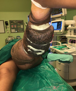
This is a case report of a 29yr female, from Kerala with h/o swelling of the left lower limb since 16 years was admitted in our centre in the month of August 2019.
On assessment, patient had chronic primary lymphedema with secondary grade 7 skin changes [WHO grading] [figure 1].

With multiple fissures over the affected limb. She also had foul smelling serous discharge form the limb. Patient underwent three application of chitosan[figure 2]
Along with a multimodal treatment of the affected limb including regular limb washes, limb elevation, compression dressings, over a period of 10 days following which the inspection of the limb revealed complete healing of the superficial ulcerations with no further discharge from the wounds [figure 3].

Chronic lymphedema is a significant problem with limited treatment options. Lymphedema occurs clinically when the capacity of the lymphatic system to drain protein-rich interstitial fluid is exceeded due to blockage or absence of lymphatic vessels. Lymphedema can occur as a hereditary congenital disorder (primary lymphedema) or as an acquired disorder (secondary lymphedema) as a result of injury, surgery, malignancy, or infection. It is defined as chronic when the condition persists for more than three to six months [3]. Worldwide, the most common cause of this condition is infection with parasitic nematode worms, commonly referred to as filariasis [4]. Patients with extremity lymphedema are at increased risk for soft tissue infections, impairment of limb function, and lymphangiosarcoma, a rare malignancy with an abysmal prognosis [5].
One of the major causes of infection is the difficulty in accessing those areas of the limb where there are significant skin changes including fissure formation, ulceration through which entry of microorganisms occurs. In the presence of infection, no active intervention for the treatment of the disease may be done.
The infection leads to further thickening of the skin. This goes on as a vicious cycle.
To treat ulceration and fissure various dressing materials are used along with the maintenance of hygiene. One of the dressing materials that is very promising is Chitosan due to its various unique properties.
Chitosan is the second most available bio polymer after cellulose derived from shrimp shell. It is a linear polysaccharide composed of β--linked D-glucosamine and N- acetyl-D-glucosamine[6]. It has various modes of action include aiding in wound healing[6], as a drug carrier[7]. The uses are not just limited to use in the biomedical field. There are various studies that have shown its use in agriculture, horticulture, as catalyst, electronics, non-linear optics [8].
Chitosan’s properties of binding with red blood cells allow it to rapidly clot blood, and it has recently gained regulatory approval in the USA for use in bandages and other hemostatic agents [9, 10]. In addition, chitosan modulates the functions of inflammatory cells and subsequently promotes granulation and organization [11]. As a semipermeable biological dressing, it maintains a sterile wound exudate beneath a dry scab, preventing dehydration and contamination of the wound, to optimize conditions for healing. Furthermore, chitosan is a polymer with a number of basic amino groups and hence possesses an overall cationic charge, especially at acidic pH. Chitosan has pronounced antimicrobial effects due to destabilization of the outer membrane of gram-negative bacteria and permeabilization of the microbial plasma membrane.
In a study conducted by Sudheesh et al it the chitin/nanosilver composite scaffolds were found to be anti-bactericidal against E. coli and S. aureus and showed good blood clotting ability as well. In addition, -chitin/nanosilver composite scaffolds were evaluated for their cell adhesion properties using epithelial cells (Vero cells)[12].
Our use of chitosan has shown good improvement in the amount of discharge from the lesions and the healing of the fissures. This has further reduced the risk of secondary infection by helping in the treatment of the raw area[13] and also its effect on micro-organisms which possibly prevented further infection.
Chitosan may be used as part of the multimodal treatment of lymphoedema. However further research is needed to understand the exact mechanism of action of chitosan in lymphedema.
Ours is a single case study and hence, further larger studies are required to definitively establish the use of chitosan as a part of the armamentarium available for the treatment of lymphoedema.
Dear Editorial Team, Clinical Medical Reviews and Reports. My experience with the journal was highly positive. The peer-review process was rigorous, constructive, and completed in a timely manner. The reviewers provided valuable comments that helped improve the quality and clarity of our manuscript. The editorial office was professional, responsive, and supportive throughout all stages of the publication process. Communication was clear and efficient, and any questions were addressed promptly. Overall, I found the journal to maintain high scientific standards and an excellent publication workflow. I would be pleased to consider submitting future work to this journal. Best wishes from, Elena Popa.

It was my pleasure to submit my testimonial concerning the Reviewer Board of our Scientific Journal “Brain and Neurological Disorders”. The Reviewers focused on some modifications and their contribution was helpful. The ladies of our Editorial Office were also supported my efforts. It was my honor to have such a co-operation and I am looking forward for more collaboration.

Dear Grace Pierce, Editorial Coordinator of Journal of Clinical Research and Reports, Thank you for the speedy and efficient peer review process. I appreciate the fact that your peer reviewers do not take months to respond like with some other journals. I would also like to thank the editorial office for responding quickly to my questions. It is an excellent journal. I plan to submit more manuscripts in the future. Best wishes from, Robert W. McGee

Dear Grace Pierce, Editorial Coordinator of Journal of Clinical Research and Reports, Working with you and your team on our recent publication in JCRR has been a truly wonderful and enjoyable experience. The responses were prompt, and the reviewers were patient, constructive, and highly professional. One reviewer in particular gave me the feeling that a professor was carefully reading and commenting on my coursework, which was deeply touching. The entire process was straightforward and hassle‑free, with no tedious online forms to complete. I highly recommend this journal. Best wishes from, DR Aibing Rao, Head of R&D

I Appreciate the Opportunity to Share my Experience with the Journal of Clinical Research and Reports. The peer review process was timely and constructive, and the feedback provided helped improve the quality of our manuscript. The editorial office was professional, responsive, and supportive throughout the process, ensuring smooth communication and efficient handling of the submission. Overall, it was a positive experience collaborating with your team.

Dear Mercy Grace, Editorial Coordinator of Obstetrics Gynecology and Reproductive Sciences, We would like to express our gratitude for your help at all stages of publishing and editing the article. The editors of the magazine answer all the necessary questions and help at every stage. We will definitely continue to cooperate and publish other works in the Obstetrics Gynecology and Reproductive Sciences! Best wishes from, Alla Konstantinovna Politova,
