
Review Article | DOI: https://doi.org/10.31579/2641-0419/171Copyright
*Corresponding Author: P. Syamasundar Rao, MD, Professor and Emeritus Chief of Pediatric Cardiology, UT-Houston McGovern Medical School, 6410 Fannin, UTPB Suite # 425, Houston, TX. 77030. Phone: 713-500-5738; Fax: 713-500-5171.
Citation: Rao PS (2021) Role of Balloon Aortic Valvuloplasty in the Management of Aortic Stenosis. J. Clinical Cardiology and Cardiovascular Interventions, 4(12); Doi:10.31579/2641-0419/171
Copyright: © 2021 P. Syamasundar Rao, This is an open-access article distributed under the terms of the Creative Commons Attribution License, which permits unrestricted use, distribution, and reproduction in any medium, provided the original author and source are credited.
Received: 23 April 2021 | Accepted: 03 June 2021 | Published: 15 June 2021
Keywords: aortic stenosis; balloon aortic valvuloplasty; restenosis; aortic insufficiency; long-term results
Balloon aortic valvuloplasty (BAV) provides an excellent alternative to surgical intervention and has become the preferred intervention for initial palliation for aortic stenosis in neonates, infants, children, adolescents, and young adults. The elderly patients with calcific aortic stenosis do not benefit from BAV. With the exception of neonates, most patients can be discharged home within 24-hours of the procedure. Although there is definitive evidence for pressure gradient relief immediately after as well as at follow-up and postponement of surgical intervention following BAV, the progression of aortic insufficiency at late follow up remain a major concern. In the neonatal population, severe aortic insufficiency may develop requiring surgical intervention. Despite these limitations, balloon aortic valvuloplasty is currently considered as therapeutic procedure of choice in the management of congenital aortic stenosis in the pediatric and young adult population. Careful follow-up to detect recurrence of stenosis and development of significant aortic insufficiency is recommended.
Aortic stenosis (AS) is generally an isolated lesion although it may be seen in association with other defects such as coarctation of the aorta and Shone's syndrome. The prevalence of valvar AS is 5% to 6% of all congenital heart defects (CHDs). Its prevalence is higher in males than in females. Although the pathology of stenosis is variable, it is most commonly a bicuspid valve with commissural fusion. Unicuspid aortic valves are more often seen in neonates with critical stenosis while bicuspid valves are common in children and adults. Concentric left ventricular (LV) hypertrophy proportional to the degree of aortic valve obstruction and dilatation of the ascending aorta, independent of the degree of the obstruction are present [1-3]. The pathologic, pathophysiologic, clinical, X-ray, electrocardiographic (ECG), echo-Doppler, and angiographic features of AS were reviewed by the author elsewhere [1-6] and will not be repeated here. Surgical aortic valvotomy has been a standard management approach for this lesion until the techniques of Dotter [7] and Gruntzig [8] were applied successfully to treat aortic valve stenosis in the early 1980s [9,10]. In this chapter role of balloon aortic valvuloplasty in the management of aortic stenosis will be reviewed.

Retrograde Femoral Arterial Approach [1,10,16-19]
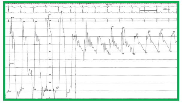
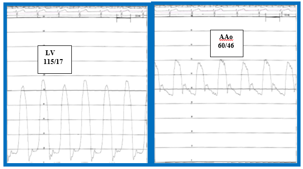
In this, most commonly used method, a #4 to #7-F sheath is placed percutaneously into the femoral artery and a #4- to 7-F multipurpose or right coronary artery catheter is advanced into the ascending aorta. With the help of a floppy-tipped coronary guide wire (in infants), a 0.035-inch straight Benston guide wire (Cook) or similar wires, the catheter is advanced into the left ventricle across the stenotic aortic valve. Other types of catheters and guide wires may be used if there is difficulty in crossing the aortic valve. Peak to peak systolic pressure gradient is determined by pressure pullback across the aortic valve (Figure 3) and cardiac output measurements performed. If feasible, simultaneous recording of pressures from both the LV and aorta are recorded (Figure 4). However, if there is marked difficulty in crossing the aortic valve, no pressure pullback is performed; instead, previously recorded aortic pressure is used to determine the peak to peak systolic pressure gradient across the aortic valve (Figure 5). Cineaortography and left ventriculography (Figure 6) are performed and a final diagnosis is made. Cine projections (most commonly left anterior oblique and right anterior oblique) should be chosen to best highlight the aortic valve stenosis and any additional subvalvar and supravalvar anomalies.
Historical Aspects
Following successful application of Gruntzig’s technique [8] to relieve obstructions caused by coarctation of the aorta by Sos [11], Singer [12], Sperling [13], and their associates and pulmonary valve stenosis by Kan and her colleagues [14], Lababidi et al. [9,10] extended the technique of balloon dilatation to aortic valve stenosis. Lababidi was also the first investigator to use this technique to the neonate with critical aortic valve stenosis [15]. Subsequently, a large number of papers on acute and medium-term follow-up results of balloon aortic valvuloplasty, extensively referenced elsewhere [1,16-20], were published. The author's group was among the first to examine causes of restenosis after balloon aortic valvuloplasty [20] and call attention to the development of aortic insufficiency during follow-up [17].
Indications for Balloon Aortic Valvuloplasty
It is generally agreed that indications for percutaneous, transcatheter therapy including AS should be same as those used for surgical intervention. Indications for balloon aortic valvuloplasty (BAV) are a peak-to-peak systolic pressure gradient across the aortic valve ≥ 50 mmHg (during cardiac catheterization) with a normal cardiac index and with either symptoms or electrocardiographic ST-T wave changes indicative of myocardial perfusion abnormality or a peak-to-peak systolic pressure gradient in excess of 70 mmHg irrespective of the symptoms or ECG changes [1,16-19]. While the calculated aortic valve area may be more accurate in evaluating the degree of aortic valve obstruction, most cardiologists use peak-to-peak systolic pressure gradients for assessment of severity of AS [1,10,16-20].
At the present time, the majority of percutaneous interventional procedures in children are performed under general anesthesia and the AS gradients are lower with general anesthesia than those with conscious sedation. Consequently, the catheter-measured gradient criteria alluded to above are not necessarily applicable. Therefore, the Doppler gradients are usually used in making a decision on the need for BAV. It was initially thought that peak instantaneous and/or mean Doppler gradients reflect the peak-to-peak catheter-measured gradients [22] but, because of factors related to pressure recovery phenomenon [23,24], the Doppler gradients are not necessarily accurate in predicting catheter gradients. I use an average of peak instantaneous and mean Doppler gradients as an alternative to calculate pressure recovery.
Neonates with very severe aortic valve stenosis with high gradient across the aortic valve, congestive heart failure and/or ductal-dependent systemic circulation, designated as critical AS, will require administration of prostaglandin E1 (PGE1) initially followed by BAV. However, high gradients may not be present because of low cardiac output in some babies with critical AS and therefore, low gradients should not preclude urgent BAV [25].
Adolescents and adults with moderate to severe AS with the above described pressure gradient criteria are also candidates for BAV. Given the enthusiasm which many centers are exhibiting for transcatheter aortic valve replacement (TAVR), it should be emphasized that the TAVR should be reserved for calcific AS of the elderly and the non-calcific AS in adolescents and adults should be addressed by the less invasive BAV [20].
Recurrence of stenosis after prior surgical aortic valvotomy is not a contraindication for balloon dilatation. Significant aortic insufficiency is a contraindication for BAV because of concern for further increasing aortic valve insufficiency [1,16,18,19].
Technique of Balloon Aortic Valvuloplasty
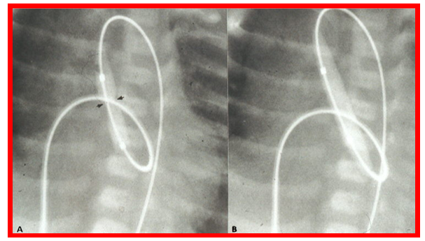
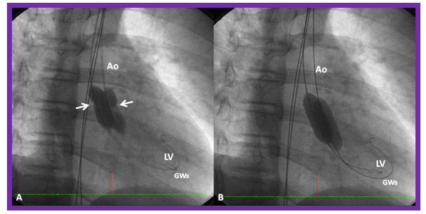
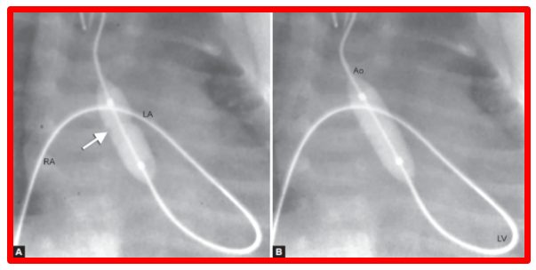
After securing informed consent, cardiac catheterization and selective cineangiography are performed to confirm the clinical and echocardiographic diagnosis. At the present time most pediatric interventionalists perform the procedure under general anesthesia with elective endotracheal intubation. In the past, conscious sedation (with a mixture of meperidine, promethazine and chlorpromazine, Midazolam and/or Ketamine) was routinely used. Conscious sedation is generally used in adult subjects. By and large, the method of sedation is largely institutional dependent. Once the venous and arterial access is achieved, 100 units/kg of heparin (maximum 3,000 units) are administered intravenously and activated clotting times (ACTs) monitored and maintained above 200 sec [1,17-19]. Percutaneous femoral arterial route (Figure 1) is the most commonly used approach for performing BAV; however, because of concern for femoral artery injury [26,27], particularly in neonates, infants and young children, alternative methods such as carotid arterial [28], axillary arterial [29], umbilical arterial [30],subscapular arterial [31], anterograde femoral venous [32,33], and umbilical venous [34,35] (Figure 2) approaches have been used. Each of these methods will be reviewed.

A J-tipped extra-stiff Amplatz guide wire (Cook, Bloomington, IN) [or an apex guide wire (Cook) in the older children and adults] is positioned in the left ventricular apex, through the catheter already in place. The chosen balloon should have a diameter 80% to 100% of the aortic valve annulus and should not exceed the aortic valve annulus. The aortic valve annulus is measured both in the echocardiogram performed prior to cardiac catheterization and from the left ventricular angiography during the procedure. The balloon length varies depending on the size of the patient: neonates and young infants – 2 cm; older infants and young children – 3 cm; older children, adolescents and adults – 4 to 5.5 cm [1,16,18,19]. There is a tendency for ejection of the balloon during balloon inflation and therefore, we prefer to use longer balloons. Others use Adenosine induced transient cardiac standstill [36] or rapid right ventricular pacing [37] to achieve stable position of the balloon during valvuloplasty. More recently, Nucleus balloons (NuMed) with a “barbell” configuration and hourglass-shaped V8 aortic valvuloplasty balloons (Venus Medtech) have been employed to help keep the balloon across the aortic valve. Using stiff guide wires and long balloons were found to be adequate in the majority of our patients [1,16-18,21] with rare need for rapid right ventricular pacing.
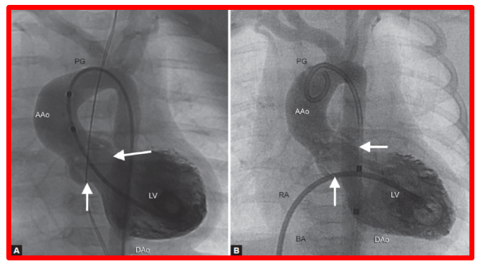
The selected balloon is placed across the aortic valve over the guide wire already in place using landmarks on the scout film and keeping with the same camera angulations. The balloon is inflated (Figure 1) with diluted contrast solution (1 in 4) to a pressure not exceeding the catheter manufacturer stated burst pressure. The recommendation is to perform two to four balloon inflations for a duration of five seconds each, five minutes apart. In the case of an aortic valve annulus that is too large to dilate with a commercially available single balloon or when the balloon catheter size is very large that there is high probability of femoral arterial damage, a double-balloon technique in which two balloons are simultaneously inflated across the aortic valve (Figures 7 and 8) is used [1,19]. Effective balloon diameter may be calculated by the following formula [26], which again, should not exceed the aortic valve annulus diameter:

Post intervention pressure pullback tracings across the aortic valve (Figure 9), cardiac output measurements and left ventricular and/or aortic root angiography are performed fifteen minutes following the valvuloplasty [1,19].

Balloon aortic valvuloplasty in the neonates [15,39-41] may also be performed in a similar manner but, as mentioned above, due to concerns for femoral artery injury in the neonatal period [26,27], alternative arterial routes such as carotid [28], axillary [29], umbilical [30], and subscapular [31] arterial, anterograde femoral venous [32,33], and umbilical venous [34,35] approaches have been attempted. These approaches will be briefly reviewed.
Balloon Aortic Valvuloplasty Via Carotid Artery
Isolation of the right carotid artery is performed by either the pediatric cardiologist or the pediatric cardiovascular surgical colleague depending on institutional practices. A 4-F sheath is placed with a purse string suture. The remainder of the procedure is performed using the above described femoral arterial access method. Due to the straight catheter course, it is easier to position the catheter/guidewire across the aortic valve into the left ventricle [28]. At the end of the procedure, the catheters and sheaths are removed, the purse string suture is tightened and the skin incision sutured.
Balloon Aortic Valvuloplasty Via Axillary and Subscapular Arteries
The procedure is similar to the above two methods with the exception of catheter entry; most often, the arterial access is by surgically exposing the axillary or subscapular arteries [29,31].
Transumbilical Arterial Balloon Aortic Valvuloplasty
A 4-French multi-A2 catheter (Cordis) is used to replace the previously existing umbilical arterial catheter and advanced in a retrograde fashion into the ascending aorta [25,30]. With the help of a floppy-tipped coronary guide wire or a similar soft-tipped guide wire, the catheter is advanced into the left ventricle across the stenotic aortic valve. If there is difficulty in crossing aortic valve other catheters and wires may be used. At this juncture, left ventricular angiography is performed and balloon dilatation is performed as in the previously described femoral arterial access method [25].
Transumbilical Venous Balloon Aortic Valvuloplasty
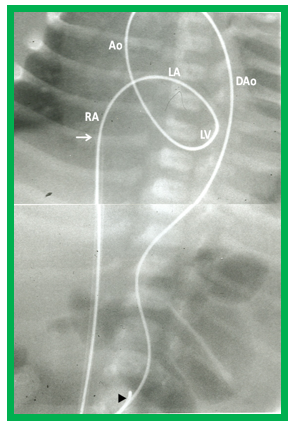
We encourage our neonatology colleagues to place an umbilical venous catheter (as soon as a cardiac baby is identified) and position the tip of the catheter in the right atrium, prior to anticipated ductus venosus closure. During balloon aortic valvuloplasty procedure, the umbilical venouscatheter is exchanged over a guidewire with a 5-F sheath and the tip of the sheath is located in the low right atrium [25,34,35]. After recording the routine hemodynamic data and left ventricular cineangiography (Figure 6), the aortic annulus diameter is measured in several views. This information supplements echocardiographic diameter to estimate of aortic annulus diameter. A #4-F multipurpose catheter(Cordis) with a slightly curved tip (special order) or a similar catheter is introduced into the umbilical venous sheath and advanced into the left atrium across the patent foramen ovale (PFO) and then via the mitral valve into the LV. With the aid of a J-shaped and/or a straight, soft-tipped 0.035" Benston guide wires (Cook), the multipurpose catheter is advanced into the ascending aorta and if possible, the catheter tip is negotiated into the proximal descending aorta. At this time, the guidewire is replaced with a 0.018" or 0.021" J-tipped guidewire, suited to accommodate the selected balloon angioplasty catheter. The multipurpose catheter is removed and a 6–8 mm diameter Tyshak II (Braun) or ultrathin (Meditech) balloon dilatation catheter (The diameter of the balloon selected should be 0.8 to 1.0 times the aortic valve annulus.) is advanced over the guidewire from the umbilical vein, inferior vena cava, right atrium, left atrium, LV and aorta, while maintaining a wide loop of the guidewire in the left ventricle. Once the balloon is placed across the aortic valve, the balloon is inflated with diluted contrast material; the pressure of inflation should be up to the manufacturer’s suggested pressure, or until waist of the balloon is eliminated (Figures 2 and 10). We usually inflate the balloon one or two more times to assure adequate valvuloplasty. The balloon catheter is exchanged with a #4-F multipurpose catheter and the guidewire is removed. Aortic root angiography is performed and pullback pressures across the aortic valve are recorded. Cineangiogram from the LV may be performed as deemed appropriate. Heparin is administered at the beginning of the procedure and ACTs monitored. Vancomycin is given for antibiotic prophylaxis; this is because of extensive handling of the umbilical area during the procedure [25,34,35].
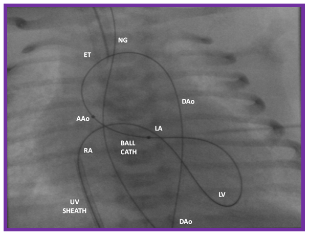
If the guidewire could not be maneuvered into the descending aorta or the balloon catheter could not be positioned across the aortic valve, a gooseneck micro-snare (Microvena, White Bear Lake, MN) may be placed in the aorta either through the umbilical or femoral artery and snare the tip of the anterogradely placed guidewire and pull it down into the descending aorta and held in place. With this, umbilical venous-to-umbilical/femoral arterial wire “rail” is established (Figure 11); a gentle traction on the umbilical/femoral artery component of the rail (while preserving the wire loop in the LV), the balloon catheter may be more easily positioned across the aortic valve, facilitating balloon aortic valvuloplasty. Once the procedure is completed the guidewire is released from the snare and withdrawn via the umbilical vein; positioning a catheter over the whole course of the guidewire within the heart prevents injury of the intracardiac structures [34].
Given the availability of better-tracking balloon valvuloplasty catheters (Figure 12) such as Tyshak II (Braun), the above described maneuvers may not be necessary in most cases [19].
Antegrade Femoral Venous Balloon Aortic Valvuloplasty
In this method, initially described in 1993 [32,33], a #5-F sheath is used to achieve femoral venous access. The remaining procedure is performed in a manner similar to the above described umbilical venous access method; however, it should be mentioned that the transumbilical venous balloon aortic valvuloplasty [34,35] is patterned after the anterograde femoral venous method [32,33].
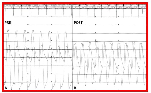
There is an acute reduction in the peak to peak systolic pressures across the aortic valve (Figures 9, 13-15) along with a reduction in the left ventricular peak systolic and end diastolic pressures without significant change in cardiac index. There is approximately 60% reduction in the gradient compared to the pre-valvuloplasty gradients (Figure 15). The degree of aortic insufficiency does not worsen as a general rule (Figures 16; pre vs. post). Some improvement is seen in some patients; this suggests better coaptation of the aortic valve leaflets after balloon dilatation. With the exception of neonates, most patients are discharged home within 24 hours of the procedure [17,19,21].


In the first series of 23 consecutive patients with valvar aortic stenosis, reported by Lababidi and associates [10], the peak to peak systolic gradient across the aortic valve decreased from 113 ± 48 mmHg to 32 ± 15 mmHg (p<0>
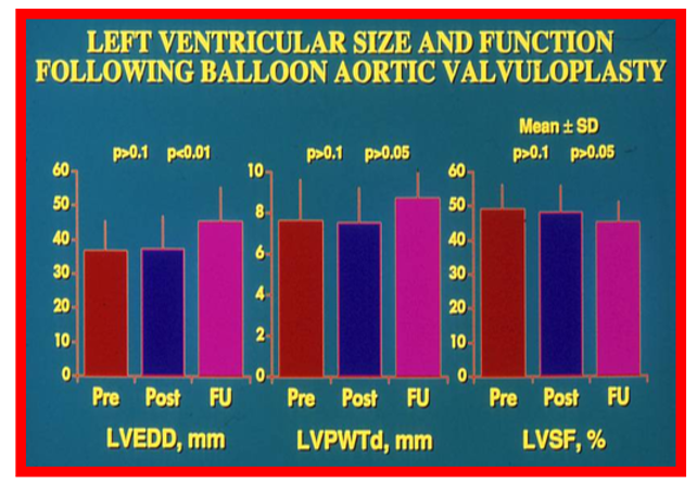
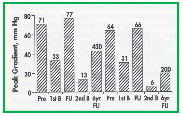
Immediate results of balloon aortic valvuloplasty were presented by the author [16,21] in late-1980s with subsequent publication of immediate results in a larger group of patients [1,17]. In the first sixteen patients, reduction of peak-to-peak systolic pressure gradients across the aortic valve (72 ± 21 vs. 28 ± 13 mmHg; p < 0> 0.1) in cardiac index [21]. The gradients were generally reduced by 60% of pre-valvuloplasty gradients (Figure 15). Similar reduction in peak-to-peak systolic pressure gradients were observed (Figure 17) in the second cohort consisting of 26 patients [17]. These acute results are similar to those observed by other workers, as tabulated elsewhere [1]. The prevalence of significant (3+ or more) aortic insufficiency did not change for the group as a whole (Figure 16); in some patients the aortic insufficiency actually improved, suggesting a better coaptation of aortic valve leaflets following BAV. By echocardiogram, the LV end-diastolic dimension (36 ± 9 vs. 35 ± 10 mm; p > 0.1), LV posterior wall thickness in diastole (7.2 ± 2.1 vs. 7.5 ± 1.9 mm; p > 0.1), and LV shortening fraction (50 ± 8 vs. 47 ± 8%; p > 0.1) did not change after BAV (Figure 18). However, the Doppler flow velocity across the aortic valve (4.0 ± 0.05 vs. 3.0 ± 0.8 m/s; p < 0>


At intermediate-term follow up (defined as ≤ 2 years), peak-to-peak systolic pressure gradients across the aortic valve by repeat cardiac catheterization (Figure 17) and Doppler peak instantaneous gradients (Figure 19) either remain unchanged or increased slightly compared to immediate post-intervention values but continued to be significantly lower than pre-valvuloplasty values [17]. Peak instantaneous Doppler gradients in all 26 patients 16 ± 11 months after BAV were 31 ± 15 mmHg; these gradients were similar (p > 0.1) to post-valvuloplasty gradients and continue to be lower (p < 0> 0.1) at follow-up (Figure 18). However, when results of individual patients were examined, restenosis defined as a peak to peak gradient of greater than or equal to 50 mmHg, was found in 6 (23%) children (Figure 20). Four of these children in our early experience underwent surgical valvotomy and two repeat balloon valvuloplasty at a median interval of 9 months following the first BAV. The degree of aortic insufficiency remained stable during intermediate-term follow-up [17]. Intermediate-term follow up results reported by other investigators were similar to ours and were tabulated elsewhere [1] for the interested reader.

Restenosis and Predictors of Restenosis
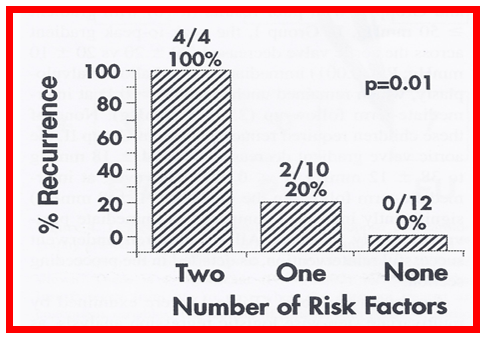
As mentioned in the preceding section, restenosis following BAV does occur (Figure 20). The causes of restenosis after BAV were investigated by scrutinizing the follow-up results of 16 children [21]. Based on the intermediate-term follow-up results, these 16 patients were divided into two groups: Group I with good results (N=12) with aortic valve gradients < 50 N=4)> 0.1) at intermediate-term follow-up (26 ± 10 mmHg). (Figure 21). None of these children required re-intervention. In group II, the aortic valve gradient decreased from 79 ± 20 mmHg to 42 ± 13 mmHg (p < 0>
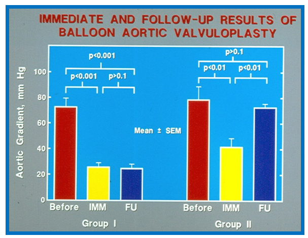
Figure 21: Bar graph showing immediate (IMM) and follow-up (FU) results of balloon aortic valvuloplasty in Group I with good results (left panel) and in Group II with poor results (right panel). In Group I with good results, the aortic valve gradient decreased significantly (p < 0> 0.1) at follow-up. Mean + standard error of mean (SEM) are shown. Reproduced from Rao PS. Pediatric Cardiology: How It Has Evolved Over The Last 50 Years. Cambridge Scholars Publishing, New Castle upon Tyne. 2020:231-256.
Sholler et al [44] investigated the influence of various technical and morphological factors on the immediate results of balloon aortic valvuloplasty but no statistical significance was demonstrated on any factors tested. Other investigators, as reviewed elsewhere [17-19], investigated causes of recurrence of stenosis after BAV, but were not able to detect any factors responsible for restenosis. There was a claim that double balloon technique is better than single balloon valvuloplasty [45]; but detailed analysis of these data [46] did not validate such interpretation. Balloon/annulus ratios and aortic valve morphology may be important determinants of restenosis; however, the range of variability seen in our study and those of others could not be demonstrate statistically significant differences; perhaps studies in larger groups of patients may uncover the causes [17-19].
In may be concluded that age ≤ 3 years and immediate post-balloon aortic valve peak-to-peak gradient ≥ 30 mmHg may be predictive of restenosis and, avoiding or minimizing risk factors may help reduce recurrence after BAV. Since the immediate post-valvuloplasty aortic valve peak-to-peak systolic pressure gradient ≥ 30 mmHg is an alterable risk factor, the author advocated use of larger balloons, large enough to reduce the gradient to < 30>
Repeat Balloon Valvuloplasy for Restenosis after Prior BAV
As indicated above, recurrence of aortic stenosis after BAV was observed. We have studied the feasibility and effectiveness of repeating balloon dilatation in relieving the recurred obstruction following prior balloon procedures for pulmonary stenosis, aortic stenosis and coarctation of the aorta [47]. In the aortic stenosis group, twenty-six children underwent BAV between 1983 and 1993 with reduction in aortic valve peak gradients from 71 ± 20 mmHg to 26 ± 12 mmHg (p < 0>
Although there are several reports on immediate and intermediate-term results of BAV for the relief of congenital aortic valve stenosis in infants and children, reports of long-term results are few. We reported long-term follow-up results of 25 patients followed for 3 to 10 years (median 6.7 ± 1.7 years); 22 of these patients were followed for longer than 5 years [17]; details are presented in the ensuing paragraphs.
Residual Gradients [17]
The long-term follow-up gradients are excellent with very low (27 ± 17 mmHg) residual Doppler-derived gradients (Figure 19); these gradients were lower than pre-valvuloplasty gradients (p < 0> 0.1) to both immediate post-valvuloplasty and intermediate-term follow up gradients.
Re-interventions and Actuarial Event-Free Rates
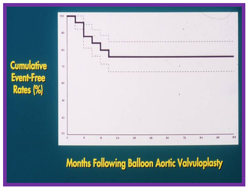
A total of eight (31%) children, including six at intermediate follow-up, were found to have restenosis; they were successfully treated with either surgical valvotomy or repeat BAV. One child required a left ventricular apex-to-descending aortic conduit for severe left ventricular mid-cavitary obstruction. Seven (27%) children developed severe aortic insufficiency (will be discussed in detail in a later section of this chapter) at long-term follow-up (Figure 16), and two of these children required the Ross procedure. Event-free rates suggested 80%, 76%, 76% and 60% probability of freedom from re-intervention at 1-, 2-, 5- and 10- year follow-up respectively (Figure 24) [17].
Ventricular Dimensions and Function [17,50]
TheLV end-diastolic dimension (45.4 ± 9.9 mm) was larger (p < 0> 0.05) (Figure 18).
Aortic Insufficiency
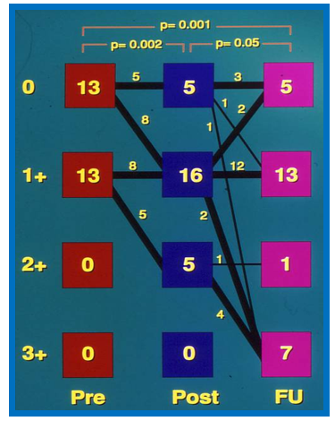
Ratio of aortic insufficiency (AI) jet width to width of the LV outflow tract was used to grade the degree of AI [17]. This type of grading at last follow-up demonstrated that the number of patients with 3+ aortic insufficiency increased (Figures 16 and 25) significantly (p < 0>
Long-Term Results by Other Investigators
Long-term results of BAV reported by other interventional cardiologists were extensively reviewed in several of the author's publications [18-20,49.50] for the interested reader; these will also be presented in a tabular form (Table-1).
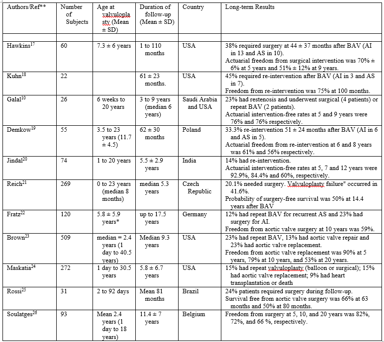
AI, Aortic insufficiency; AS, Aortic stenosis; BAV, Balloon aortic valvuloplasty; SD, Standard deviation.
Reproduced from Rao PS. Indian Heart Journal 2016; 68:592-5.
Summary of Long-Term Results
In summary, the long-term results of BAV indicate continuance of relief of obstruction for the group as a whole with indication for
minimal additional restenosis, progressive increase of AI, enlargement of the left ventricle and relatively high re-intervention rates
[17,19,20].
As indicated above, significant aortic insufficiency (AI) was seen at long-term follow-up after BAV (Figures 16 and 25). Most studies including ours show a trend toward increase in the degree of AI with time; longer the follow up, the greater the AI. Significant AI was reported in 24 to 38% patients with requirement for aortic valve replacement in 8 to 14% patients, as tabulated elsewhere [18] and in the above table.
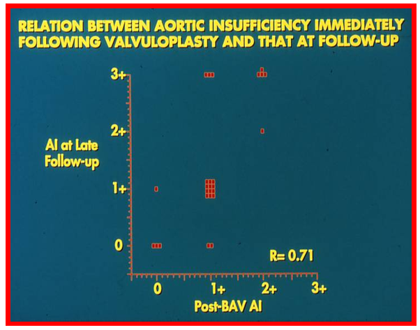
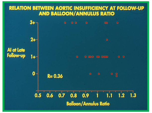
The author sought to investigate if the reason for development of AI could be discerned [17].The study subjects were divided into two groups: Group A, 19 patients without significant AI (grade 2+ or less) and Group B, 7 patients with 3+ AI. Fifteen biographic, anatomic, physiologic, and technical data (Table II of reference 17) were examined by multivariate logistic regression analysis to identify factors producing AI [17]. This analysis identified several factors that were statistically different between groups (Table IV of reference 17); these are Doppler quantitated AI both prior to and immediately following BAV and the procedure performed during the latter half of our experience with BAV. These three variables were entered into a multivariate logistic regression model with all possible combinations. A model that includes post-BAV Doppler AI fits the data best. The addition of pre-BAV Doppler AI and procedural experience to the model already including post-BAV Doppler AI did not significantly improve its predictive power [17]. Therefore, it was concluded that immediate post-BAV grade of AI is predictive of late onset of significant AI; the relationship between these two is illustrated in Figure 26. Large balloons (1.2 to 1.5 times the valve annulus) in animal models and in clinical models with intra-operative balloon dilatation as referenced elsewhere [18] do produce injury to and tears of the aortic valve causing AI. Therefore, we plotted the degree of AI at follow-up against the balloon valve annulus ratio (Figure 27) and found no relationship between the balloon size and degree of AI. The reasons for progression of AI following BAV are not well understood. The hypotheses put forward by several investigators include greater relief of gradient immediately following BAV [51], Doppler-quantified AI both prior to and immediately following BAV [17], unicommissural aortic valves [44], aortic valve prolapse [52], poor valve morphology [17] and large balloon/annulus ratio [44,52,53] but, none of these seem to have evidence to support their role in causing AI. Our data [17] indicated that the degree of aortic insufficiency immediately after balloon aortic valvuloplasty is predictive of development significant late aortic insufficiency (Figure 26). We speculated that a combination of poor valve morphology and liberal sized balloons [17-20] may eventually prove to be responsible for aortic valve insufficiency at late follow-up after BAV. Additional studies to investigate these and other causes for development of late AI and devise methods to prevent AI were recommended.
Balloon Valvuloplasty in Specific Age Groups
The above review included discussion of BAV which are mostly focused on infants, children, adolescents and young adults. In the ensuing sections the results of BAV in the fetus, neonates and premature infants with critical aortic stenosis and aortic stenosis in the elderly adults will be reviewed.
Aortic Stenosis in the Fetus
Prenatally diagnosed critical AS carries a poor prognosis [1]. It is generally believed that critical AS in the fetus progressively develops into hypoplastic left heart syndrome (HLHS). The extent to which simple BAV in the fetus prevents this progression is not clearly understood. The rationale behind using BAV is to augment ventricular filling, improve LV diastolic function, and increase normal division of myocardial cells through the remaining fetal life, thus positively altering post-natal outcomes [54,55].
The procedure of BAV is usually performed between 21 to 29 weeks of gestation. Maternal general anesthesia is usually used. Although general anesthesia imposes certain risk to the mother, it facilitates re-positioning the fetus to an appropriate lie to facilitate performing BAV. Fetal anesthesia and paralysis are induced by fetal intramuscular injection of atropine, vecuronium, and fentanyl. The technique of accessing the fetus is similar to that used by Daffos and associates [56] for chorionic villous sampling and subsequently applied to fetal BAV by Maxwell at al. [57]. A 19 gauge cannula is introduced trans-cutaneously via the maternal abdominal wall and uterus and then across the fetal chest into the LV cavity. A floppy-tipped 0.014" coronary guidewire is used to cross the aortic valve. Once the position is confirmed by fetal ultrasound, a coronary balloon angioplasty catheter with a balloon diameter 10% smaller than aortic valve annulus is positioned across the aortic valve and the balloon inflated at the manufacturer's recommended pressure. If the percutaneous route is not successful, the uterus is exposed with a mini-laparotomy [57-62].
Fetal BAV to address AS was first reported in 1991 [57]. Of the two fetuses that they attempted BAV procedure, they were successful in performing the procedure in one of them. Even this baby required repeat post-natal BAVs and eventually died. However, these initial attempts demonstrated that the BAV can be performed during fetal life. During the next decade only 11 cases were reported to have BAV [58]. Subsequently, a large number of investigators reported their respective experiences with fetal BAV [59-70]. Technical success, defined as performing BAV in the fetus has improved over time. In the initial attempts, 1 of the first 4 attempts (25%) was technically successful [54]; the technical success rate improved with additional experiences [54,55,61,64,68], and the most recent experience suggests a technical success rate of 94% [68]. Similarly, fetal demise has decreased to 4% [68]. Achieving biventricular circulation occurred in 50% patients, but the experience since 2009 puts it at 66% [68].
Establishing selection criteria for performing fetal BAV have been debated and preventing development of HLHS appears to be prime objective. It was suggested that mid-gestation AS fetuses with reversed flow in both the transverse aortic arch and foramen ovale, monophasic mitral inflow, and LV dysfunction are likely to develop HLHS [55]. When these criteria were applied to 107 cases of fetal AS in an European multicenter retrospective study, substantial proportion these fetuses attained biventricular circulation without any treatment [71]. Given the complexity of the fetal BAV procedure and risk for the mother and fetus, though small, more appropriate criteria for performing BAV in the fetus must be developed.
Critical Aortic Stenosis in the Neonate
Critical aortic stenosis is a term used to describe babies who have very severe aortic valve obstruction with a very high peak-to-peak systolic pressure gradient across the aortic valve, have signs and symptoms of congestive heart failure, have ductal dependent systemic circulation, and/or a combination thereof. Because of poor LV function, the pressure gradient across the aortic valve may not be high in some patients, however. As reviewed in the section on Technique of Balloon Aortic Valvuloplasty, because of concern for femoral artery injury [26,27], particularly in neonates, alternative methods such as carotid arterial, axillary arterial, umbilical arterial, subscapular arterial, anterograde femoral venous and umbilical venous approaches [28-35] have been used in the neonates. The author's preference is to utilize anterograde, transumbilical venous route [34,35]. If that is not successful, retrograde femoral arterial entry is used. Retrograde transumbilical arterial, anterograde femoral venous and carotid artery cut down are the other available options.
The peak-peak systolic pressure gradient across the aortic valve decreases and clinical improvement occurs in the vast majority of the babies. The balloon aortic valvuloplasty results from the initial seven studies involving neonates were presented elsewhere [1]; the interested reader may review the said publication. Impressive reduction in gradients, similar to that reported for children, as reviewed in the section on Immediate Results, was also noted in the neonates. These studies suggested that balloon valvuloplasty is beneficial in the treatment of ill neonates with critical aortic valve stenosis. However, complications including death and necessity for surgery secondary to onset of severe aortic valve insufficiency were reported in the neonates [1,18]. Poor results appear to be due to either technical issues or to abnormal anatomic substrate (aortic valve dysplasia, aortic valve annular hypoplasia, hypoplastic left ventricle, mitralvalve abnormalities and endocardial fibroelastosis). More recent availability of miniaturized balloon dilatation catheters, the procedural difficulties have been to a large extent resolved. In neonates with less severe obstruction, BAV may be performed at a later time, past the neonatal period [1,18,19,25].
A comparison of anterograde and retrograde balloon aortic valvuloplasty techniques was made by Magee et al [72]. They found the results to be similar with regard to feasibility and pressure gradient reduction. But, a higher mortality, more severe aortic insufficiency and arterial complications occurred in the retrograde when compared to anterograde technique. However, a more recent evaluation of this issue suggests that large balloon/annulus ratio is likely to be the causative factor for the aortic insufficiency instead of the route of entry of balloon catheter.
A comparison of surgical and balloon valvuloplasty procedures, both single institutional and multi-institutional studies [41,73-76] suggested that pressure gradient reduction and rates of freedom from re-intervention are similar. Nevertheless, high mortality and re-operation rates seen with surgical aortic valvotomy tend to support balloon valvuloplasty as an attractive alternative to surgical intervention in the newbornwith critical aorticvalve stenosis.
Aortic Stenosis in the Premature Infant
Premature babies with critical AS should also undergo BAV similar to that of full-term neonates. To the best of the author's knowledge, Tometzki and associates [77] were the first to report BAV in a premature infant with AS. They performed BAV in an 8-day-old, 28-week gestational-age preterm infant weighing 1.08 kg using a 5-mm diameter balloon carried on a #4.3F catheter introduced via the femoral artery. The procedure reduced peak systolic gradient from 90 mmHg to 20 mmHg. However, evulsion of the femoral artery ensued requiring surgical reconstruction [77]. Cursory search of the PubMed revealed that a number of other cardiologists reported on their respective experiences with BAV in the premature infants [78-83]. Some of these investigators used trans-carotid approach [80], anterograde transvenous route [78,79] or a hybrid method (surgical exposure of LV apex [81] or ascending aorta [82]) to avoid femoral arterial access for performing BAV. In another report [83], trans-femoral approach was used in 5 premature infants with gestational ages of 32 to 36 weeks and birth weights of 1.4 to 1.9 kg at postnatal ages of 2 to 10 days. They used 4.5 to 6 mm sized balloons for BAV; the peak-to-peak systolic gradients across the aortic valve fell by more than 50% in each baby. One baby developed severe aortic insufficiency, presumably related to unicuspid aortic valve and underwent Ross operation at the age of two months. Another baby required repeat BAV at the age of 6 months for recurrence of severe obstruction. Two other patients required a Ross operation at 5 and 7 years of age respectively. Only one patient did not need any re-intervention through the age of 9 years. Thus, this small series demonstrated procedural success with relief of obstruction, but required re-intervention in 80% of the babies [83].
Aortic Stenosis in Adults
Following the description of BAV by Lababidi et al [9,10], the technique was extended to adults with calcific aortic stenosis with the initial impression that the technique is valuable in the management of elderly with calcific aortic stenosis, as reviewed elsewhere [1,84-87]. Subsequently, however, the relief of obstruction was found to be temporary and transient [88,89] and at the current time, the elderly patients with calcific aortic stenosis are candidates for transcatheter aortic valve replacement (TAVR) [90]. Discussion of TAVR is beyond the scope of the objectives of the chapter and the interested reader is referred to other reviews and AHA/ACC recommendations [90-92]. The results of BAV of non-calcific aortic stenosis in adolescents and adults is similar to that of seen in children (see Table in the section on "Long-Term Results by Other Investigators"), as reviewed elsewhere [20,93].
Comparison with Surgery
Comparison of results of BAV with surgical aortic valvotomy is fraught with problems, similar to those seen with pulmonary stenosis because: a. few or no studies exist that compare concurrent balloon and surgical procedures nor are there any randomized studies to address this issue, b. problems in comparing “older” historical surgical results with “current” BAV, c. short duration of follow-up after BAV, and d. smaller number of transcatheter patients available for follow up compared to surgical patients. In the early 1990s, the author examined the outcomes of surgery from 10 papers [1]. The investigators of these 10 publications followed 41 to 179 patients for 0.3 to 26 years after surgery. Operative mortality for children varied from 0% to 4%. Late mortality varied from 4% to 22%. In the natural history study [94], these rates were lower; the operative mortality rate was a 1.2% and late mortality was 1.9%. Development of restenosis of the aortic valve was seen in 16% to 78% of patients and aortic insufficiency in 6% to 65% patients. Surgical re-intervention to relieve restenosed aortic valve or to repair/replace incompetent aortic valve was necessary in 16% to 39% patients [1]. These surgical results are worse than BAV results [1]. Gatzoulis et al [95] found no significant difference in mortality, morbidity or the need for re-intervention within 12 months of the procedure between the surgical and balloon groups. McCrindle and associates [76], comparing surgical and balloon groups in neonates, found that the two modes of therapy have similar rates of freedom from re-intervention at five years following the procedure. More recent studies, detailed and referenced elsewhere [18,19] found no significant difference in mortality, morbidity or need for re-intervention between surgical and balloon groups and have similar rates of freedom from re-intervention at five years following either procedure. Consequently, significant prevalence of early and late mortality and the need for re-operation associated with surgical valvotomy would make BAV an attractive alternative to surgical approach [1,18,19].
Complications Associated with BAV
Complication observed immediately following BAV and those at follow-up will be separately reviewed.
Immediate Complications
Immediate complications include transient bradycardia, premature beats and a fall in systemic pressure during balloon inflation; these return to baseline following balloon deflation, thus reiterating the previously suggested balloon inflation time of ≤ 5 seconds [1]. Other reported complications are blood loss requiring transfusions; femoral artery thrombosis requiring heparin, streptokinase or thrombectomy [96]; and rhythm disorders including transient left bundle branch block [1], right bundle branch block, transient prolongation of QTc interval [97], transient atrioventricular block, supraventricular and ventricular dysrhythmias [1,98], and cardiac arrest [99]. Transmural tears with vessel or ventricular wall perforation [96,100]; balloon rupture [10,101]; balloon dislodgement [53]; aortic or mitral valve tears [53,102]; myocardial perforation; occlusion of the right coronary artery; transient myocardial ischemia [98]; cerebrovascular accidents [103]; and development of subvalvar obstructions [104], although rare have been reported. Aortic valve tears have been seen with animal models with large balloon sizes,1.2 to 1.5 times the valve annulus [18,105] and therefore, large balloons should not be used. Death associated with balloon dilatation has also been reported [44,51,102,106,107]; these are associated with aortic rupture, occlusion of extreme critical obstruction, perforation or avulsion of aortic valve cusp, exsanguinations from torn iliac/femoral vessels, and ventricular fibrillation. Sudden unexplained death is also recorded, but is extremely rare [107].
Complications at Follow up
Complications at follow up were femoral artery occlusion [16,26,27], aortic valve insufficiency and recurrence of obstruction; the latter two were discussed in the preceding section. Issues related to femoral artery occlusion following BAV, including other transfemoral artery balloon dilatations will be reviewed in this section. Early on, when we evaluated this issue [1,16], of the 32 infants and children who had follow-up catheterization following a prior trans-femoral artery balloon dilatation including BAV, three femoral arteries were found to be occluded (complete in two and partial in one) (Figure 28), but all of them had good collateral blood flow (Figure 28B). Arterial occlusion after femoral artery catheterization even during diagnostic studies has been well documented [108-115]; the reported incidence of arterial occlusion varied between 3% and 40%. Given the need for using larger diameter catheters for trans-femoral artery balloon dilatations, it is not unexpected to have a higher prevalence of femoral arterial occlusions with transfemoral artery balloon dilatations than with diagnostic catheterizations.

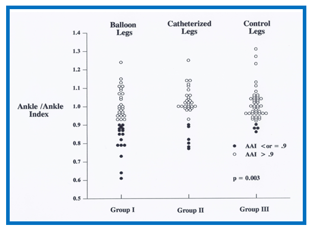
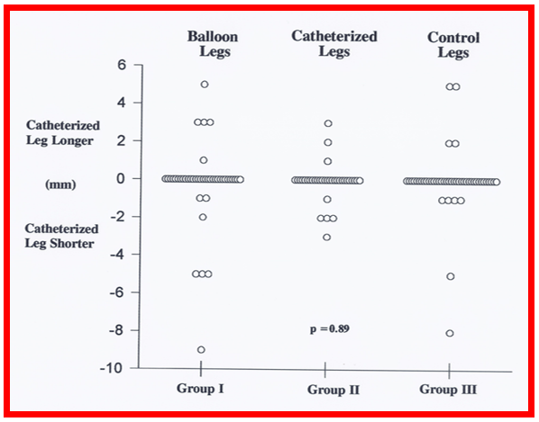
In a subsequent study we sought to evaluate the prevalence of superficial femoral artery (SFA) compromise and its effect on limb growth in children who had transfemoral artery balloon dilatations [27]. Data on 43 consecutive patients (1 day to 15.5 years old at the time of balloon dilatation) seen on follow-up (42 ± 23 months) (group I) were compared with those of 35 patients undergoing retrograde femoral arterial catheterization (group II) and 47 control patients (group III). Interventional ankle/control ankle blood pressure index (AAI), ratio of interventional/control lower limb length (LLI), and leg length difference (LLD) were measured. Ages and weights at study were similar in all three groups, as were the ages and weights at intervention and duration of follow-up in groups I and II. The AAI was lower (P = 0.023) in group I (0.95 ± 0.13) than in groups II (1.0 ± 0.1) and III (1.01 ± 0.09) (Figure 29). The prevalence of subjects with AAI ≤ 0.9 was higher (P = 0.003) in group I than in the other two groups. The LLI and LLD were similar (P > .1) in all three groups (Figure 30). AAI and LLD in the balloon group are not significantly associated with age and weight at intervention, duration of follow-up, or size of the balloon or balloon catheter shaft [27].
It was concluded that transfemoral artery balloon dilatation procedures produce SFA compromise, but there was no significant limb growth retardation at a 3.5-year mean follow-up, which may be related to development of collateral circulation. We suggested that a study of a larger number of children at a longer follow-up interval may be necessary to further confirm or refute these observations.
Other issues related BAV, not discussed previously, will be reviewed in this section.
Development of Subvalvar Stenosis
Subaortic obstruction following BAV, similar to infundibular stenosis following balloon pulmonary valvuloplasty [116-118] is rare and was seen in one (4%) of 24 patients from our study subjects [1]. A 9-year-old child whose peak-to-peak gradient across the aortic valve was reduced from 112 to 36 mmHg after BAV was found to have marked subaortic hyperactivity with a peak Doppler velocity of 5.5 m/s on the day following BAV. There was typical triangular pattern of the Doppler curve suggestive of subaortic obstruction. A similarly low prevalence of development subaortic obstruction after BAV was observed by other cardiologists [104]; they found this phenomenon in three (9%) of 33 BAVs. The subaortic obstruction may be secondary to unmasking of proximal obstruction following relief of distal aortic valve obstruction by BAV due to the phenomenon of forced vibration [1,119] and is likely to resolve with time [104].
Balloon Types Used during BAV and Their Characteristics
The author examined the role of technical factors in the results of BAV [1,16,21,46] and found that balloon diameter (balloon/annulus ratio), number of balloons used (one vs. two), pressure, duration and number of balloon inflations used during BAV (table III of reference 21) did not have any influence on the outcome of the procedure at intermediate-term follow-up. The findings from other investigators, as reviewed and referenced elsewhere [1,21,46] are generally similar to our observations.
Mechanism of Valvuloplasty
The mechanism by which BAV produces relief of aortic valve obstruction has been reviewed in multiple publications in the past [1,16,120,121] and will only be summarized here. On the basis of direct inspection of the aortic valve leaflets at surgery or at postmortem examination and indirect observation on echocardiography or angiography, splitting of the valve commissures, tearing of valve leaflets and avulsion of the valve leaflets are thought to be the mechanisms by which aortic valve stenosis is relieved by BAV. The radial dilating forces of balloon inflation are likely to rupture/tear the fused valve commissures, the weakest part of the valve mechanism. However, if the fused commissures are too strong to be torn, valve cusp tears and even avulsion of valve leaflets may occur. The latter may result in severe AI. The mechanism for relief of obstruction in adult patients with calcific aortic stenosis is likely to be fracture of nodular calcifications and improved leaflet mobility [1,121]. For a more detailed discussion of mechanism of BAV, the reader is referred to detailed discussion presented elsewhere [121].
Cardiac catheterization was used initially to assess the follow-up results of BAV. After showing efficacy of echo-Doppler studies in quantifying the residual gradients, echo-Doppler studies were almost exclusively used by most investigators in the assessment of results of BAV at follow-up and cardiac catheterization was used only when catheter re-intervention is planned. Peak Doppler flow velocity was used to calculate peak instantaneous Doppler gradient using modified Bernoulli equation. Although, peak instantaneous and/or mean Doppler gradients were initially thought to reflect the peak-to-peak catheter gradients [22], because of pressure recovery phenomenon [5,23,24], the Doppler gradients are not accurate in estimating the catheterization-derived gradients. The author generally uses an average of peak instantaneous and mean Doppler aortic valve gradients to estimate the catheterization gradients. The author reported results of follow-up echo-Doppler studies of children who had BAV [1,16-18,21]. Doppler flow velocities, Doppler gradients, LV dimensions, LV posterior wall thickness, LV shortening fraction and the degree of AI immediately after BAV and at intermediate-term and long-term follow-up were reviewed (Figures 18,19,25,26) in the respective sections above and will not be repeated. It is concluded that echo-Doppler studies are useful in the assessment of results of BAV [1,16-18,21].
Summary and Conclusions
Following the description by Lababidi and associates in 1983 of balloon aortic valvuloplasty, it has been adopted by several groups of workers for relief of aortic valve stenosis. The indications for the procedure are peak-to-peak systolic pressure gradients in excess of 50 mmHg with symptoms or ECG changes or a gradient greater than 70 mmHg irrespective of the symptoms or ECG changes. One or more balloon dilatation catheters are placed across the aortic valve percutaneously, over extra-stiff guide wire(s) and the balloon(s) inflated until waist produced by the stenotic valve is abolished. A balloon/annulus ratio is 0.8 to 1.0 is recommended. While trans-femoral arterial route is the most commonly used for balloon aortic valvuloplasty, trans-umbilical arterial or venous or trans-venous routes are preferred in neonate and young infants to avoid femoral arterial injury.
Reduction of peak-to-peak systolic pressure gradient along with a fall in left ventricular peak systolic and end-diastolic pressures is seen after balloon aortic valvuloplasty in the majority of patients. Significant aortic insufficiency, though rare, may develop, particularly in the neonate. At intermediate-term follow-up, peak-to-peak gradients, at repeat cardiac catheterization and noninvasive Doppler gradients remain low for the group as a whole. Nevertheless, restenosis, defined as peak-to-peak gradient ≥ 50 mmHg may develop in nearly one quarter of the patients. Predictors of restenosis are age ≤ 3 years and an immediate post-valvuloplasty aortic valve gradient ≥ 30 mmHg. The restenosis may be addressed by repeat balloon valvuloplasty or surgical valvotomy. Feasibility and effectiveness of repeat balloon valvuloplasty in relieving restenosis has been demonstrated. Long-term follow-up data suggests low Doppler peak instantaneous gradients, minimal additional restenosis beyond what was observed at intermediate-term follow-up and progression of aortic insufficiency in nearly one-quarter of patients. Event-free rates are in mid 70s and low 60s respectively at 5 and 10-years after initial balloon valvuloplasty. A number of complications have been reported, but are rare. Comparison with surgical results is fraught with problems, but overall, the balloon therapy appears to carry less morbidity.
Immediate, intermediate and long-term-term follow-up data following balloon aortic valvuloplasty suggest reasonably good results, avoiding/postponing the need for surgical intervention. However, late follow-up data indicate that significant aortic insufficiency with left ventricular dilatation may develop, some require surgical intervention and are of concern. Current recommendations favor balloon valvuloplasty as first line therapeutic procedure for relief of aortic valve stenosis.
Dear Editorial Team, Clinical Medical Reviews and Reports. My experience with the journal was highly positive. The peer-review process was rigorous, constructive, and completed in a timely manner. The reviewers provided valuable comments that helped improve the quality and clarity of our manuscript. The editorial office was professional, responsive, and supportive throughout all stages of the publication process. Communication was clear and efficient, and any questions were addressed promptly. Overall, I found the journal to maintain high scientific standards and an excellent publication workflow. I would be pleased to consider submitting future work to this journal. Best wishes from, Elena Popa.

It was my pleasure to submit my testimonial concerning the Reviewer Board of our Scientific Journal “Brain and Neurological Disorders”. The Reviewers focused on some modifications and their contribution was helpful. The ladies of our Editorial Office were also supported my efforts. It was my honor to have such a co-operation and I am looking forward for more collaboration.

Dear Grace Pierce, Editorial Coordinator of Journal of Clinical Research and Reports, Thank you for the speedy and efficient peer review process. I appreciate the fact that your peer reviewers do not take months to respond like with some other journals. I would also like to thank the editorial office for responding quickly to my questions. It is an excellent journal. I plan to submit more manuscripts in the future. Best wishes from, Robert W. McGee

Dear Grace Pierce, Editorial Coordinator of Journal of Clinical Research and Reports, Working with you and your team on our recent publication in JCRR has been a truly wonderful and enjoyable experience. The responses were prompt, and the reviewers were patient, constructive, and highly professional. One reviewer in particular gave me the feeling that a professor was carefully reading and commenting on my coursework, which was deeply touching. The entire process was straightforward and hassle‑free, with no tedious online forms to complete. I highly recommend this journal. Best wishes from, DR Aibing Rao, Head of R&D

I Appreciate the Opportunity to Share my Experience with the Journal of Clinical Research and Reports. The peer review process was timely and constructive, and the feedback provided helped improve the quality of our manuscript. The editorial office was professional, responsive, and supportive throughout the process, ensuring smooth communication and efficient handling of the submission. Overall, it was a positive experience collaborating with your team.

Dear Mercy Grace, Editorial Coordinator of Obstetrics Gynecology and Reproductive Sciences, We would like to express our gratitude for your help at all stages of publishing and editing the article. The editors of the magazine answer all the necessary questions and help at every stage. We will definitely continue to cooperate and publish other works in the Obstetrics Gynecology and Reproductive Sciences! Best wishes from, Alla Konstantinovna Politova,
