

Research Article | DOI: https://doi.org/10.31579/jscp.2026/002
Fashion Institute Dalian Polytechnic University China.
*Corresponding Author: Chung Gon Kim, Fashion Institute Dalian Polytechnic University China.
Citation: Chung Gon Kim, Chol Ju Jong, Chang Hyok Kim, Jin Wi Hwang, Yong Ju Pak, (2026), Research on the Extraction of Human Body Outline Comparison Values Based on 3D Image Integration, Computer Science and Programming, 2(1); DOI:10.31579/jscp.2026/002
Copyright: © 2026, Chung Gon Kim. This is an open-access article distributed under the terms of the Creative Commons Attribution License, which permits unrestricted use, distribution, and reproduction in any medium, provided the original author and source are credited.
Received: 29 June 2026 | Accepted: 16 July 2026 | Published: 28 July 2026
Keywords: clothing customization; image integration; space margin; body silhouette
The rapid development of digital technology has also brought about many changes to the traditional method of custom-made clothing. This study analyzed the three-dimensional integration of photo images, and calculated the differences in body(Height: 151-180 cm, Age: 20-50 years old)part areas (area residuals) and distances (distance residuals) between the standard female body contour and any arbitrary female body contour. This research is beneficial for fashion designers to understand consumers' demands for a beautiful figure, and it can provide a reference for solving the technical problems in custom-made clothing related to compensating for body deficiencies.
The pursuit of an attractive figure is a universal aspiration among women. To cater to this consumer demand, a wide range of shapewear has been developed with diverse fabric compositions and silhouette designs.
Judging from the functional characteristics of commercially available women’s shapewear, such products reshape the human figure by compressing body tissues. This type of shapewear delivers limited shaping efficacy for women with weak physical constitutions, particularly those with unstable pelvic support. Under such circumstances, it is critical to opt for custom-fitted shapewear that compensates for individual figure imperfections. To address the aforementionedlimitations, this study adopts a three-dimensional image fusion method. The contour differences between an arbitrary female body and the ideal standard figure are analyzed based on subjects ranging from 151 cm to 180 cm in height and 20 to 50 years of age; corresponding feature values are calculated to resolve technical bottlenecks in custom women’s apparel manufacturing.
The core focusof this research lies in three key procedures: extracting body contours from photographic images, computing contourcomparison metrics (distance margin and spatial margin) after fusing two sets of body contour images, and applying the derived parameters to the custom design of women’s garments.
The rapid advancement of information and digital technologies is profoundly transforming the custom apparel industry. Traditional manual anthropometry, a fundamental body measuring method, relies on contact measurement conducted by measurers using low-cost tools to capture customers’ body dimensions. The measured data are susceptible to the measurer’s proficiency and experience, resulting in low measurement efficiency[1].
The integration of information technology is particularly vital for personalized apparel customization, giving rise to intelligent anthropometric solutions.
At present, 3D body scanning technology delivers the highest accuracy and efficiency for anthropometric measurement [2]. This technology enables rapid acquisition of high-precision three-dimensional body data and detailed feature data. Furthermore, calculation and analysis can be performed based on the figure parameters extracted from 3D human body models, resolving drawbacks of traditional apparel measurement such as low operational efficiency, repeated pattern revision, and poor compatibility with personalized customization requirements. Nevertheless, scanned raw data requires extensive post-processing operations including denoising, registration and rectification, which are time-consuming and demand professional expertise. In addition, high-precision 3D body scanners carry high procurement costs, hindering their widespread popularization.
Photogrammetric body measurement allows users to capture and upload front and side human body photos via mobile phones or other mobile devices, and extract anthropometric data with dedicated body measurement models and algorithms[3]. The core principle of photogrammetric body measurement algorithms lies in in-depth analysis of front and side body images. In this process, key feature information correlated with dimensional data of each body part is first extracted from images, and human body dimension values are then calculated based on such feature information.
Body contour extraction serves as a critical step in photogrammetric body measurement algorithms,whose precision largely determines the accuracy of subsequent dimensional measurement of individual body segments.
Liu Hai[3] verified through comparative experiments that the Canny operator achieves optimal performance in human body contour detection. It can not only detect real image edges with high precision, but also mitigate the interference of noiseon edges to a greater extent, so as to realize accurate edge localization.
Feature lines are extracted after body contour extraction. Feature lines refer to boundary lines that divide different body parts, including body centerline, bust line, waistline, hip line and so on. The extraction of feature lines relies on analysis of diverse body shapes and joint landmark positions. For instance, the waistline is determined according to the spatial relationship between hip and waist landmarks combined with the variation trend of body contour curves. The bust line is formed by fitting shoulderjoint landmarks and extreme points on the thoracic contour. Geometric algorithms are adopted to fit and smooth feature points, generating anatomicallyconsistent body feature lines[3]. The main limitation of the current algorithm lies in its poor adaptability to complex shooting environments such as cluttered backgrounds and unevenillumination. In addition, it presents certain difficulties when measuring subjects with extreme body types.
With the rapid development of e-commerce, online shopping has become a vital channel for daily consumption, and consumers increasingly pursue personalized, differentiated and niche apparelproducts. Against this backdrop,users can downloadan apparel customization APP on their smartphones and enter the system interface to browse variousstyles, fabrics and prices freely. After selection, customers only need to capture one front-view and one side-view body photo via mobile phones to complete the custom ordering process[4]. Nevertheless, the measurement accuracy derived from photos taken under ordinary shooting environments will decline due to the interference of lightingand background conditions. This study proposes a contour detection algorithm to mitigate adverse interferences such as uneven lighting, on the basis of which image fusion and spatial information calculation are carriedout.
Thirty standardized idealfemale body models with diverse body dimensions (height: 151–180 cm; age: 20–50 years old) were adopted in this thesis. The 30 ideal female body models exist as contour silhouettes and have been binarized and stored in a computer database.
The binarized contour images of ideal female figures can be effectively utilized when calculating the spatial margin information between the contour of any female subject and the ideal body contour.Specifically, the spatial disparity is calculated by matching the contour curve of a given female body to the most similar contour among the 30 ideal body silhouettes in the dataset. The database stores ideal body images in four views: front, back, left side and right side. Accordingly, images of each test subject’s body must be captured in these four corresponding perspectives.
2.1 Detect body contours from photographic images Extensive research has been conducted on
detecting human body contours from ordinary photographic images. The SUITS system launched by Japan’s ZOZO Company captures full-body multi-angle images of users via smartphone cameras and calibrates data with dedicated body-measuring shapewear to effectively restrain measurement errors[5]. This system collects multi-angle full-body photographs of users through smartphone cameras and performs data calibration using custom measuring shapewear to keep measurement errors under control. Its innovation lies in replacing costly conventional 3D scanning equipment at an extremely low cost while boosting measurement efficiency. Accordingly, this thesis proposes a contour detection method based on skin regions extracted from images captured by smartphone cameras. At this stage, subjects are required to wear tight swimwear.
2.1.1 Skin regiondetection
The fundamental factors degrading the performance of skin regiondetection in photographic images are detection errors stemming from illumination, ethnic characteristics, and skin color space[3].
RGB color space and YCbCr color space enable diversified skin region detection basedon distinct color space models. However, detection performance tends to degrade under varying illumination conditions, which constitutes a major drawback of such methods. The key approach to improve skin detection performance is to mitigate the interference of illumination conditions in photographic images. To eliminate illumination interference, the LIP (Logarithmic Image Processing) model theory has been proposed in previous studies. This thesis extends the LIP model and puts forward the LUX (Logarithmic eXtended Color) color space to reduce the adverse impact of illumination on photographic images. The LUX color space proposed in this paper is illumination-invariant and delivers superiorskin region detection performance for photographic images with a relatively large proportion of skin regions.
From a practical deployment perspective, the number of operations must be minimized to guarantee real-time performance of skin region detection. In other words, computational complexity is determined by mathematical operations; therefore, the volume of mathematical calculations should be reduced to ensure the real-time capability and practical applicability of skin region detection.
2.1.2 Determination of Human Body Region and Central Axis
Once the skin region is determined, the human body central axis can be acquired accordingly. The front and back images of the subject are bilaterally symmetric, and paired side-view images also feature symmetry. Such symmetry information facilitates contour detection.
To be specific, after confirming the spatial region occupied by the human body in the photographic image as well as the body central axis, the symmetric body contour curve can be accurately estimated.
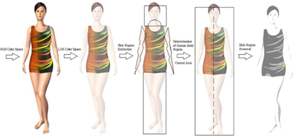
Figure 1: Determination of human body region and central axis as well as skin region removal.
2.1.3 Contour Estimation
Contour enhancement is performed on the determined human body region, and the Wavelet Transform is then applied to estimate body contours. Figure 2 presents the contour estimation results.
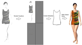
Figure 2: Contour estimation results
Figure 3: illustrates the algorithm for estimating human body contours from photographic images
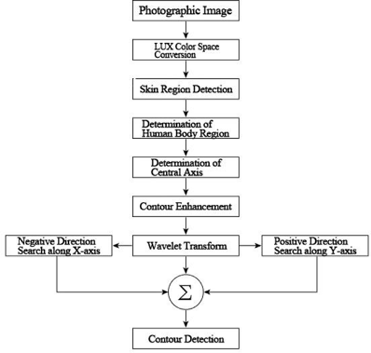
Figure 3: Human body contour detection algorithm
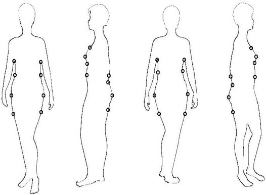
Figure 4: Integrated factors defined in this thesis
2.2 Optimized 3D Image Integration Based on Integrated Factors
2.2.1 Definition and Estimation of Integrated Factors
It is impossible to achieve optimized integration with the ideal body shape image for arbitrary human body images merely relying on visual observation and empirical judgment. Therefore, it is necessary to estimate ideal body shapes and reasonable integration factors established based on a database. The front, back, left and right photographic images of any female subject covered in this study correspond to the body contours stored in the database. For instance, in terms of front-view images, despite spatial positional differences of all human body components, partial correspondences exist in the overall skeletal structure. In this thesis, based on the integrated factors defined in Figure 4, the body shape of the measured subject is integrated with database body shapes, and spatial margin values are derived from the integration results.
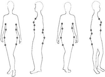
Figure 4: Integrated factors defined in this thesis
As shown in Figure 5, when searching along the arrow direction of the X-axis, a pixel point is judged as a feature integration point if the adjacent pixel values are white [RGB(255, 255, 255)] and black [RGB(0, 0, 0)]. The integrated factors for the left and right sides can also be inferred by searching left and right outward from the central axis.
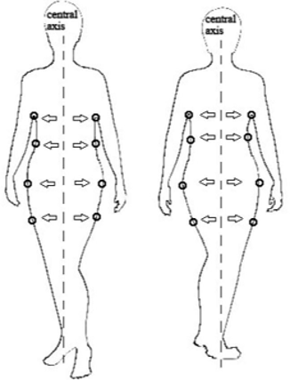
Figure 5: Estimation of integrated factors (Front and Back Views)
2.2.2 Optimized Image Integration Based on Integrated Factors
The core foundation of this study lies in integrating the input female body shape with the closest matching ideal body shape. This paper realizes optimized integration of human body contour curves via the defined integrated factors, adopting length ratio integration and angle integration as implementation methods.
① Integration Using Squared Error of Length Ratio
The length ratio integration method calculates the length ratios of corresponding integrated factors between the input human body and the database human body, then selects the body shape with the minimum ratio error. The integrated factors used for ratio error calculation are illustrated in Figure 6.
The ratio calculation is performed separately for the front and back views. For side-view images, one side profile is fixed, and calculations are carried out on the other side image to compute the ratio errors of all ideal body shapes stored in the database. Based on the calculated length ratio errors, the body contour curve of the measured input subject is matched to the most similar body contour curve among the database ideal body shapes, and the integration results are obtained accordingly.
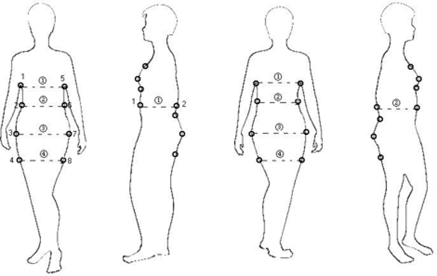
Figure 6: Integrated factors for ratio error calculation
② Integration Using Squared Error of Deflection Angle To improve integration efficiency, this paper organically combines the integration method based on the squared error of deflection angle with the integration method based on the squared error of length ratio to achieve superior integration efficiency.
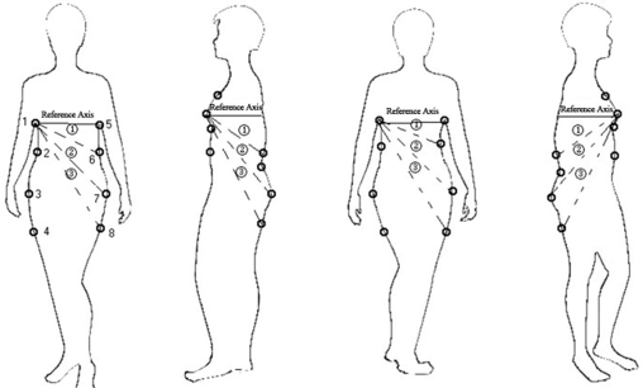
Figure 7: Integrated factors for calculating squared error of deflection angle
The deflection angles relative to the reference axis are calculated on each side-view image, followed by error computation. From the candidate sets obtained via the previously calculated squared error of length ratio and squared error of deflection angle, the ideal body shape with the minimum overall error rank is selected as the closest match to the measured body shape. The body contour integration method proposed in this paper leverages coordinate information (x-axis coordinate information, y-axis coordinate information) of integrated factors extracted from the four multi-view images to enhance integration efficiency.
The previous section has extracted body contour curves from images and selected the most similar matching contour curve among the ideal body curves stored in the database. The distance and area margins between these two curves are calculated through 3D image integration. Generalized margin information calculation is implemented to satisfy the design requirements of custom apparel for women with vulnerable pelvises.
3.1 Definition of Body Contour Curve Set
The specific cross-sections that directly affect apparel design include the chest cross-section, abdomen cross-section, waist cross-section, hip cross-section and lower hip cross-section. Spatial margin information for apparel design can be obtained by generating body contour curves and cross-section curves of these body parts.
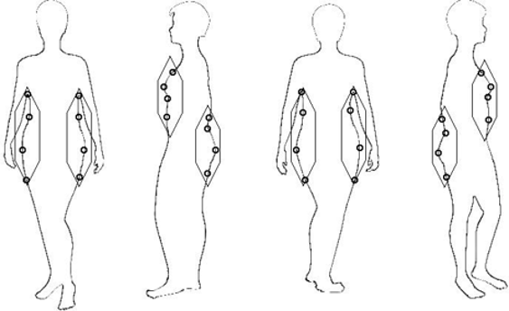
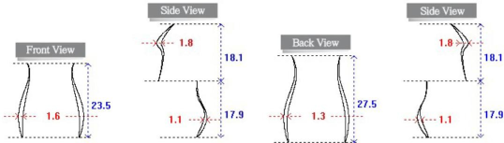
Figure 8: Body partial curve set for distance margin calculation
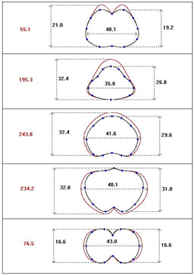
Figure 8 illustrates the partial body contour curve set used for distance margin calculation. The human body curve set for distance margin calculation consists of 8 curves and 32 integrated factors. Figure 9 presents the cross-section curves adopted for area margin calculation.
The cross-section curve set for area margin calculation consists of 5 cross-section curves and 10 integrated factors.
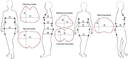
Figure 9: Area margin calculation cross-section curve set
3.2 Body Contour Curve Generation
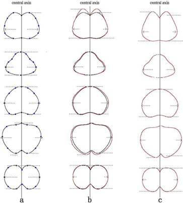
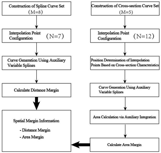
Thirty-two integrated factors estimated from the four photographic images of the measured subject and the ideal body contours stored in the database can be applied to generate body contour curves. Consider a planar auxiliary curve expressed as (P(t)=(x(t), y(t))), which is partitioned such that (0 The auxiliary variable spline separately constructs interpolating splines for (x(t)) and (y(t)) using two sets ({ti,xi}) and ({ti, yi}). The auxiliary curve obtained at this stage, (sk(t)=(s{k,x}, s{k,y})), is the body contour curve to be calculated in this paper. First, a generation scheme for eight partial body curves used in distance margin calculation is proposed. Figure 10 demonstrates the process of generating body contour curves for the measured subject and database body curves respectively via auxiliary variable splines. Figure 10: Based on generation results of human body curves via auxiliary variable splines Secondly, a generation scheme for five specific cross-section curves used to calculate area margin is proposed. A large number of interpolation points are required to generate these five specific cross-section curves (Figure 9), as cross-section curves feature sharp bends and relatively long lengths. This paper sets 12 interpolation points for generating the specific cross-section curves. Subplot a in Figure 11 marks the 12 interpolation points for constructing specific cross-section curves; subplot c presents the specific cross-section curves of the ideal body shape; subplot b illustrates the cross-section comparison between the measured subject’s curves and the ideal body curves. Figure 11: Twelve interpolation points for generating specific cross-section curves 3.3 Spatial Calculation of Distance and Area Subplot b of Figure 10 and Figure 11 allows visual identification of the spatial margin between the measured subject’s body contour and the ideal human body contour. Accurate estimation of spatial margin information serves as a method to approximate the measured subject’s body contour to the ideal body shape. The maximum distance margin on the closed curve shown in Figure 10 can be easily calculated. Specifically, measurements are taken from the upper and lower ends of the closed curve, and the margin corresponding to the maximum width between the two curves is defined as the distance margin. Figure 12 presents the quantitative evaluation of distance margins computed from human body curves generated via auxiliary variable splines. Figure 12: Distance margin calculated based on human body curves The distance margin information illustrated in the above figure provides the most practical approach to matching the body shape of the measured subject to the ideal body contour. Figure 13: Area margin In addition, from a practical perspective, it also indicates the specific implementation guidelines regarding the shape, size of attached materials and processing methods for custom underwear, so as to shape the body of measured subjects with weak pelvic support. The distance margin corresponds to mathematical analysis performed along the X-axis direction, while the area margin is analyzed along the Y-axis direction. To calculate the area margin, the area difference between the two cross-sections shown in subplot b of Figure 11 needs to be computed. In this study, the cross-sectional area of the measured subject’s body is always smaller than that of the ideal body shape. This is because the algorithm is designed to automatically select the fullest and closest matching contour curve from the 30 ideal body curves stored in the database for the measured subject’s body contour. This paper proposes a method for calculating the area enclosed by closed curves generated via auxiliary variable splines. The spatial margin information extraction algorithm proposed in this paper is as follows: Figure 14: Proposed spatial margin information extraction algorithm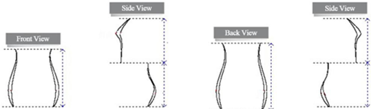
The computer system is Windows, and the algorithm language is C++.
Four photographic images of the measured subject (front view, two side views, rear view) are input. Integrated factors are estimated separately to achieve optimized matching with the ideal human body contours stored in the database.
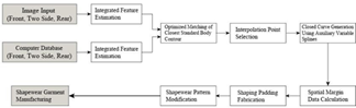
Figure 15: Computer simulation experiment structure diagram
The database adopts a fixed structural framework. To reduce computational complexity, the spatial coordinates of body feature parameters are precomputed and stored in tabular format. The system
searches for the optimal matching body contour curve from the database that best aligns with the contour extracted from the input photographic images.
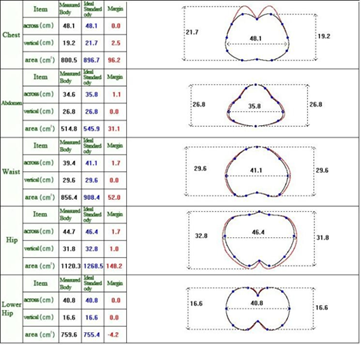
Figure 16: Calculation Results of Spatial Margin (Distance Margin, Area Margin)
Interpolation points are selected from the two contour curves. Spatial position data are adopted to generate body shape curves and designated cross-section curves. Curve generation is realized via auxiliary variable spline interpolation. The distance margin and area margin are calculated on the generated closed curves, and shaping fabrics are designed according to the calculated results.
Based on subjective and objective evaluations of shaping fabric fabrication and fitting trial effects, the calculation of excess margin data is performed to derive all potential ideal body shapes that the measured subject can approximate. The variation of excess margin data can be set to adjustable values as required. This adjustable margin variation technology underpins a custom apparel service solution that leverages 30 standard body contours built upon ergonomic principles and empirical apparel design data, enabling pelvic-support deficient body types to meet aesthetic criteria. Figure 16 presents measurement results of a female subject with an 80 cm bust circumference, 72 cm waist circumference, and 86 cm hip circumference.
This study mainly completes the following research works:
(1) This paper investigates the technical scheme proposed to realize 3D image matching of key contour points between contour images of women with weak pelvic support and standard ideal body contour images stored in the database. Front, rear, left and right photographic images of the measured subject are adopted to extract the key curve points forming the human body contour for analyzing body shape characteristics; comparative analysis is then carried out against contour points of the closest matching ideal standard body shape.
(2) Based on the 3D image matching results, the spatial margins (area margin and distance margin) required to approximate the ideal body contour are calculated. The computed values of area margin and distance margin serve as essential calculation indicators for custom garment production to adjust the measured subject’s body shape toward the ideal standard figure.
Dear Editorial Team, Clinical Medical Reviews and Reports. My experience with the journal was highly positive. The peer-review process was rigorous, constructive, and completed in a timely manner. The reviewers provided valuable comments that helped improve the quality and clarity of our manuscript. The editorial office was professional, responsive, and supportive throughout all stages of the publication process. Communication was clear and efficient, and any questions were addressed promptly. Overall, I found the journal to maintain high scientific standards and an excellent publication workflow. I would be pleased to consider submitting future work to this journal. Best wishes from, Elena Popa.

It was my pleasure to submit my testimonial concerning the Reviewer Board of our Scientific Journal “Brain and Neurological Disorders”. The Reviewers focused on some modifications and their contribution was helpful. The ladies of our Editorial Office were also supported my efforts. It was my honor to have such a co-operation and I am looking forward for more collaboration.

Dear Grace Pierce, Editorial Coordinator of Journal of Clinical Research and Reports, Thank you for the speedy and efficient peer review process. I appreciate the fact that your peer reviewers do not take months to respond like with some other journals. I would also like to thank the editorial office for responding quickly to my questions. It is an excellent journal. I plan to submit more manuscripts in the future. Best wishes from, Robert W. McGee

Dear Grace Pierce, Editorial Coordinator of Journal of Clinical Research and Reports, Working with you and your team on our recent publication in JCRR has been a truly wonderful and enjoyable experience. The responses were prompt, and the reviewers were patient, constructive, and highly professional. One reviewer in particular gave me the feeling that a professor was carefully reading and commenting on my coursework, which was deeply touching. The entire process was straightforward and hassle‑free, with no tedious online forms to complete. I highly recommend this journal. Best wishes from, DR Aibing Rao, Head of R&D

I Appreciate the Opportunity to Share my Experience with the Journal of Clinical Research and Reports. The peer review process was timely and constructive, and the feedback provided helped improve the quality of our manuscript. The editorial office was professional, responsive, and supportive throughout the process, ensuring smooth communication and efficient handling of the submission. Overall, it was a positive experience collaborating with your team.

Dear Mercy Grace, Editorial Coordinator of Obstetrics Gynecology and Reproductive Sciences, We would like to express our gratitude for your help at all stages of publishing and editing the article. The editors of the magazine answer all the necessary questions and help at every stage. We will definitely continue to cooperate and publish other works in the Obstetrics Gynecology and Reproductive Sciences! Best wishes from, Alla Konstantinovna Politova,
