
Case Report | DOI: https://doi.org/10.31579/2643-6612/005
Department of Oral Rehabilitation, Faculty of Dentistry, Pakistan.
*Corresponding Author: Mohammed M Alhajj, Department of Oral Rehabilitation, Faculty of Dentistry , Pakistan.
Citation: Mohammed M Alhajj, Removal of foreign objects from root canals- A Case Report, J Dentistry and Oral Maxillofacial Surgery. DOI: 10.31579/2643-6612/005
Copyright: © 2018 Mohammed M Alhajj, This is an open-access article distributed under the terms of the Creative Commons Attribution License, which permits unrestricted use, distribution, and reproduction in any medium, provided the original author and source are credited.
Received: 30 November -0001 | Accepted: 31 July 2018 | Published: 10 August 2018
Keywords: foreign bodies, root canal treatment, sewing needle, endodontics
Foreign bodies in root canals are rarely seen, and usually objects are accidently lodged and broken in root canals by the patients themselves. The occurrence of a foreign body, such as a metal screw, staple pin, sewing needle, pencil lead, bead or toothpick in the root canal system, makes the eradication of microorganisms impossible. Due to difficulties of eradicating microorganisms, foreign bodies may become sources of infection. These objects must be removed. This case report describes a rare clinical case in which a sewing needle, inserted into the root canal by the patient, was removed the orthograde approach with the aid of ultrasonic devices.
Introduction
The ideal outcome of root canal treatment is the eradication of microorganisms from the root canal system, or at least their significant reduction to levels compatible with periradicular tissue healing [1]. The occurrence of a foreign body, such as a metal screw, staple pin, sewing needle, pencil lead, bead or toothpick [2] in the root canal system, makes the eradication of microorganisms impossible. Cases of foreign bodies in the root canal system are rare in literature. The presence of foreign bodies may be asymptomatic and revealed accidentally or during the radiographic examination [3], but foreign bodies often cause pain and infection.
This case report describes a rare clinical case of an immature maxillary anterior tooth with sewing needles inside root canal and its non-surgical retreatment.
Case report
A 13-year-old female patient with a history of unsuccessful endodontic treatment of a maxillary central incisor was referred to the Department of Endodontics. According to the patient's history, the crown of both maxillary central incisors had been fractured four years earlier as a result of trauma. The patient had applied to a general dentist the same day. The general dentist had performed endodontic treatment with two visits. After the second appointment the patient had not gone to a dentist. Six months before applying to our department, the patient had accidentally broken a darning needle in her left maxillary central incisor.
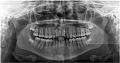
In the clinical examination the left maxillary central incisor was found to be grossly decayed with an open pulp chamber. Also clinical inspection revealed a metallic object in the entrance of the root canal. A periapical radiograph was taken and showed the presence of an unusual metallic radiopaque object with a round head at one end and a sharp end at the other (Figures 1 and 2). It was located close to the root apex of the left maxillary central incisor. The tooth was asymptomatic and the patient had no discomfort.
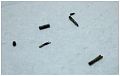
The tooth was isolated with a rubber dam and the access was modified. Attempts to bypass the metallic object failed and the decision was made to use an ultrasonic unit. The application of the ultrasonic tip directly against the exposed end of the metallic object resulted in the breakage of the metallic object. With a second application of the ultrasonic tip directly against the exposed end of the metallic object, breakage occurred again (Figure 2). After loosening and removing most of the metallic objects using the ultrasonic tip, the rest of the object was retrieval by irrigation, using 5.25% sodium hypochlorite (NaOCl).
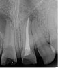
Three months later at the control visit, the tooth was fully asymptomatic and the patient had no discomfort. There was no pathology in the periapical area (Figure 3).
If the period between preparation and root canal filling is extended, as in this case, some unexpected complications may appear. If the root canal is open, the patient may try to clean the obstructing food substances from the canal with various objects that may break and get lodged in the pulp space.
Foreign objects in the root canal can be a source of infection [4] and should be removed. This may sometimes be very difficult because of the shape, size and position of the foreign body. The degree of difficulty depends also on the time that has elapsed since the foreign body was inserted in to the root canal.
Technical equipment should not be considered the only factor influencing the success or failure of removal procedures; the operator's experience and skill as well as the patient's anatomical factors are also important. The removal of foreign objects is sometimes difficult and the success rate has been reported to be 55% to 79% [5].
Many methods are described for removing broken instruments or objects within root canals, such as hand instrumentation, ultrasonic devices, the Masserann Kit, the Canal Finder System or even surgical methods [6]. Most recently, the use of ultrasonic tips has been found to be the most effective method for removing separated instruments from root canals without sacrificing a great deal of sound dentin [7].
If much time has elapsed with a metallic foreign body in the root canal there is the possibility of corrosion as in this case. Corroding metallic bodies can become more fragile and, as in this case, may break when retrieval is attempted.
Foreign bodies in the root canal system should be removed for successful endodontic treatment. These objects can clearly cause infection and pain. Non-surgical endodontic treatment should be tried first, but in some cases endodontic surgery may be required.
In the literature, removal of foreign objects from root canals has been widely discussed and various techniques have been suggested. Foreign bodies in the root canal system should be removed for successful endodontic treatment. These objects can clearly cause infection and pain. With appropriate diagnostic and treatment tools, as well as good patient cooperation, management of foreign object removal from root canals can be quite straightforward. Non-surgical endodontic treatment should be tried first, but in some cases endodontic surgery may be required.
Dear Editorial Team, Clinical Medical Reviews and Reports. My experience with the journal was highly positive. The peer-review process was rigorous, constructive, and completed in a timely manner. The reviewers provided valuable comments that helped improve the quality and clarity of our manuscript. The editorial office was professional, responsive, and supportive throughout all stages of the publication process. Communication was clear and efficient, and any questions were addressed promptly. Overall, I found the journal to maintain high scientific standards and an excellent publication workflow. I would be pleased to consider submitting future work to this journal. Best wishes from, Elena Popa.

It was my pleasure to submit my testimonial concerning the Reviewer Board of our Scientific Journal “Brain and Neurological Disorders”. The Reviewers focused on some modifications and their contribution was helpful. The ladies of our Editorial Office were also supported my efforts. It was my honor to have such a co-operation and I am looking forward for more collaboration.

Dear Grace Pierce, Editorial Coordinator of Journal of Clinical Research and Reports, Thank you for the speedy and efficient peer review process. I appreciate the fact that your peer reviewers do not take months to respond like with some other journals. I would also like to thank the editorial office for responding quickly to my questions. It is an excellent journal. I plan to submit more manuscripts in the future. Best wishes from, Robert W. McGee

Dear Grace Pierce, Editorial Coordinator of Journal of Clinical Research and Reports, Working with you and your team on our recent publication in JCRR has been a truly wonderful and enjoyable experience. The responses were prompt, and the reviewers were patient, constructive, and highly professional. One reviewer in particular gave me the feeling that a professor was carefully reading and commenting on my coursework, which was deeply touching. The entire process was straightforward and hassle‑free, with no tedious online forms to complete. I highly recommend this journal. Best wishes from, DR Aibing Rao, Head of R&D

I Appreciate the Opportunity to Share my Experience with the Journal of Clinical Research and Reports. The peer review process was timely and constructive, and the feedback provided helped improve the quality of our manuscript. The editorial office was professional, responsive, and supportive throughout the process, ensuring smooth communication and efficient handling of the submission. Overall, it was a positive experience collaborating with your team.

Dear Mercy Grace, Editorial Coordinator of Obstetrics Gynecology and Reproductive Sciences, We would like to express our gratitude for your help at all stages of publishing and editing the article. The editors of the magazine answer all the necessary questions and help at every stage. We will definitely continue to cooperate and publish other works in the Obstetrics Gynecology and Reproductive Sciences! Best wishes from, Alla Konstantinovna Politova,
